Practice Essentials
Cervical spine (C-spine) injuries are the most feared of all spinal injuries because of the potential for significant deleterious sequelae. Correlation is noted between the level of injury and morbidity/mortality (ie, the higher the level of the C-spine injury, the higher the morbidity and mortality). Craniocervical junction injuries are the deadliest.
As many as 10% of unconscious patients who present to the emergency department following a motor vehicle accident (MVA) have C-spine pathology. MVAs and falls are responsible for the bulk of C2 (axis) fractures. [1] The clinical manifestations range from absence of any symptoms to frank paralysis. Combined C1-C2 fractures may occur but are not common; when they do occur, they are associated with a higher frequency of neurologic deficits than isolated fractures of C1 or C2. [2]
This article focuses on the uniqueness of and the most common types of traumatic C2 fractures.
Anatomy
The unique features of C2 anatomy and its articulations make assessment of its pathology challenging. The odontoid process (dens) is a vertical projection that lies just posterior to the anterior arch of C1 (atlas), has ligamentous attachments to the skull base, and articulates with C1.
The C1-C2 (atlantoaxial) articulation is made up of three joints, including the central atlantoaxial joint and the paired lateral atlantoaxial joints. These joints allow rotation of C1 on C2.
The transverse ligament of the atlas stabilizes the central atlantoaxial joint, and, together with the odontoid process, acts as a restraint against horizontal displacement of the atlas. The dentate ligament attaches the apex of the odontoid process to the clivus and the paired alar ligaments, which originate from the transverse ligament and attach to the anterolateral rim of the foramen magnum. These ligaments provide rotational and translational stability.
The lateral atlantoaxial joints articulate at the superior articular facets of C2 and the inferior articular facets of C1. C2 also is composed of inferior facets, pedicles, transverse processes, and a spinal process.
Pathophysiology
Odontoid fractures
The incidence of odontoid fractures approaches 15% of all C-spine fractures. Usually, these fractures are secondary to MVAs or falls. [3] When an odontoid fracture is suspected, it is important to rule out concomitant associated C-spine injuries. For example, C1 anterior ring fractures are not an uncommon finding, and a prevertebral soft-tissue shadow of more than 10 mm on plain films is highly suggestive of such a fracture. Anderson and D'Alonzo classified odontoid fractures according to the anatomic location of the fracture (see the image below).
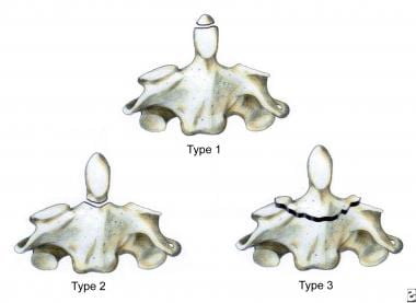 Three types of C2 odontoid fractures: type I is an oblique fracture through the upper part of the odontoid process; type II is a fracture occurring at the base of the odontoid as it attaches to the body of C2; type III occurs when the fracture line extends through the body of the axis.
A type I fracture (< 5% of cases) is an oblique fracture through the upper part of the odontoid process. This type of fracture occasionally is associated with gross instability due to traction forces applied to, and subsequent injury of, the apical and/or alar ligaments. This is an avulsion injury to the tip of the odontoid and usually is stable.
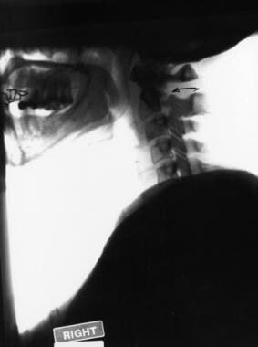
A type II fracture (>60% of cases) is a fracture occurring at the base of the odontoid as it attaches to the body of C2 (see the image below).
A type III fracture (30% of cases) occurs when the fracture line extends through the body of the axis. The fracture line can extend laterally into the superior articular facet for the atlas.
Another type of odontoid process fracture is a vertical fracture through the odontoid process and body of the axis (< 5% of cases). This type of fracture often is considered a variant of a traumatic spondylolisthesis of C2, which is discussed below.
The precise mechanism of odontoid fractures is unknown. However, the mechanism most likely includes a combination of flexion, extension, and rotation. In addition to pain and inability to move the neck actively, most patients complain of a sensation of instability, described as a feeling of the head being unstable on the spine. Patients may present by holding their head with their hands to prevent any motion. Clinical findings range from quadriplegia with respiratory center involvement to minimal upper-extremity motor and sensory deficits secondary to loss of one or more cervical nerve roots. Radiographic findings are based on the type of fracture.
C2 lateral-mass fractures
An isolated C2 lateral-mass fracture is extremely rare and usually is found serendipitously when evaluating for other C2 traumatic pathology. If a C2 lateral-mass fracture is found, other C-spine pathology must be sought. The mechanism of this fracture is axial compression with concomitant lateral bending.
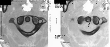
Signs and symptoms of concomitant C-spine pathology tend to dominate the clinical picture. The isolated fracture may present with high neck pain and a normal neurologic examination. Radiographic findings include impaction of the C2 component of the atlantoaxial articulation surface, asymmetry of C2 lateral-mass height, and lateral tilting of the arch of C1. Atlanto-occipital and atlantoaxial dissociation can be seen in the image below.
C2 extension teardrop fractures
C2 extension teardrop fractures are avulsion fractures with an intact anterior longitudinal ligament displacing and anteriorly rotating the anteroinferior vertebral-body fragment. The avulsed fragment's vertical dimension equals or exceeds its transverse dimension, and focal and minimal prevertebral soft-tissue swelling usually is present. This type of fracture tends to occur in elderly patients with osteoporotic bone. As the name implies, this type of fracture is the result of extension forces.
C2 extension teardrop fractures tend to be stable, and usually are not directly responsible for spinal cord injury. These fractures are extremely rare and differ in many aspects from the flexion teardrop fractures that are more common in the lower C-spine. Although lower C-spine teardrop fractures can result from extension forces, they usually result from severe flexion forces.
These fractures are unstable and are associated with anterior cord syndrome (quadriplegia; loss of pain, touch, and temperature sensations, but with retention of posterior column functions—proprioception and vibration), secondary to impingement of the cord by the hyperkyphotic vertebral segment or, more commonly, by retropulsion of C2 into the canal. C2 extension teardrop fractures are associated with traumatic spondylolisthesis of C2.
Traumatic spondylolisthesis of C2 (hangman fracture)
A great deal of confusion surrounds the exact pathology of this fracture. The modern origin of this confusion seems to be in a paper published by Schneider in 1964, wherein the term "hangman's fracture" was given to fractures sustained in MVAs that had radiographic similarities to fractures resulting from judicial hangings.
In 1964, Garber suggested the term traumatic spondylolisthesis for the more common injury pattern usually seen as a result of falls and MVAs. The injury pattern seen secondary to judicial hangings is a fracture-dislocation of C2. More precisely, it is a bilateral pedicle fracture of C2, along with distraction of C2 from C3 secondary to complete disruption of the disk and ligaments between C2 and C3. The injury pattern seen secondary to MVAs and falls also is a bilateral pedicle fracture, but it differs from the judicial hanging injury pattern with respect to varying degrees of disk and ligamentous disruption and secondary C2/C3 distraction.
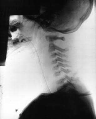
The mechanism of a hangman fracture (see the image below) is a combination of hyperextension and distraction. The mechanism of the traumatic spondylolisthesis fracture pattern primarily is hyperextension with varying degrees of axial compression and lateral flexion. Without the primary distraction force, there are varying degrees of disk and ligamentous disruption and secondary C2 displacement.
Traumatic spondylolisthesis is a common C-spine injury in fatal MVAs, and only occipitoatlantal dissociations occur more often. As many as one third of patients sustaining this injury have additional C-spine pathology, and as many as 10% have noncervical spinal pathology. Craniofacial and vertebral artery injuries are not uncommon findings. It is noteworthy that neurologic sequelae from spinal pathology are not as common as might be expected. This is because of the autodecompression of the spinal canal that takes place secondary to bilateral pedicle fractures.
Epidemiology
A study by Berkay et al used data from the National Electronic Injury Surveillance System (NEISS) database to determine the incidence rate (IR) of C2 fractures in the United States between 2002 and 2021. [4] The total number of patients presenting with these fractures over this period was estimated at 72,764. For the purposes of comparison, patients were split into three groups by age: children and young adults (0-64 y), older adults (65-79 y), and elderly individuals (≥80 y). The overall IR was 1.17 per 100,000 patient-years at risk (PYR). Elderly individuals had the highest IR, at 15.9/100,000.
Prognosis
Follow-up is critical for any patient who has sustained a C2 fracture. In addition to the clinical examination, repeat imaging studies are warranted. Generally, the various treatment modalities used for C2 fractures are quite successful. The available outcome data have focused on surgical treatment of type II odontoid fractures. A meta-analysis showed that single screw odontoid fixation using the anterior approach yielded better results than those found with transarticular fusion, multiple screws, or closed reduction with halo vest immobilization.
Parker et al retrospectively reviewed the records of 167 patients who underwent posterior cervical fusion with either C2 pedicle (PD) or C2 translaminar (TL) screw fixation and found that breach of PD screws occurred more frequently. [5] In total, 152 TL screws and 161 PD screws were placed. Thirty-one cases of axial cervical fusion (C1-C2 or C1-C3) were performed with TL (16 cases) or PD (15 cases) screw fixation, and 136 cases of subaxial cervical fusion (C2-caudal) were performed with TL (66 cases) or PD (70 cases) fixation.
In this study, 11 (7%) PD screws breached the pedicle (none requiring acute revision), and two (1.3%) TL screws breached the C2 lamina (one requiring acute revision). [5] At 1 year after operation, pseudarthrosis or screw pullout requiring reoperation occurred in four (6.1%) patients with TL screws but no patients with PD screws. The authors noted that the 1-year durability of TL screws may be inferior to PD screws for subaxial fusions but equally effective for axial fusions.
Omeis et al found that C2 fractures in the elderly (>70 y) can be treated surgically with both anterior and posterior approaches with acceptable morbidity and mortality and that the majority of patients can be mobilized early and return to their previous levels of independence. [3] Of 29 patients who had undergone surgical treatment for C2 fractures, 25 (86.2%) were able to return to their previous environment.
Chen et al retrospectively compared operative and nonoperative treatment of closed C2 fractures without spinal cord injury (SCI) in elderly patients. [6] The primary outcomes were 30-day mortality and complication rates, with length of hospital stay and long-term survival measured as secondary outcomes. The authors concluded that elderly patients faced high morbidity and mortality regardless of the type of treatment and that they should not be excluded from the surgical treatment option solely on the basis of their age.
Nizare et al performed a retrospective review of the management of 70 patients with various upper C-spine injuries. [7] They concluded, on the basis of the good radiologic and clinical improvement observed in these patients, that early management of cervical spine injuries could optimize the final outcome.
Stulik et al analyzed the management and prognosis of pediatric patients with unstable upper C-spine injuries. [8] The majority of these injuries (91.3%) were treated operatively, with the posterior approach used in about two thirds of the patients and the anterior in the remaining one third. The key finding was that even though these pediatric patients had a relatively high frequency of neurologic deficits, they had a better prognosis than adults did; this was especially the case for the youngest children with mild deficits. The authors recommended that therapy for these children be strictly individualized to maximize positive outcomes.
Honda et al evaluated the risk of early mortality in elderly patients with unstable isolated odontoid fractures (N = 891) who were treated with halo-vest immobilization (n = 463), anterior spinal fixation (n = 74), or posterior spinal fixation (n = 354). [9] The treatment type was not found to be significantly associated with in-hospital mortality; however, male sex and a Charlson comorbidity index of 3 or higher were found to be independent risk factors.
-
Odontoid type II fracture
-
Atlantooccipital and atlantoaxial dissociation
-
Hangman fracture
-
Three types of C2 odontoid fractures: type I is an oblique fracture through the upper part of the odontoid process; type II is a fracture occurring at the base of the odontoid as it attaches to the body of C2; type III occurs when the fracture line extends through the body of the axis.


