You are in: eMedicine Specialties > Orthopedic Surgery > KNEE Plica SyndromeArticle Last Updated: Aug 18, 2008AUTHOR AND EDITOR INFORMATIONAuthor: Tracy Lee Bigelow, DO, Fellow, Department of Orthopedic Surgery, University of Florida Tracy Lee Bigelow is a member of the following medical societies: American Academy of Orthopaedic Surgeons, American Osteopathic Association, and American Society for Surgery of the Hand Coauthor(s): Charles T Mehlman, DO, MPH, Director, Musculoskeletal Outcomes Research, Associate Professor, Division of Pediatric Orthopedic Surgery, Cincinnati Children's Hospital Medical Center Editors: Miguel A Schmitz, MD, Consulting Surgeon, Department of Orthopedics, Klamath Orthopedic and Sports Medicine Clinic; Francisco Talavera, PharmD, PhD, Senior Pharmacy Editor, eMedicine; Thomas M DeBerardino, MD, Director, John A Feagin, Jr, Sports Medicine Fellowship at West Point, Associate Professor of Orthopedic Surgery, Uniformed Services University of the Health Sciences and Keller Army Community Hospital; Dinesh Patel, MD, FACS, Associate Clinical Professor of Orthopedic Surgery, Harvard Medical School; Chief of Arthroscopic Surgery, Department of Orthopedic Surgery, Massachusetts General Hospital; Carlos J Lavernia, MD, FAAOS, Adjunct Clinical Professor, Department of Orthopedic Surgery, University of Miami School of Medicine; Medical Director, Orthopedic Institute at Mercy Hospital Author and Editor Disclosure Synonyms and related keywords: plica syndrome, medial synovial shelf, medial shelf, synovial chorda, medial pleat, Iino's band, Iino band, Aoki's ledge, Aoki ledge, medial intraarticular band, meniscus of the patella, mediopatellar pseudomeniscus, plica synovialis mediopatellaris, plica synovialis suprapatellaris, superomedial plica, medial suprapatellar plica, plica alaris elongata, ligamentum mucosum, plica synovialis patellaris, plica synovialis patellae, infrapatellar plica, infrapatellar fold, infrapatellar septum, knee pain INTRODUCTIONPlica syndrome of the knee is a constellation of signs and symptoms that occur secondary to injury or overuse. An otherwise normal structure, a plica can be a significant source of anterior knee pain. Once an inflammatory process is established, the normal plical tissue may hypertrophy into a truly pathologic structure (see Image 1). This article provides an overview of pertinent anatomy as well as diagnosis and treatment of plica syndrome of the knee. During embryonic development, the knee is divided initially by synovial membranes into 3 separate compartments. By the third or fourth month of fetal life, the membranes are resorbed, and the knee becomes a single chamber. If the membranes resorb incompletely, various degrees of septation may persist. These embryonic remnants are known as synovial plicae. Four types of synovial plicae of the knee have been described in the literature.1 The suprapatellar plica, or plica synovialis suprapatellaris, divides the suprapatellar pouch from the remainder of the knee. Rarely, this plica may initiate a suprapatellar bursitis or perhaps chondromalacia, and symptoms secondary to these conditions may be present.2 Anatomically, the suprapatellar plica can be complete or in the form of a porta, which only partially separates the compartments. It courses from the anterior femoral metaphysis or the posterior quadriceps tendon to the medial wall of the joint. The suprapatellar plica most commonly begins proximal to the superior pole of the patella but may begin anywhere. The mediopatellar plica is the most frequently cited cause of plica syndrome. It lies on the medial wall of the joint, originating suprapatellar, and courses obliquely down to insert on the infrapatellar fat pad. This plica, sometimes known as a shelf, lies in the coronal plane. The rare and poorly documented lateral synovial plica is a wider and thicker band than the medial plica. It is located along the lateral parapatellar synovium, inserting on the lateral patellar facet. The lateral plica has been argued to be derived from the parapatellar adipose synovial fringe rather than being a vestigial septum. The plica found to be the least symptomatic of all, the infrapatellar plica or ligamentum mucosum, is, ironically, the most commonly encountered plica. Some authors even claim this plica is never responsible for plica syndrome. This bell-shaped remnant originates in the intercondylar notch, widens as it sweeps through the anterior joint space, and attaches to the infrapatellar fat pad. This plica's ability to obscure portal entry sites or interfere with visualization during arthroscopy is touted as its only significance. For excellent patient education resources, visit eMedicine's Foot, Ankle, Knee, and Hip Center, Arthritis Center, and Bone Health Center. Also, see eMedicine's patient education article Knee Pain. History of the ProcedureBoth the normal and pathologic aspects of various synovial plicae readily became apparent to orthopedic surgeons with the advent of arthroscopy. Arthroscopy is a Japanese innovation that changed the face of orthopedics forever. Kenji Takagi (1888-1963) and Masaki Watanabe and others in Japan drove its early development, and early adopters in North America, such as Robert Jackson and Lanny Johnson, helped popularize this technology.3, 4 The word plica comes from the Latin word meaning fold. This term is simply descriptive in nature, as there is no empirical evidence that true folding of the synovial lining ever occurs. The medial parapatellar plica has been referred to by some as the Aoki ledge or Iino band. The plica tends to course from the medial region of the parapatellar synovial cavity down to the infrapatellar fat pad region. These eponymic associations bear further testimony to the Japanese surgeons who were the early pioneers of arthroscopy. Problem
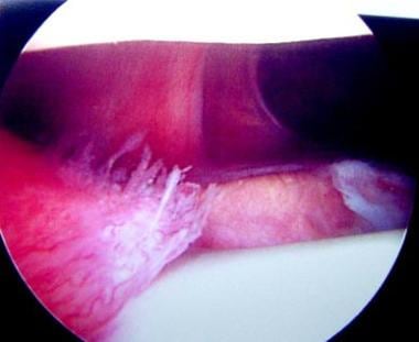
Synovial plicae are normal structures found in many knees. Under normal circumstances, they are not associated with any painful conditions. However, with the right combination of events they can become quite problematic. These events almost certainly include a somewhat exuberant plical shelf at baseline combined with an inciting event (either discrete macrotrauma or repeated microtrauma). Once a painful and, at times, hypertrophic inflammatory cycle has been established, an athlete may be faced with finding a way to tolerate the knee pain or give up the particular inciting sporting activity. FrequencyThe precise incidence of plica syndrome is a source of ongoing controversy. The incidence of symptomatic or pathologic plica versus asymptomatic plica is even more debatable. Some authors contend that distribution is regional. Distribution differences based on race or ethnicity may exist. The incidence of suprapatellar plica has been reported to be as low as 11%; however, one study reported a rate of 89% on autopsy. Medial plica has an 18-60% reported incidence rate. The incidence of medial plica in anatomic studies is similar to that in arthroscopic studies. The most commonly cited incidence rate in the literature is approximately 20-25%. Little literature exists on lateral plica. Most of this literature is Japanese, and few cases have been reported in English.5 Incidence of this kind of plica is less than 1%. Dupont et al found some type of inferior plica in 65% of their 200 dissected cadaveric knees. They stated it was the most frequently found plica. In their review of the literature, infrapatellar plica was most common in some studies but not in others.1 EtiologyThe etiology of symptomatic plica is unclear. Potential causes of inflammation include repetitive stress, a single blunt trauma, loose bodies, osteochondritis dissecans, meniscal tears, or other aggravating knee pathology. The most common symptomatic plica is medial plica; occasionally, suprapatellar plica may also be symptomatic.6, 7, 8, 9 A popular theory for the initiation of inflammation is that the plica is converted to a bowstring, which causes it to contact the medial femoral condyle. During flexion of the knee, the plica causes an abrasion to the condyle, resulting in symptoms. Others contend that a plica need not contact the femoral condyle to cause symptoms. One study found the onset of symptoms was usually delayed until adolescence. Possible explanations include a decrease in tissue elasticity with age, and a biomechanical change resulting from a growth spurt. PathophysiologyNot all synovial plicae are symptomatic. For those that are, the etiology may not always be apparent. Inflammation leads to edema, thickening, and decreased elasticity of the plica. The plica may develop irregular edges and may snap over the femoral condyle, leading to a secondary synovitis and chondromalacia. Loose areolar fatty tissue appears to become gristlelike, and when plicae are soft, wavy, and vascular with synovial-covered edges, they are not pathologic. Numerous studies describe pathologic plicae as thick, fibrotic, white, and inelastic. Histologically, fibrosis, hyalinization, and calcification are present. ClinicalThe spectrum and diversity of symptoms can make this syndrome difficult to pinpoint. Often, symptoms resemble or overlap with those of other pathology.10, 11 Reported symptoms include anterior or anteromedial knee pain; intermittent or episodic pain; clicking; high-pitched snapping; occasional giving way; locking (really pseudolocking) and catching; and aggravation of symptoms by activity, by stair climbing, or by prolonged standing, squatting, or sitting. Meniscal tears, patellar tendinitis, Osgood-Schlatter disease, Sinding-Larsen-Johansson disease, and patellar instability are the most commonly found concomitant conditions. On physical examination, the patient typically has tender points along the medial and inferior aspect of the patella (see Image 2); in some instances, a painful, hypertrophied membrane is palpable. The inferomedial quadrant (see Image 3) is the most consistently painful region. Occasionally, a medial apprehension test of the patella elicits a positive response, but careful evaluation reveals that it is due to direct tenderness to palpation in the region of the plica and is not true patellar instability.
In addition to the history and physical examination, a radiograph is recommended to exclude other causes of knee pain. As stated previously, plica syndrome is difficult to differentiate from other pathology and remains a diagnosis of exclusion. Other adjunctive diagnostic studies include contrast pneumoarthrography and double-contrast arthrography, yet arthroscopy remains the standard for definitive diagnosis.13 Most plicae are found incidentally during knee arthroscopy. Munzinger classified the mediopatellar plica into 4 types based on appearance, as follows14:
Other authors believe that to differentiate between symptomatic and asymptomatic plicae, the following criteria must be met upon arthroscopic examination: plica must appear, one must visualize impingement, and chondromalacia must be present in the areas of impingement. INDICATIONSThe most common symptomatic plica is medial parapatellar plica. Surgical intervention for plica syndrome should be considered when symptoms have persisted and efforts at nonoperative management have failed for a period typically approaching 6 months. Permanent modification of athletic activity is usually another option, but patients rarely find this option acceptable. Moreover, surgical treatment offers such predictable results that withholding treatment could be argued to be ethically questionable. RELEVANT ANATOMYThe anatomy of the various plicae about the knee is described in the Introduction. Key anatomic points relative to common plicae are reiterated here. The ligamentum mucosum is a synovial fold that extends from the region of the anterior cruciate ligament to the anterior synovial lining of the knee. Kim et al have classified ligamentum mucosum (ie, infrapatellar plica) into 5 groups, as follows15:
The lateral parapatellar plica extends from the lateral synovial lining distally toward the infrapatellar region. The medial parapatellar plica extends from the medial synovial lining distally to the infrapatellar fat pad (ie, Hoffa fat pad). In a clinical study of 400 knees in more than 350 patients, Kim and Choe found suprapatellar plicae in 87%, mediopatellar plicae in 72%, infrapatellar plicae in 86%, and lateral plicae in 1.3%.16 CONTRAINDICATIONSPatients with absolute contraindications for surgical treatment include those who are considered unfit for surgery from a medical standpoint. Active soft-tissue infection around the knee that precludes the use of standard arthroscopic portals is another contraindication. Relative contraindications include hypercoagulability syndromes that predispose the patient to thrombotic complications, as well as excessive risk for reflex sympathetic dystrophy (RSD; see Complications). WORKUPImaging Studies
Diagnostic Procedures
Histologic FindingsThe histology of a symptomatic plica is typically that of synovial tissue (type A macrophagelike cells and type B fibroblastlike cells) immersed in an inflammatory reaction.19 In other instances, the histology may show dense fibrotic tissue that only secondarily impinges upon articular surfaces to produce pain. Kasim and Fulkerson found "fibrosis, vascular proliferation, and small nerves with deceased myelin (neuromata)" on histologic analysis of specimens from their plica patients.20 StagingJee et al stage medial parapatellar plicae according to how far the plica extends into the region of the patellofemoral joint.18 Their system is as follows:
TREATMENTMedical therapyMedical treatment of plica syndrome has been driven largely by empirical evidence. A structured program of stretching and strengthening exercises often leads to some improvement. This may include short-arc quadriceps extension exercises (terminal approximately 20° of extension). These exercises are aimed at optimizing patellofemoral biodynamic relationships in an effort to control symptoms. A patellar knee sleeve worn during sporting activities (usually a neoprene-type brace) may also be a useful adjunct for many athletes. In addition, nonsteroidal anti-inflammatory medications are a time-tested and confirmed aid for many athletes with plica syndrome. Surgical therapySurgical therapy for plica syndrome is virtually always arthroscopic. The arthroscopic surgeon needs to exclude other potential intra-articular causes of knee pain and then address any pathologic plicae. Plica resection may be performed with arthroscopic hand instruments, a motorized soft-tissue resector, or certain commercially available electrothermal devices. Preoperative detailsThe preoperative phase of treatment involves optimizing the patient's knee strength and flexibility in an effort to streamline postoperative rehabilitation. Preoperative preparation of the patient also involves education and appropriate goal setting. For instance, the patient should understand that therapeutic exercises typically begin shortly after surgery (hours to days) and that a full return to sports can be realized soon thereafter (days to weeks). Patients who know this in advance tend to achieve these goals quite readily. Intraoperative detailsAfter arthroscopic evaluation establishes that no other intra-articular abnormalities need to be addressed, the plica can be resected. Using whatever tools work best in the surgeon's hands, the plica should be resected back to a point where it no longer impinges on articular structures. With beefy synovitic plicae that extend into the patellofemoral joint space (typically 50% or more) (see Image 5), this may require extensive debridement (see Image 6). With tough, fibrotic plicae that drape over the medial femoral condyle, this may involve little more than disruption of the tight band. At times, even a suprapatellar plica may lead to symptoms. Strover et al reported on an arthroscopic technique demonstrating the pathomechanics of such suprapatellar plicae.21 They recommended that the arthroscope should be inserted through a lateral suprapatellar portal. Proximal visualization is then optimized. In those patients in whom the suprapatellar plica is symptomatic, progressive flexion of the knee results in the plical tissue becoming taut. It also makes contact with the medial femoral condyle and even becomes entrapped between the quadriceps tendon and medial femoral condyle.21 Postoperative detailsPostoperatively, the patient is started on a structured course of therapeutic exercise that initially emphasizes reestablishment of active quadriceps control and firing. This progresses to regaining full range of motion and then full strength. The patient concludes therapeutic recovery by gradually performing more and more sport-specific exercises until a controlled reentry to the sport is achieved. Follow-upFollow-up care focuses on confirmation that symptoms have abated. True recurrence of the original plical pathology is quite rare and is more likely to represent either an incomplete resection or entirely new knee pathology. Continued use of a patellar stabilizing-type brace is preferred by many patients. COMPLICATIONSComplications of surgical treatment of plica syndrome are really complications associated with arthroscopic surgery of the knee. These include septic arthritis, neurapraxias or neuromas, and synovial fistulae. Reflex sympathetic dystrophy may also occur following such surgery. The rate of each of these complications is extremely small (<1% in most cases). Only patients with particular risk factors (eg, diabetes, steroid dependence, history of RSD) may be at a significantly higher risk. OUTCOME AND PROGNOSISThe outcome of surgical treatment for well-selected patients with plica syndrome is very good.22, 23, 24 A clinical trial conducted by Johnson et al in England demonstrated a success rate of more than 80%.25 In this same study, nearly 50% of patients in the control group experienced continued symptoms severe enough that they later returned for definitive arthroscopic resection of their plicae. In a predominantly adult population (average age 25 y, age range 11-56 y), Kasim and Fulkerson reported 88% moderate-to-substantial improvement at an average of more than 4 years following resection of localized segments of painful retinacula (ie, plicae) about the knee.20 FUTURE AND CONTROVERSIESSynovial plicae within the knee are clearly normal anatomic structures that can become potent pain generators.26 Therefore, little debate exists as to whether they may become pathologically involved.27 Future treatment of plica syndrome may be supplemented further by improved brace designs and pharmacological treatments. Earlier recognition of patients with the syndrome might facilitate syndrome resolution by such means, in contrast to patients who experience years of symptoms. MULTIMEDIA
REFERENCES
Article Last Updated: Aug 18, 2008 | ||||||||||||||||||||||||||||||||||||||||||