Continually Updated Clinical Reference
 
 
  All Sources     eMedicine     Medscape     Drug Reference     MEDLINE
 
eMedicine - Solitary Osteochondroma : Article by

Quick Find
Authors & Editors
Introduction
Indications
RELEVANT ANATOMY
Contraindications
Workup
Treatment
Complications
Outcome And Prognosis
Future And Controversies
Multimedia
References


Patient Education
Click here for patient education.


Author: Ian D Dickey, MD, FRCSC, Adjunct Professor, Department of Chemical and Biological Engineering, University of Maine; Consulting Staff, Adult Reconstruction, Orthopedic Oncology, Department of Orthopedics, Eastern Maine Medical Center

Ian D Dickey is a member of the following medical societies: American Academy of Orthopaedic Surgeons, British Columbia Medical Association, Canadian Medical Association, and Royal College of Physicians and Surgeons of Canada

Editors: Lynn A Crosby, MD, FACS, Chief of Shoulder Division, Professor, Department of Orthopedic Surgery, Wright State University School of Medicine; Francisco Talavera, PharmD, PhD, Senior Pharmacy Editor, eMedicine; Sean P Scully, MD, PhD, Professor, Department of Orthopedics, University of Miami; Dinesh Patel, MD, FACS, Associate Clinical Professor of Orthopedic Surgery, Harvard Medical School; Chief of Arthroscopic Surgery, Department of Orthopedic Surgery, Massachusetts General Hospital; Harris Gellman, MD, Consulting Surgeon, Broward Hand Center, Voluntary Clinical Professor of Orthopedic Surgery and Plastic Surgery, Departments of Orthopedic Surgery and Surgery, University of Miami School of Medicine

Author and Editor Disclosure

Synonyms and related keywords: solitary osteochondroma, osteocartilaginous exostosis, solitary osteocartilaginous exostosis, solitary OCE, bone tumor, bone lesion, bone cancer, chondrosarcoma

Osteochondroma is the most common benign bone tumor. The tumor is often diagnosed as an incidental finding. Osteochondromas account for approximately 35% of benign bone tumors and 9% of all bone tumors. Most are asymptomatic, but they can cause mechanical symptoms depending on their location and size.

As benign lesions, osteochondromas have no propensity for metastasis. In fewer than 1% of solitary osteochondromas, malignant degeneration of the cartilage cap into secondary chondrosarcoma has been described and is usually heralded by new onset of growth of the lesion, new onset of pain, or rapid growth of the lesion.1, 2, 3

Related Medscape topics:
Specialty Site Orthopaedics
Orthopaedics News

Related eMedicine topics:
Chondrosarcoma
Non-neoplastic Conditions Simulating Bone Tumors
Bone Metastases
Lymphoma, Bone

History of the Procedure

Historically and currently, most osteochondromas are incidental findings and are treated solely with observation. If they remain asymptomatic, they can be ignored. Lesions that create mechanical symptoms, become painful, begin to enlarge, or cause growth disturbance have historically been treated with surgical removal, and this remains the mainstay of treatment.

Problem

Osteochondroma is a benign, cartilaginous neoplasm that is found in any bone that undergoes enchondral bone formation in its development. The World Health Organization (WHO) defines osteochondroma as a cartilage-capped bony projection on the external surface of a bone. It is found most commonly around the knee and the proximal humerus; however, it can occur in any bone. The osteochondroma may have a stalk (see Images 1-2) and be defined as pedunculated, or it may have a broad base of attachment and be considered sessile in nature (see Image 3). Whether sessile or pedunculated, the medullary canal of the stalk and the bone are in continuity by definition.

Osteochondroma is a hamartoma, and patients most commonly present in the second decade of life. Osteochondromas grow until skeletal maturity; growth generally stops once the growth plates fuse.4 Slow growth from the cap may continue over time, as described by Virchow, but this usually stops by age 30 years.

Frequency

The actual frequency of osteochondromas is unknown because many are not diagnosed. Most are found in patients younger than 20 years (see Image 4). The male-to-female ratio is 3:1.

Osteochondromas can occur in any bone that undergoes enchondral bone formation, but they are most common around the knee.

Etiology

Although the exact etiology of these growths is not known, a peripheral portion of the physis is thought to herniate from the growth plate.5 This herniation may be idiopathic or may be the result of trauma or a perichondrial ring deficiency. Whatever the cause, the result is an abnormal extension of metaplastic cartilage that responds to the factors that stimulate the growth plate and thus results in exostosis growth.

This island of cartilage organizes into a structure similar to the epiphysis (see Workup, Histologic Findings, below). As this metaplastic cartilage is stimulated, enchondral bone formation occurs, developing a bony stalk. The histology of the cartilage cap reflects the classic, defined zones observed in the growth platenamely, a zone of proliferation, columniation, hypertrophy, calcification, and ossification.

This theory is thought to explain the classic finding of the osteochondroma associated with a growth plate and growing away from the physis while maintaining its medullary continuity. The theory is also thought to explain the clinical behavior of the exostosis growing only until skeletal maturity.

Genetic karyotyping has suggested that reproducible genetic abnormalities are associated with these benign growths and that they may actually represent a true neoplastic process, not a reactive one.6, 7 This research is in the early stages, and further investigation is necessary.8, 9, 10, 11

Related eMedicine topic:
Histology of Bone

Pathophysiology

Osteochondromas are located adjacent to growth plates and develop away from the growth plate with time because they are essentially isolated growth plates. They are affected by, and respond to, various growth factors and hormones in the same manner as epiphyseal growth plates; thus, growth of an osteochondroma should cease at skeletal maturity.

Clinical

Osteochondromas are the most common benign bone tumors. They represent 35% of all benign tumors and 9% of all bone tumors. Most are diagnosed in patients younger than 20 years. A marked predilection for males exists; the male-to-female ratio is 3:1.

Although they can be located almost anywhere in the skeleton, almost half of osteochondromas are found around the knee, in either the distal femur or the proximal tibia (see Image 4).

Osteochondromas are most commonly diagnosed incidentally on radiographs obtained for other reasons. The second most common presentation is a mass, which may or may not be associated with pain. Most of these lesions do not need to be treated, and asymptomatic lesions can be safely ignored. When painful, however, they need to be evaluated properly.

Pain is usually caused by a direct, mechanical, mass effect of the osteochondroma on the overlying soft tissue. This can result in an associated sac or bursitis over the exostosis. Irritation of surrounding tendons, muscles, or nerves can result in pain. Pain can also result from fracture of the stalk of the osteochondroma from direct trauma. The bony cap of the stalk may infarct or undergo ischemic necrosis.


Asymptomatic lesions require no treatment and can be monitored initially with radiographs and subsequently by clinical examination. Further investigation is indicated if the patient presents with a painful lesion or develops pain or an increase in size of a preexisting lesion. Such changes may represent either a new mechanical symptom or malignant degeneration. MRI is very useful for investigating these changes. The most common causes of pain are bursa formation, impingement, fracture of the stalk, and malignant degeneration.12, 13, 14

Excision is the treatment of choice for symptomatic lesions. As with all lesions of muscle and bone, the physician must be confident of the diagnosis and well versed in the care of tumors, should the lesion in fact be malignant. If the surgeon has any doubt about the diagnosis of the lesion or the management of a potential malignancy, patient referral is the most appropriate course of action.

In excising the lesion, it is important to avoid leaving any remnants of cartilage from the cap or any perichondrium, because this can allow recurrence. The reported rate of local recurrence is less than 2-5%.15 The risk of recurrence is thought by some to be higher in the skeletally immature; therefore, resection might best be delayed until skeletal maturity is reached. Great care must be exercised with lesions close to the physeal plate in the immature patient, because of the risk of growth plate arrest and subsequent deformity.


Osteochondromas can occur in many different locations in the body. Thus, a complete understanding of local anatomy is paramount to ensure that local structures are not harmed during surgical resection. Because these lesions arise from the metaphysis, particular care must be taken to avoid damage to the growth plate in the skeletally immature patient.


No frank contraindications to removal exist, but the surgeon should be aware that a large osteochondroma may in fact be a chondrosarcoma and should exercise appropriate caution. Removal by a surgeon who is not well versed in dealing with orthopedic malignancies may be a relative contraindication.


Imaging Studies

  • Plain radiography is the mainstay of imaging for osteochondroma. Good quality radiographs should be obtained in 2 perpendicular planes to characterize the lesion fully. Classic radiographic features include orientation of the lesion away from the physis and medullary continuity.16
  • In certain bones, such as the pelvis and the scapula, CT scanning is a useful adjunct to localize the lesion (see Images 1-2 and Image 5). CT localization can be useful when planning resection (see Images 6-7).17
  • MRI is needed only in cases in which malignancy is a concern or in which relevant soft-tissue anatomy needs to be delineated. MRI is the modality of choice to assess cartilage cap thickness (see Image 8). While not an absolute indication, cartilage cap thickness is related to malignancy. Thick cartilage caps (>4 cm) are suggestive of malignant degeneration, especially when they are associated with pain.
  • Bone scans, as a rule, are not useful in the workup of osteochondromas or for preoperative planning for resection.18

Histologic Findings

Grossly, the stalk is contiguous with the intramedullary marrow. By definition, the medullary canal of the affected bone and the canal of the tumor are connected. The stalk is made up of mature bone. The cartilage cap, which tops the lesion and can be quite thick in children, is replaced by enchondral bone formation in maturing patients (see Images 9-10).

On microscopic examination, the cartilage cap can exhibit varying amounts of cellularity. The cap has an overlying fibrous layer that contains mesenchymal cells, which are thought to be responsible for the lesion's growth.19 The cells in the cartilage are orientated vertically, as is found in a growth plate. In skeletally immature patients, the cells undergo enchondral bone formation (see Images 11-12). While no specific cartilage cap thickness is an absolute indicator of risk for malignancy, less than 4 cm generally is thought to be in the range of normal. Further, the cap should not thicken in persons older than 30 years.

Staging

Osteochondromas are benign lesions and can be staged under the Musculoskeletal Tumor Society (MSTS) staging for benign lesions, as follows:

  • Stage I - Inactive or static lesions
  • Stage II - Actively growing lesions
  • Stage III - Actively growing lesions that are locally destructive/aggressive

Most osteochondromas are stage I or II. However, significant deformity secondary to mass effect can occur in areas such as the radioulnar joint and tibiofibular joint. While classification is not perfect, such lesions could be considered stage III lesions (see Image 13). These cases likely represent a pressure erosive process, rather than a truly invasive process (as the staging for benign lesions is defined), a subtle but distinct biologic process.

In the rare case of malignant degeneration of the cartilage cap, the lesion is usually a low-grade chondrosarcoma that would be graded a low-grade extracompartmental lesion, MSTS stage IB.

Related eMedicine topic:
Cartilage Tumors, Low Grade


Medical therapy

No medical therapy currently exists for osteochondromas. The mainstay of nonoperative treatment is observation because most lesions are asymptomatic. Lesions found incidentally can be observed, and the patient can be reassured.

Surgical therapy

The treatment for symptomatic osteochondromas is resection. Care must be taken to ensure that none of the cartilage cap or perichondrium is left in the resection bed; otherwise, there may be a recurrence. Ideally, the line of resection should be through the base of the stalk; thus, the entire lesion is removed en bloc with its fibrous covering. Atypical or very large lesions should be investigated fully to exclude the remote possibility of malignancy. MRI is useful in assessing cartilage cap thickness.

In the skeletally immature patient, care must be taken to avoid damage to the growth plate during the exposure and resection of the lesion.

Preoperative details

Local anatomic constraints must be considered carefully so that the approach and resection do not damage nearby structures. CT scanning and MRI can be useful for lesions that arise from flat bones or that are located in difficult areas, such as lesions around the hip or scapula.

Intraoperative details

Once the osteochondroma is exposed, dissection is limited to the base of the lesion so that an osteotome can be used to shear off the base at the level of the host bone cortex. Care is required to ensure that the resection neither violates normal host cortex by straying too deep nor leaves residual lesion by staying too shallow. The overlying bursa should be left intact, and the loose adhesive tissue should be dissected away so that the lesion and the bursa are removed en bloc.

The resected surface of the host bone can be rasped smooth, and if needed, bone wax can be packed on the cut surface to stop bleeding.

Once the specimen is removed and pathologic confirmation is received, the wound should be irrigated well and a surgical drain can be placed if needed. The drain should exit in line with the wound.

Postoperative details

Most osteochondromas allow the patient to return to activity as tolerated. However, after resection of a large sessile lesion, restriction of activities should be considered because the stress riser created by the violation of the cortex may increase the risk of fracture.

Follow-up

The local recurrence rate after resection of osteochondroma is about 1.8%.20 Once the wound is healed, follow-up on an as-needed basis is reasonable if no associated bone deformity or potential growth-arrest concerns exist.


Complications after surgical resection of osteochondromas are rare. Considerations include physeal disturbance or growth arrest, fracture, recurrence, incorrect diagnosis, and hematoma formation.


For solitary osteochondromas, the outcome and prognosis after surgery are excellent, with excellent local control and a local recurrence rate of less than 2%.21, 22 The process is a benign one; thus, the prognosis is usually one of complete recovery. Poorer outcomes usually are related to the morbidity associated with the exposure required to remove the lesion or associated with secondary bone deformity, but the latter is usually observed in the multiple hereditary form of the disease.23, 24, 25, 26, 27, 28, 29


Genetic karyotyping suggests that reproducible genetic abnormalities are associated with these benign growths and that they may represent a true neoplastic process, not a reactive one.6, 7 Research is in the early stages, and further investigation is needed.8, 9, 10, 11 Biologic therapies for osteochondromas may be possible in the future.

Related Medscape topic:
Resource Center Biologic Therapies in Cancer


Media file 1:  Solitary osteochondroma. Anteroposterior radiograph of a pedunculated osteochondroma of the distal femur.
Click to see larger pictureClick to see detailView Full Size Image
Media type:  X-RAY

Media file 2:  Solitary osteochondroma. Lateral radiograph of a pedunculated osteochondroma of the distal femur. Orientation is away from the growth plate, and medullary continuity is clear.
Click to see larger pictureClick to see detailView Full Size Image
Media type:  X-RAY

Media file 3:  Solitary osteochondroma. Lateral radiograph of a sessile osteochondroma of the distal femur.
Click to see larger pictureClick to see detailView Full Size Image
Media type:  X-RAY

Media file 4:  
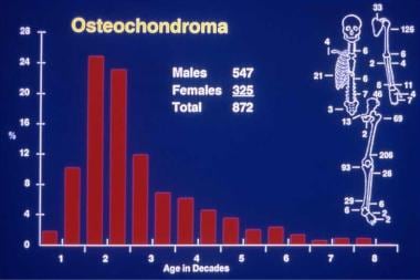
Solitary osteochondroma. Anatomic and age distribution of solitary osteochondromas.
Click to see larger pictureClick to see detailView Full Size Image
Media type:  Graph

Media file 5:  Solitary osteochondroma. CT scan of the pelvis depicting a massive solitary osteochondroma.
Click to see larger pictureClick to see detailView Full Size Image
Media type:  CT

Media file 6:  Solitary osteochondroma. Anteroposterior radiograph of sessile osteochondroma of the humerus.
Click to see larger pictureClick to see detailView Full Size Image
Media type:  X-RAY

Media file 7:  Solitary osteochondroma. CT scan of the same sessile osteochondroma of the humerus as in Image 6.
Click to see larger pictureClick to see detailView Full Size Image
Media type:  CT

Media file 8:  Solitary osteochondroma. MRI of sessile osteochondroma of the femur demonstrating the thickness of the cartilage cap.
Click to see larger pictureClick to see detailView Full Size Image
Media type:  MRI

Media file 9:  Solitary osteochondroma. Gross osteochondroma specimen at the time of resection. Bone stalk and overlying membrane on cartilage cap.
Click to see larger pictureClick to see detailView Full Size Image
Media type:  Photo

Media file 10:  Solitary osteochondroma. Cut surface of surgical osteochondroma specimen. Cartilage cap and underlying bone with medullary continuity.
Click to see larger pictureClick to see detailView Full Size Image
Media type:  Photo

Media file 11:  Solitary osteochondroma. Histology of cut osteochondroma specimen. Cartilage cap and orientation of enchondral bone formation.
Click to see larger pictureClick to see detailView Full Size Image
Media type:  Photo

Media file 12:  Solitary osteochondroma. High-power view of benign cartilage cells arranged in vertical growth plate pattern.
Click to see larger pictureClick to see detailView Full Size Image
Media type:  Photo

Media file 13:  Solitary osteochondroma. Radiograph demonstrating the deformation of the distal tibiofibular joint in a patient with a solitary osteochondroma.
Click to see larger pictureClick to see detailView Full Size Image
Media type:  X-RAY


  1. Garrison RC, Unni KK, McLeod RA, Pritchard DJ, Dahlin DC. Chondrosarcoma arising in osteochondroma. Cancer. May 1 1982;49(9):1890-7. [Medline].
  2. Staals EL, Bacchini P, Mercuri M, Bertoni F. Dedifferentiated chondrosarcomas arising in preexisting osteochondromas. J Bone Joint Surg Am. May 2007;89(5):987-93. [Medline].
  3. Akahane T, Shimizu T, Isobe K, Yoshimura Y, Kato H. Dedifferentiated chondrosarcoma arising in a solitary osteochondroma with leiomyosarcomatous component: a case report. Arch Orthop Trauma Surg. Jan 12 2008;[Medline].
  4. Nogier A, De Pinieux G, Hottya G, Anract P. Case reports: enlargement of a calcaneal osteochondroma after skeletal maturity. Clin Orthop Relat Res. Jun 2006;447:260-6. [Medline].
  5. D'Ambrosia R, Ferguson AB Jr. The formation of osteochondroma by epiphyseal cartilage transplantation. Clin Orthop. Nov-Dec 1968;61:103-15. [Medline].
  6. Hameetman L, Szuhai K, Yavas A, Knijnenburg J, van Duin M, van Dekken H, et al. The role of EXT1 in nonhereditary osteochondroma: identification of homozygous deletions. J Natl Cancer Inst. Mar 7 2007;99(5):396-406. [Medline].
  7. Hecht JT, Hogue D, Strong LC, et al. Hereditary multiple exostosis and chondrosarcoma: linkage to chromosome II and loss of heterozygosity for EXT-linked markers on chromosomes II and 8. Am J Hum Genet. May 1995;56(5):1125-31. [Medline].
  8. Coughlan B, Feliz A, Ishida T, Czerniak B, Dorfman HD. p53 expression and DNA ploidy of cartilage lesions. Hum Pathol. Jun 1995;26(6):620-4. [Medline].
  9. Legeai-Mallet L, Margaritte-Jeannin P, Lemdani M, et al. An extension of the admixture test for the study of genetic heterogeneity in hereditary multiple exostoses. Hum Genet. Mar 1997;99(3):298-302. [Medline].
  10. Legeai-Mallet L, Munnich A, Maroteaux P, et al. Incomplete penetrance and expressivity skewing in hereditary multiple exostoses. Clin Genet. Jul 1997;52(1):12-6. [Medline].
  11. Park KJ, Shin KH, Ku JL. Germline mutations in the EXT1 and EXT2 genes in Korean patients with hereditary multiple exostoses. J Hum Genet. 1999;44(4):230-4. [Medline].
  12. Karasick D, Schweitzer ME, Eschelman DJ. Symptomatic osteochondromas: imaging features. AJR Am J Roentgenol. Jun 1997;168(6):1507-12. [Medline].
  13. Murphey MD, Choi JJ, Kransdorf MJ, Flemming DJ, Gannon FH. Imaging of osteochondroma: variants and complications with radiologic-pathologic correlation. Radiographics. Sep-Oct 2000;20(5):1407-34. [Medline].
  14. El-Khoury GY, Bassett GS. Symptomatic bursa formation with osteochondromas. AJR Am J Roentgenol. Nov 1979;133(5):895-8. [Medline].
  15. Day FN, Ruggieri C, Britton C. Recurrent osteochondroma. J Foot Ankle Surg. Mar-Apr 1998;37(2):162-4; discussion 173. [Medline].
  16. Robbin MR, Murphey MD. Benign chondroid neoplasms of bone. Semin Musculoskelet Radiol. 2000;4(1):45-58. [Medline].
  17. Hudson TM, Springfield DS, Spanier SS, et al. Benign exostoses and exostotic chondrosarcomas: evaluation of cartilage thickness by CT. Radiology. Sep 1984;152(3):595-9. [Medline].
  18. Lange RH, Lange TA, Rao BK. Correlative radiographic, scintigraphic, and histological evaluation of exostoses. J Bone Joint Surg Am. Dec 1984;66(9):1454-9. [Medline].
  19. Robinson D, Hasharoni A, Oganesian A, et al. Role of FGF9 and FGF receptor 3 in osteochondroma formation. Orthopedics. Aug 2001;24(8):783-7. [Medline].
  20. Humbert ET, Mehlman C, Crawford AH. Two cases of osteochondroma recurrence after surgical resection. Am J Orthop. Jan 2001;30(1):62-4. [Medline].
  21. Chin KR, Kharrazi FD, Miller BS, Mankin HJ, Gebhardt MC. Osteochondromas of the distal aspect of the tibia or fibula. Natural history and treatment. J Bone Joint Surg Am. Sep 2000;82(9):1269-78. [Medline].
  22. Florez B, Mönckeberg J, Castillo G, Beguiristain J. Solitary osteochondroma long-term follow-up. J Pediatr Orthop B. Mar 2008;17(2):91-4. [Medline].
  23. Fogel GR, McElfresh EC, Peterson HA, Wicklund PT. Management of deformities of the forearm in multiple hereditary osteochondromas. J Bone Joint Surg Am. Jun 1984;66(5):670-80. [Medline].
  24. Govender S, Parbhoo AH. Osteochondroma with compression of the spinal cord. A report of two cases. J Bone Joint Surg Br. Jul 1999;81(4):667-9. [Medline].
  25. Peterson HA. Multiple hereditary osteochondromata. Clin Orthop. Feb 1989;(239):222-30. [Medline].
  26. Porter DE, Benson MK, Hosney GA. The hip in hereditary multiple exostoses. J Bone Joint Surg Br. Sep 2001;83(7):988-95. [Medline].
  27. Snearly WN, Peterson HA. Management of ankle deformities in multiple hereditary osteochondromata. J Pediatr Orthop. Jul-Aug 1989;9(4):427-32. [Medline].
  28. Wuyts W, Ramlakhan S, Van Hul W. Refinement of the multiple exostoses locus (EXT2) to a 3-cM interval on chromosome 11. Am J Hum Genet. Aug 1995;57(2):382-7. [Medline].
  29. Wicklund CL, Pauli RM, Johnston D. Natural history study of hereditary multiple exostoses. Am J Med Genet. Jan 2 1995;55(1):43-6. [Medline].
  30. Abd-el-Naby W, Hammond T, Court-Brown CM. Atypical osteochondroma of the distal femur. Orthopedics. Jul 2000;23(7):725-6. [Medline].
  31. Coenen L, Biltjes I. High radial nerve palsy caused by a humeral exostosis: a case report. J Hand Surg [Am]. Jul 1992;17(4):668-9. [Medline].
  32. Pitcher Jr, David J. PORT Notes, Lecture Syllabus. Islamorada, Fla: Florida Keyes Musculoskeletal Oncology Review; 2002:155.
  33. Porter DE, Emerton ME, Villanueva-Lopez F, Simpson AH. Clinical and radiographic analysis of osteochondromas and growth disturbance in hereditary multiple exostoses. J Pediatr Orthop. Mar-Apr 2000;20(2):246-50. [Medline].

Solitary Osteochondroma excerpt

Article Last Updated: Jul 9, 2008