Practice Essentials
Osteoarthritis is the most common type of joint disease, affecting more than 30 million individuals in the United States alone. [1] It is the leading cause of chronic disability in older adults, costing the US greater than $185 billion annually. [2] It can be thought of as primarily a degenerative disorder with inflammatory components arising from the biochemical breakdown of articular (hyaline) cartilage in the synovial joints. The current view holds that osteoarthritis involves not only the articular cartilage but the entire joint organ, including the subchondral bone and synovium. (See the image below.)
 Anteroposterior (AP) radiograph of the hip reveals severe superior migration of the femoral head (which reflects loss of articular cartilage), subchondral sclerosis, prominent osteophytes, and a large Egger cyst in the superior acetabulum. Mild flattening of the superior aspect of the femoral head is present.
Signs and symptoms
Symptoms of osteoarthritis include the following:
-
Deep, achy joint pain exacerbated by extensive use - The disease’s primary symptom
-
Reduced range of motion and crepitus - Frequently present
-
Stiffness during rest (gelling) - May develop, with morning joint stiffness usually lasting for less than 30 minutes
-
Swelling
Osteoarthritis of the hand
-
Distal interphalangeal (DIP) joints are most often affected
-
Proximal interphalangeal (PIP) joints and the carpometacarpal (cmc) joints at the base of the thumb are also typically involved
-
Heberden nodes, which represent palpable osteophytes in the DIP joints, are more characteristic in women than in men
-
Inflammatory changes are typically absent, less pronounced, or go unnoticed
See Presentation for more detail.
Diagnosis
Osteoarthritis is typically diagnosed on the basis of clinical and radiographic evidence. [3, 4, 5, 6, 7] There are no specific clinical laboratory abnormalities that are diagnostically associated with osteoarthritis.
Imaging studies
-
Computed tomography (CT) scanning - Rarely used in the diagnosis of primary osteoarthritis; however, it may be used in the diagnosis of malalignment of the patellofemoral joint or of the foot and ankle joints
-
Magnetic resonance imaging (MRI) - Not necessary in most patients with osteoarthritis unless additional pathology amenable to surgical repair is suspected; unlike radiography, MRI can directly visualize articular cartilage and other joint tissues (eg, meniscus, tendon, muscle, or effusion)
-
Ultrasonography - No role in the routine clinical assessment of patients with osteoarthritis; however, it is being investigated as a tool for monitoring cartilage degeneration, and it can be used for guided injections of joints not easily accessed without imaging
-
Bone scanning - May be helpful in the early diagnosis of osteoarthritis of the hand [9] ; bone scans also can help differentiate osteoarthritis from osteomyelitis, bone metastases, and metabolic bone diseases
Arthrocentesis
The presence of noninflammatory joint fluid helps distinguish osteoarthritis from other causes of joint pain. Other synovial fluid findings that aid in the differentiation of osteoarthritis from other conditions are negative Gram stains and cultures, as well as the absence of crystals when fluid is viewed under a polarized microscope.
See Workup for more detail.
Management
Nonpharmacologic interventions
The cornerstones of osteoarthritis therapy, nonpharmacologic interventions include the following:
-
Patient education
-
Heat and cold
-
Weight loss [10]
-
Exercise
-
Physical therapy
-
Muscle training (eg, quadriceps strengthening for knee OA)
-
Occupational therapy
-
Unloading in certain joints (eg, knee and hip)
Pharmacologic therapy
For hand osteoarthritis, the American College of Rheumatology (ACR)/Arthritis Foundation strongly recommend oral nonsteroidal anti-inflammatory drugs (NSAIDs)—although at doses as low as possible, taken for as a short time as possible—and conditionally recommend using one or more of the following:
-
Topical NSAIDs
-
Intra-articular glucocorticoid injections
-
Acetaminophen
-
Duloxetine
-
Tramadol
-
Chondroitin sulfate
For knee osteoarthritis, the ACR/Arthritis Foundation strongly recommend topical and oral NSAIDs and intra-articular corticosteroid injections, and conditionally recommend using one of the following:
-
Topical capsaicin
-
Acetaminophen
-
Duloxetine
-
Tramadol
For hip osteoarthritis, the ACR/Arthritis Foundation strongly recommend oral NSAIDs and intra-articular corticosteroid injections (which may be ultrasound guided) and conditionally recommend using 1 or more of the following for initial management:
-
Acetaminophen
-
Duloxetine
-
Tramadol
Surgery
A referral to an orthopedic surgeon may be necessary if the osteoarthritis fails to respond to a medical management plan. Surgical procedures for osteoarthritis include the following:
-
Arthroscopy
-
Osteotomy
-
Arthroplasty - Particularly with knee or hip osteoarthritis
-
Fusion
See Treatment and Medication for more detail.
Background
Osteoarthritis is the most common type of joint disease, affecting more than 30 million individuals in the United States alone (see Epidemiology). It represents a heterogeneous group of conditions resulting in common histopathologic and radiologic changes. It has been thought of as a degenerative disorder arising from biochemical breakdown of articular (hyaline) cartilage in the synovial joints. However, the current view holds that osteoarthritis involves not only the articular cartilage but also the entire joint organ, including the subchondral bone and synovium.
Osteoarthritis predominantly involves the weight-bearing joints, including the knees, hips, cervical and lumbosacral spine, and feet. Other commonly affected joints include the distal interphalangeal (DIP), proximal interphalangeal (PIP), and carpometacarpal (CMC) joints. This article primarily focuses on osteoarthritis of the hand, knee, and hip joints (see Pathophysiology). For more information on arthritis in other joints, see Glenohumeral Arthritis and Wrist Arthritis.
Although osteoarthritis was previously thought to be caused largely by excessive wear and tear, increasing evidence points to the contributions of abnormal mechanics and inflammation. In addition, some invasive procedures (eg, arthroscopic meniscectomy) can result in rapid progression to osteoarthritis in the knee joint. [11] Therefore, the term degenerative joint disease is no longer appropriate in referring to osteoarthritis. (See Pathophysiology.)
Historically, osteoarthritis has been divided into primary and secondary forms, though this division is somewhat artificial. Secondary osteoarthritis is conceptually easier to understand: It refers to disease of the synovial joints that results from some predisposing condition that has adversely altered the joint tissues (eg, trauma to articular cartilage or subchondral bone, infection, diseases such as rheumatoid arthritis, dysplasia); consequently, secondary osteoarthritis can occur in relatively younger individuals (see Etiology). [12]
The definition of primary osteoarthritis is more nebulous. Although this form of osteoarthritis is related to the aging process and typically occurs in older individuals, it is, in the broadest sense of the term, an idiopathic phenomenon, occurring in previously intact joints and having no apparent initiating factor.
Some clinicians limit the term primary osteoarthritis to the joints of the hands (specifically, the DIP and PIP joints and the joints at the base of the thumb). Others include the knees, hips, and spine (apophyseal articulations) as well.
As underlying causes of osteoarthritis are discovered, the term primary, or idiopathic, osteoarthritis may become obsolete. For instance, many investigators believe that most cases of primary osteoarthritis of the hip may, in fact, be due to subtle or even unrecognizable congenital or developmental defects.
No specific laboratory abnormalities are associated with osteoarthritis. Rather, it is typically diagnosed on the basis of clinical findings, with or without radiographic studies (see Workup).
The goals of osteoarthritis treatment include pain alleviation and improvement of functional status. Nonpharmacologic interventions are the cornerstones of osteoarthritis therapy and include the following:
-
Patient education
-
Application of heat and cold
-
Weight loss
-
Exercise
-
Physical therapy
-
Occupational therapy
-
Joint unloading, in certain joints (eg, knee and hip)
Intra-articular pharmacologic therapy includes corticosteroid injection and viscosupplementation, which may provide pain relief and have an anti-inflammatory effect on the affected joint. (See Treatment.) Oral pharmacologic therapy begins with acetaminophen for mild or moderate pain without apparent inflammation.
If the clinical response to acetaminophen is not satisfactory or the clinical presentation is inflammatory, consider nonsteroidal anti-inflammatory drugs (NSAIDs). (See Medication.) If all other modalities are ineffective and osteotomy is not viable, or if a patient cannot perform his or her daily activities despite maximal therapy, arthroplasty is indicated.
The high prevalence of osteoarthritis entails significant costs to society. Direct costs include clinician visits, medications, therapeutic modalities, and surgical intervention. Indirect costs include time lost from work.
Costs associated with osteoarthritis can be particularly significant for elderly persons, who face potential loss of social interactions and independence, leading to a need for help with activities of daily living. As populations of developed nations age over the coming decades, the need for better understanding of osteoarthritis and for improved therapeutic alternatives will continue to grow. (See Epidemiology.)
Anatomy
Joints can be classified in either functional or structural terms. A functional classification, based on movement, would categorize joints as follows:
-
Synarthroses (immovable)
-
Amphiarthroses (slightly moveable)
-
Diarthroses (freely moveable)
A structural classification would categorize joints as follows:
-
Synovial
-
Fibrous
-
Cartilaginous
Normal synovial joints allow a significant amount of motion along their extremely smooth articular surface. These joints are composed of the following:
-
Articular cartilage
-
Subchondral bone
-
Synovial membrane
-
Synovial fluid
-
Joint capsule
The normal articular surface of synovial joints consists of articular cartilage (composed of chondrocytes) surrounded by an extracellular matrix that includes various macromolecules, most importantly proteoglycans and collagen. The cartilage facilitates joint function and protects the underlying subchondral bone by distributing large loads, maintaining low contact stresses, and reducing friction at the joint.
Synovial fluid is formed through a serum ultrafiltration process by cells that form the synovial membrane (synoviocytes). Synovial cells also manufacture hyaluronic acid (HA, also known as hyaluronate), a glycosaminoglycan that is the major noncellular component of synovial fluid. Synovial fluid supplies nutrients to the avascular articular cartilage; it also provides the viscosity needed to absorb shock from slow movements, as well as the elasticity required to absorb shock from rapid movements.
Pathophysiology
Primary and secondary osteoarthritis are not separable on a pathologic basis, though bilateral symmetry is often seen in cases of primary osteoarthritis, particularly when the hands are affected. [3, 13] Traditionally, osteoarthritis was thought to affect primarily the articular cartilage of synovial joints; however, pathophysiologic changes are also known to occur in the synovial fluid, as well as in the underlying (subchondral) bone, the overlying joint capsule, and other joint tissues (see Workup). [14, 15, 16, 17]
Although osteoarthritis has been classified as a noninflammatory arthritis, increasing evidence has shown that inflammation occurs as cytokines and metalloproteinases are released into the joint. These agents are involved in the excessive matrix degradation that characterizes cartilage degeneration in osteoarthritis. [16] Therefore, it is no longer appropriate to use the term degenerative joint disease when referring to osteoarthritis.
Studies of interleukin-17 (IL-17), a proinflammatory cytokine, have found increased IL-17 levels in the synovium of osteoarthritis joints, as is seen in inflammatory arthritis (ie, rheumatoid arthritis). [18] Other inflammatory molecules that have been associated with osteoarthritis include 15‐hydroxyeicosatetraenoic acid, prostaglandin E2, IL‐1β, TNF alpha, IL‐1 receptor antagonist, and uric acid. [19]
In early osteoarthritis, swelling of the cartilage usually occurs, because of the increased synthesis of proteoglycans; this reflects an effort by the chondrocytes to repair cartilage damage. However, proinflammatory cytokines result in deterioration of chondrocyte metabolism. [20] This stage may last for years or decades and is characterized by hypertrophic repair of the articular cartilage.
As osteoarthritis progresses, however, the level of proteoglycans eventually drops very low, causing the cartilage to soften and lose elasticity and thereby further compromising joint surface integrity. Microscopically, flaking and fibrillations (vertical clefts) develop along the normally smooth articular cartilage on the surface of an osteoarthritic joint. Over time, the loss of cartilage results in loss of joint space.
In major weight-bearing joints of persons with osteoarthritis, a greater loss of joint space occurs at those areas experiencing the highest loads. This effect contrasts with that of inflammatory arthritides, in which uniform joint-space narrowing is the rule.
In the osteoarthritic knee, for example, the greatest loss of joint space is commonly seen in the medial femorotibial compartment, though the lateral femorotibial compartment and patellofemoral compartment may also be affected. Collapse of the medial or lateral compartments may result in varus or valgus deformities, respectively.
Krasnokutsky et al reported that the serum uric acid level can predict future joint space narrowing. In their study of 88 patients with medial knee osteoarthritis, over the course of 24 months, mean joint space narrowing of 0.31 mm occurred in patients with a serum uric acid level of less than 6.8 mg/dL (the solubility point for serum urate), compared with 0.90 mm in those with a serum uric acid level of 6.8 mg/dL or higher (P < 0.01). These authors suggest that serum uric acid levels may serve as a biomarker for progression of osteoarthritis. [19]
Erosion of the damaged cartilage in an osteoarthritic joint progresses until the underlying bone is exposed. Bone denuded of its protective cartilage continues to articulate with the opposing surface. Eventually, the increasing stresses exceed the biomechanical yield strength of the bone. The subchondral bone responds with vascular invasion and increased cellularity, becoming thickened and dense (a process known as eburnation) at areas of pressure. [21]
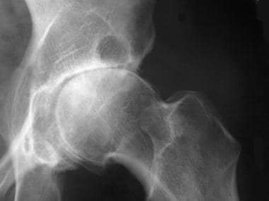
The traumatized subchondral bone may also undergo cystic degeneration, which is attributable either to osseous necrosis secondary to chronic impaction or to the intrusion of synovial fluid. Osteoarthritic cysts are also referred to as subchondral cysts, pseudocysts, or geodes (the preferred European term) and may range from 2 to 20 mm in diameter. Osteoarthritic cysts in the acetabulum (see the image below) are termed Egger cysts.
Anteroposterior (AP) radiograph of the hip reveals severe superior migration of the femoral head (which reflects loss of articular cartilage), subchondral sclerosis, prominent osteophytes, and a large Egger cyst in the superior acetabulum. Mild flattening of the superior aspect of the femoral head is present.
At areas along the articular margin, vascularization of subchondral marrow, osseous metaplasia of synovial connective tissue, and ossifying cartilaginous protrusions lead to irregular outgrowth of new bone (osteophytes). Fragmentation of these osteophytes or of the articular cartilage itself results in the presence of intra-articular loose bodies (joint mice).
Along with joint damage, osteoarthritis may also lead to pathophysiologic changes in associated ligaments and the neuromuscular apparatus. For example, lateral collateral ligament complex abnormalities are common in knee osteoarthritis.
Pain mechanisms in osteoarthritis
Pain, the main presenting symptom of osteoarthritis, is presumed to arise from a combination of mechanisms, including the following:
-
Osteophytic periosteal elevation
-
Vascular congestion of subchondral bone, leading to increased intraosseous pressure
-
Synovitis with activation of synovial membrane nociceptors
-
Fatigue in muscles that cross the joint
-
Overall joint contracture
-
Joint effusion and stretching of the joint capsule
-
Torn menisci
-
Inflammation of periarticular bursae
-
Periarticular muscle spasm
-
Psychological factors
-
Crepitus (a rough or crunchy sensation)
-
Central pain sensitization
When the spine is involved in osteoarthritis, especially the lumbar spine, the associated changes are very commonly seen from L3 through L5. Symptoms include pain, stiffness, and occasional radicular pain from spinal stenosis. Foraminal narrowing is caused by facet arthritic changes that result in compression of the nerve roots. Acquired spondylolisthesis is a common complication of arthritis of the lumbar spine.
Etiology
The daily stresses applied to the joints, especially the weight-bearing joints (eg, ankle, knee, and hip), play an important role in the development of osteoarthritis. Most investigators believe that degenerative alterations in osteoarthritis primarily begin in the articular cartilage, as a result of either excessive loading of a healthy joint or relatively normal loading of a previously disturbed joint. External forces accelerate the catabolic effects of the chondrocytes and further disrupt the cartilaginous matrix. [22, 23, 24]
Risk factors for osteoarthritis include the following [25, 26, 27, 28] :
-
Age
-
Trauma
-
Genetics (significant family history)
-
Reduced levels of sex hormones
-
Muscle weakness [32]
-
Repetitive use (ie, jobs requiring heavy labor and bending) [33]
-
Infection
-
Crystal deposition
-
Acromegaly
-
Previous inflammatory arthritis (eg, burnt-out rheumatoid arthritis)
-
Heritable metabolic causes (eg, alkaptonuria, hemochromatosis, Wilson disease)
-
Hemoglobinopathies (eg, sickle cell disease and thalassemia)
-
Neuropathic disorders leading to a Charcot joint (eg, syringomyelia, tabes dorsalis, and diabetes)
-
Underlying morphologic risk factors (eg, congenital hip dislocation and slipped femoral capital epiphysis)
-
Disorders of bone (eg, Paget disease and avascular necrosis)
-
Previous surgical procedures (eg, meniscectomy)
-
Diabetes mellitus [34]
Advancing age
With advancing age come reductions in cartilage volume, proteoglycan content, cartilage vascularization, and cartilage perfusion. These changes may result in certain characteristic radiologic features, including a narrowed joint space and marginal osteophytes. However, biochemical and pathophysiologic findings support the notion that age alone is an insufficient cause of osteoarthritis.
Senescent cells (SnCs) accumulate in many tissues with age and contribute to age-related pathologies. A study in mice by Jeon et al found that SnCs accumulated in the articular cartilage and synovium after anterior cruciate ligament transection, and selective elimination of SnCs attenuated the development of post-traumatic osteoarthritis, reduced pain, and increased cartilage development. In addition, selective removal of SnCs from in vitro cultures of chondrocytes isolated from patients with osteoarthritis undergoing total knee replacement resulted in decreased expression of senescent and inflammatory markers and increased expression of cartilage tissue extracellular matrix proteins. [35]
Obesity
Obesity increases the mechanical stress in a weight-bearing joint. It has been strongly linked to osteoarthritis of the knees and, to a lesser extent, of the hips. A study that evaluated the associations between body mass index (BMI) over 14 years and knee pain at year 15 in 594 women found that a higher BMI at year 1 and a significant increase in BMI over 15 years were predictors of bilateral knee pain at year 15. [31] The association between BMI increase and knee pain was independent of radiographic changes.
In addition to its mechanical effects, obesity may be an inflammatory risk factor for osteoarthritis. Obesity is associated with increased levels (both systemic and intra-articular) of adipokines (cytokines derived from adipose tissue), which may promote chronic, low-grade inflammation in joints. [36]
Other causes
Trauma or surgery (including surgical repair of traumatic injury) involving the articular cartilage, ligaments, or menisci can lead to abnormal biomechanics in the joints and accelerate osteoarthritis. In individuals who have sustained significant joint injuries, the risk of post-traumatic osteoarthritis ranges from about 20% to more than 50%. [37]
Insults to the joints may occur even in the absence of obvious trauma. Microtrauma may also cause damage, especially in individuals whose occupation or lifestyle involves frequent squatting, stair-climbing, or kneeling.
Muscle dysfunction compromises the body’s neuromuscular protective mechanisms, leading to increased joint motion and ultimately resulting in osteoarthritis. This effect underscores the need for continued muscle toning exercises as a means of preventing muscle dysfunction.
Valgus malalignment at the knee has been shown to increase the incidence and risk of radiographic progression of knee osteoarthritis involving the lateral compartment. [38]
Genetics
A hereditary component, particularly in osteoarthritis presentations involving multiple joints, has long been recognized. [39, 40, 41] Several genes have been directly associated with osteoarthritis, [42] and many more have been determined to be associated with contributing factors, such as excessive inflammation and obesity.
Osteoarthritis susceptibility genes (eg, ADAM12, CLIP, COL11A2, IL10, MMP3) have also been found to have differential methylation. Jefferies et al reports that hypomethylation of FURIN, which encodes a proprotein convertase, processes several ADAMTS molecules involved in osteoarthritic collagen degradation. Differential methylation among osteoarthritis susceptibility genes has been proposed as an alternative method for disruption of normal gene activity.
Additionally, Jefferies et al found evidence for hypermethylation and reduced expression of the type XI collagen gene COL11A2. Mutations involving COL11A2 have been associated with severe and early-onset osteoarthritis. Analysis by this goup has identified pathways enriched with "differentially methylated genes" that are effectors and upstream regulators seen in osteoarthritis linked with TGFB1 and ERG. [43]
Genes in the BMP (bone morphogenetic protein) and WNT (wingless-type) signaling cascades have been implicated in osteoarthritis. Two genes in particular, GDF5 (growth and differentiation factor 5) and FRZB (frizzled related protein), have been identified in the articular cartilage in animal studies and share a strong correlation with osteoarthritis. [44, 45, 46, 47]
Genome-wide association studies (GWAS) have identified an association between osteoarthritis of large joints and the MCF2L gene. This gene is key in neurotrophin-mediated regulation of peripheral nervous system cell motility. [48]
Genetic factors are also important in certain heritable developmental defects and skeletal anomalies that can cause congenital misalignment of joints. These may result in damage to cartilage and the structure of the joint.
Currently, clinical genetic testing is not offered to patients who have osteoarthritis unless they also have other anomalies that could be associated with a genetic condition. In the future, testing may allow individualization of therapeutics.
Epidemiology
United States and international statistics
Osteoarthritis affects more than 32 million individuals in the United States, though statistical figures are influenced by how the condition is defined—that is, by self-report, by radiographic or symptomatic criteria, or by a combination of these. [1, 49] On the basis of the radiographic criteria for osteoarthritis, more 50% of adults older than 65 years are affected by the disease.
Internationally, osteoarthritis is the most common articular disease. Estimates of its frequency vary across different populations but worldwide, more than 500 million people may be affected. [50] An analysis of Global Burden of Disease data found that the age-standardized incidence rate of osteoarthritis increased by approximately 9% from 1990 to 2017; the crude incidence rate rose 102% over that 28-year period, with the increase driven by the aging of the global population. [51]
Age-related demographics
Primary osteoarthritis is a common disorder of the elderly, and patients may present asymptomatic. Approximately 80-90% of individuals older than 65 years have evidence of radiographic primary osteoarthritis. [52]
Symptoms typically do not become noticeable until after the age of 50 years. The prevalence of the disease increases dramatically among persons older than 50 years, likely because of age-related alterations in collagen and proteoglycans that decrease the tensile strength of the joint cartilage and because of a diminished nutrient supply to the cartilage. [52]
Sex-related demographics
In individuals older than 55 years, the prevalence of osteoarthritis is higher among women than among men. [52] Women are especially susceptible to osteoarthritis in the DIP joints of the fingers. Women also have osteoarthritis of the knee joints more frequently than men do, with a female-to-male incidence ratio of 1.7:1. Women are also more prone to erosive osteoarthritis, with a female-to-male ratio of about 12:1.
Race-related demographics
Interethnic differences in the prevalence of osteoarthritis have been noted. [53] The disorder is more prevalent in Native Americans than in the general population. Disease of the hip is seen less frequently in Chinese patients from Hong Kong than in age-matched white populations. However, symptomatic knee osteoarthritis is extremely common in China. [54]
In persons older than 65 years, osteoarthritis is more common in Whites than in Blacks. Knee osteoarthritis appears to be more common in Black women than in other groups. Jordan et al found that in comparison with Whites, African-American men and women had a slightly higher prevalence of radiographic and symptomatic knee osteoarthritis. [55]
Prognosis
The prognosis in patients with osteoarthritis depends on the joints involved and on the severity of the condition. No proven disease-modifying or structure-modifying drugs for osteoarthritis are currently known; consequently, pharmacologic treatment is directed at symptom relief.
A systematic review found the following clinical features to be associated with more rapid progression of knee osteoarthritis [56] :
-
Older age
-
Higher BMI
-
Varus deformity
-
Multiple involved joints
Patients with osteoarthritis who have undergone joint replacement have a good prognosis, with success rates for hip and knee arthroplasty generally exceeding 90%. However, a joint prosthesis may have to be revised 10-15 years after its placement, depending on the patient’s activity level. Younger and more active patients are more likely to require revisions, whereas the majority of older patients will not. (See Treatment.)
Patient Education
Educate patients on the natural history of and management options for osteoarthritis, emphasizing the benefits of exercise and weight loss. Explain the differences between osteoarthritis and more rapidly progressive arthritides, such as rheumatoid arthritis.
Several Arthritis Foundation studies have demonstrated that education in osteoarthritis benefits the patient. Through education, patients can learn and implement strategies for reducing pain and improving joint function. Emphasize the need for physician follow-up visits.
For patient education information, see the Osteoarthritis Health Center.
-
Anteroposterior (AP) radiograph of the hip reveals severe superior migration of the femoral head (which reflects loss of articular cartilage), subchondral sclerosis, prominent osteophytes, and a large Egger cyst in the superior acetabulum. Mild flattening of the superior aspect of the femoral head is present.
-
Osteoarthritis of the elbow is not commonly seen; however, it can occur with a history of previous trauma.
-
Osteoarthritis of the elbow is not commonly seen; however, it can occur with a history of previous trauma.
-
Osteoarthritis of the elbow is not commonly seen; however, it can occur with a history of previous trauma.
-
This radiograph demonstrates osteoarthritis of the right hip, including the finding of sclerosis at the superior aspect of the acetabulum. Frequently, osteoarthritis at the hip is a bilateral finding, but it may occur unilaterally in an individual who has a previous history of hip trauma that was confined to that one side.
-
Arthroscopic view of a torn meniscus before (top) and after (bottom) removal of loose meniscal fragments.
-
Arthroscopic view of an arthritic knee.
-
Arthroscopic view of a knee after the removal of loose fragments of articular and meniscal cartilage.
-
Arthroscopic view of the removal of cartilaginous loose body.
-
Anteroposterior radiograph shows knee replacement in 1 knee and arthritis in the other, with medial joint-space narrowing and subchondral sclerosis.
-
Anteroposterior radiograph of the pelvis and hips shows an arthritic hip not treated surgically and a total hip replacement.
-
Anteroposterior radiograph obtained after knee replacement.
-
Lateral radiograph obtained after knee replacement (same patient as in the above image).
-
Oblique projection demonstrates gradual narrowing and sclerosis of the facet joints as one progresses down the lumbar spine.
-
Standing radiograph of the knee reveals narrowing of the medial and lateral femorotibial compartments with marginal osteophytes.
-
H&E stain, high power magnification showing clusters of chondrocytes (center) that is a proliferation known as "chondrocyte cloning". Image courtesy of Francis Gannon, MD.
-
H&E stain, medium power magnification showing numerous vertical slits (upper left) in the articular surface cartilage known as "fibrillation" with increase in eosinophilia of the remaining cartilage due to loss of proteoglycans. Image courtesy of Francis Gannon, MD.
-
Gross photograph of external (top) and cut surface (bottom) showing areas of complete loss of articular cartilage with peripheral osteophytes formation. Image courtesy of Francis Gannon, MD.