Background
Human fingernails, located on the dorsal aspect of the terminal 40% of the distal phalanx of each finger, are dynamic and physiologically important organs of the hand. Trauma to the fingertip and nail unit remains the most common of all hand injuries. The fingertip is one of the last anatomic structures to be pulled away when closing doors or using saws, machines, chains, or hammers. The long finger is most commonly involved, followed by the ring, index, little finger, and thumb. [1]
The distal phalanx provides bony support for the nail bed and is fractured in 50% of fingertip injuries. Loss of nail-bed integrity can produce a permanent and significantly dysfunctional deformity of the fingernail. Therefore, the fingernail unit must be appreciated, and primary anatomic repair of nail-bed injuries must be provided whenever possible. [2, 3, 4, 5, 6, 7]
The nail bed lies protected between the nail plate and the distal phalanx. Blunt or sharp trauma to the nail compresses the nail bed and can result in lacerations and more complex crush injuries. Avulsion injuries are common and usually signify a greater level of trauma [8, 9, 10] (see the image below).
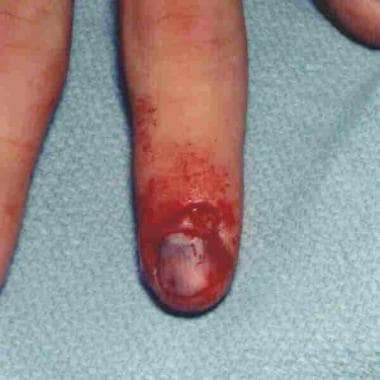 Significant nailbed injuries can occur from nail root avulsions.
See 15 Fingernail Abnormalities: Nail the Diagnosis, a Critical Images slideshow, to help identify conditions associated with various nail abnormalities.
Regrowth of the nail plate after an injury is influenced by the integrity of the nail bed and the nail folds. A smooth nail bed is essential for regrowth of the normal-appearing nail. Inadequate or unrepaired nail-bed lacerations may form granulation tissue or scars, which result in an area of nonadherence or other nail-plate deformities. Damage to the germinal matrix can result in absent or poor nail formation, a split-nail deformity, [11] or synechial adhesions of the nail fold to the nail bed.
A comprehensive awareness of fingernail anatomy, the dynamics of nail regeneration and adhesive properties, and the sequelae of inadequate initial management sets the stage for the appropriate treatment of nail-bed injuries.
Nail Anatomy and Physiology
The fingernail enhances the ability of the hand to manipulate and grasp fine objects. Loss of the nail results in a measurable decrease of fingertip sensitivity. [12, 13, 14] The two-point discrimination of the fingertip is enhanced because of the counterforce or buttress effect of the nail plate on the distal finger pad. The rigid nail also protects the vulnerable distal fingertip and distal phalanx from trauma.
The entire fingernail unit, or onyx, consists of the following [3, 13, 15, 16, 17, 18] :
-
Nail plate
-
Proximal nail fold (eponychium)
-
Lateral nail fold (perionychium)
-
Distal nail fold (hyponychium)
-
Germinal and sterile matrices
The nail plate is a multilayered stacked sheet of cornified cells derived from anucleate onychocytes that arise from the germinal matrix epithelium of the nail bed. [4, 19] The eponychial fold covers the softer and less cornified proximal nail plate. The cells of this proximal plate are not completely anucleate and project a white color because of the nuclear parakeratosis, which occurs only at the level of the germinal matrix. [13, 19, 20]
The lunula, a white semicircle just distal to the eponychial fold, represents the parakeratotic proximal nail plate where the germinal matrix extends just beyond the nail fold. [21] The sterile matrix epithelium does not undergo parakeratosis; therefore, the nail plate takes on the color corresponding to the underlying well-vascularized nail bed.
The epithelia of the germinal matrix, sterile matrix, and eponychial fold contribute to the production of the nail plate through three modes of keratinization, as follows:
-
The sterile matrix epithelium produces a semirigid keratin through a process known as onycholemmal keratinization; this semirigid keratin, sometimes referred to as the horny solehorn, increases the overall thickness of the nail and also acts as a superglue adhesive for the nail plate to maintain its adherence to the nail bed [13]
-
The external sheen of the healthy nail plate is a product of epidermoid keratinization from the dorsal roof of the eponychial fold; the cuticle, hyponychium, and lateral nail folds also contribute, in a minor degree, to the surface epidermoid keratinization of the nail plate [13]
The hyponychium represents the junction of the terminal nail bed with the glabrous skin of the fingertip. The nail plate becomes nonadherent at this level and extends for a variable distance over the tip of the finger. Keratin, polymorphonuclear cells, lymphocytes, and debris build up along the undersurface of the distal nail at this transition from nail bed to fingertip skin. [23]
Therefore, the hyponychium serves as a mechanical and important immunologic protective barrier at the distal end of the nail bed. The importance of the hyponychium cannot be overstated, considering the variety of potentially contaminated areas with which the fingertip comes in contact during activities of normal daily living.
The nail bed is an extremely vascular, longitudinally ridged structure. The most superficial layer of the nail bed is derived from the germinal matrix and glides as a unit with the nail plate as it grows distally. [5, 24] The remarkable adherence of the nail plate to the nail bed is the result of the adhesive properties of the solehorn layer and of the parallel ridging on the undersurface of the nail plate that interdigitates with the ridges of the underlying nail bed, which is intimately attached to the periosteum of the distal phalanx. [13, 20]
The nail bed is intimately anchored directly to the periosteum of the tuft of the distal phalanx, and retaining ligaments attach it to the bone at the proximal nail fold and distal nail groove, respectively. [25] The nail bed consists of fibroconnective tissue and is devoid of adnexa. It receives its vascularity from the ulnar and radial digital arteries. Branches of these vessels course around the distal phalanx and penetrate fibrous septae to form a proximal and distal anastomotic arcade within the basal layer of the nail bed. [26, 27]
Multiple arteriovenous anastomosis and myoneural glomus units are present within the nail bed. [28] Capillaries are abundant in the germinal matrix. An arborizing arcade of vessels in the distal nail bed communicates with a similar arcade from the palmar pulp vessels. Two large venous channels emerge from the lateral aspect of the nail bed and proceed dorsally to converge with the dorsal venous drainage system of the finger just distal to the distal interphalangeal joint.
The lymphatic vessels follow the same route and have a similar network. The nerve supply to the nail bed is from the radial and ulnar digital nerves, which parallel the course of the arterial supply to the nail bed.
Nail growth
The nail plate is approximately 0.5 mm thick in females and 0.6 mm thick in males, and its thickness tends to increase with age.
The nail grows at a rate of approximately 1.8-4.5 mm/month or 0.1 mm/day [29] ; thus, the average nail can regrow completely in 6-9 months. A number of factors can affect the rate of nail growth. [6, 29, 30, 31, 32, 33, 34, 35, 36] Periods of stress or illness can detrimentally inhibit nail growth, whereas biting or short trimming of the nail can accentuate growth.
The hardness of the nail is dependent on the onychocyte bands, matrix proteins, and the hydration level of the nail. [37] The water content of fingernails ranges from 10% to 30%. Brittle nail plates have lower water contents.
Nail-plate regeneration
Hand surgeons who are acquainted with the anatomy and physiology of the nail and its adnexa understand the predictable pattern of nail-plate regeneration after injury. [38, 39] A traumatic nail-plate avulsion exposes the underlying nail bed, rendering it susceptible to the harsh environmental conditions. Blood and plasma exudate create a scab over this exposed nail bed. The surface epithelium and keratinaceous solehorn invariably remain adherent to the avulsed nail plate.
The lateral nail folds and hyponychium provide the reparative epidermis that migrates under the scab to cover the nail bed. [5, 38] This new layer is hyperplastic and somewhat hyperkeratotic but provides protection to the bed. The vast neural and vascular supply to the nail bed makes this freshly exposed area somewhat hypersensitive after initial avulsion of the nail plate. A new nail plate begins to regenerate within the next 2-3 weeks. This delay produces a proximal thickening or rolling front of the advancing nail plate.
Replacement of the reparative epidermis with bed epithelium immediately precedes, or is synchronous with, the progression of the new nail plate. Both the epithelium and the nail plate are products of the germinal matrix. [4, 19, 26]
The nail plate conforms to the constraints of the lateral nail folds, the eponychial fold, and the contours of the nail bed on the distal phalanx. These structures are important to the ultimate shape of the nail plate. [40, 41] The proximal lateral nail migrates to the level of the hyponychium and, at this point, is no longer adherent to the nail bed.
Acute Management of Nail Injuries
Traumatic nail deformities
Appropriately managing the nail-bed wound at the time of the initial injury is easier than reconstructing secondary nail deformities. The importance of anatomic alignment of the nail bed following injury cannot be overstated.
A poorly coapted nail-bed laceration that heals with significant scar tissue cannot maintain the adherence of the regenerating nail. [42] The result is a loss of continuity between the nail plate and the nail bed at the level of the scarring. The nail continues to grow away from the nail bed at this level and can become problematic as it catches on clothing and other objects. The fingertip is traumatized repeatedly as a consequence. [43, 44]
Nail-bed injuries should be treated with the respect that all hand injuries demand. An appropriate patient history and physical examination should be performed. Knowledge of the mechanism of injury helps the surgeon be aware of other potential injuries to the finger or hand. Radiographs are obtained because distal phalangeal fractures are extremely common findings in patients with fingertip injuries. Most of these injuries can be treated in the emergency department.
The patient is placed in the supine position, and the entire hand is prepared with a sterilizing solution. The hand is draped in an appropriate fashion, so as to allow easy manipulation of the entire hand while maintaining sterility. Digital block anesthesia is used, and the finger is exsanguinated with a Penrose drain, much as an Esmarch bandage is used for other upper-extremity cases. Loupe magnification is required to appropriately visualize the nail bed and anatomically repair lacerations.
The nail plate or remnant of nail is removed with careful dissection using a Freer periosteal elevator or curved sharp-point scissors. The instrument is placed between the nail plate and the nail bed and gently swept from side to side to release the plate from the nail bed and lateral nail folds. The eponychial fold is elevated off the nail plate in a similar fashion.
The undersurface of the removed nail plate is cleaned of any solehorn layer and debris. The nail plate, when present, is placed in a sterile saline solution and is replaced into the nail fold at the end of the procedure. At this point, the exact nature and extent of the injury to the nail bed can be visualized and appropriately managed surgically.
Subungual hematoma
A subungual hematoma is the result of an injury to the sterile or germinal matrix, causing bleeding beneath the nail plate and its subsequent separation from the nail bed. The hematoma places pressure of the nail bed and causes a marked throbbing after injury. [45]
Some degree of nail-bed injury is necessary to cause subungual bleeding. The salient concern is determining when the degree of injury warrants removal of the nail plate and repair of the nail matrix. The authors have advocated removing the nail plate and examining the nail matrix when 50% or more of the visible nail is undermined by hematoma. Other authors of a small series have demonstrated a low incidence of late nail deformity when contained subungual hematomas were treated by observation alone.
If the hematoma has been decompressed by disruption of the hyponychium, perionychium, or eponychium, removal of the nail plate and repair of the matrix are warranted. The choice of method of repair must be individualized. Exploration and primary nail-bed repair should yield an excellent result but may delay the return of normal activity.
The results of a properly treated acute nail-bed wound are much better and more cost-effective than the results of a secondary reconstruction. Subungual hematomas involving less than 50% of the visible nail are often treated conservatively with simple hand elevation.
Intense pain or throbbing warrants evacuation of the hematoma. A small opening in the nail plate over the hematoma allows the blood that is trapped under pressure to be released. The finger is prepared with a sterile solution. The hole in the nail plate is made with a hand-held disposable cautery device, a heated paper clip, or a No. 11 scalpel blade. Once the instrument penetrates the nail plate, the hematoma ejects from the hole. Care must be taken to not project the instrument into the extremely sensitive nail bed.
The wound may continue to ooze for some time. A light dressing is applied for 24-45 hours, and then the wound may be left open. Residual subungual hematomas migrate distally over 4-9 weeks as the nail grows.
Soyuncu and Bektas reported a case in which nail-bed injury was detected by means of ultrasonography. [46] Others have suggested that point-of-care ultrasonography shows promise as a means of detecting nail-bed injury in patients who have sustained distal finger trauma. [47]
Simple and stellate laceration
These two types of injuries are categorized together because they are treated in essentially the same way. Simple and stellate lacerations of the nail bed usually result from a localized blow to the fingernail. The nail bed is lacerated or crushed between the nail plate and the distal phalanx. [48] A relatively localized force striking the nail causes a linear compression of the nail bed and a relatively straight laceration. A stellate laceration is caused by compression of the nail bed between the nail and the bone, resulting in an exploding type of injury. [1]
The nail bed must be irrigated thoroughly, and devitalized tissue must be debrided carefully. The irregular edges should be approximated as accurately as possible with minimal trimming to prevent tension on the repair. A nail bed repaired under tension promotes scarring and poor adhesion of the nail plate as it regenerates.
To achieve accurate coaptation, the nail bed may have to be undermined approximately 1-2 mm on each side of the wound edges. The 7-0 chromic on a microspatula, RR, GS-9 needle (Ethicon, Inc, Sommerville, NJ) is minimally traumatic to the nail bed while still providing appropriate strength for the repair.
Repair of a stellate laceration can be more time-consuming because of the diffuse nature of the laceration; however, because no loss of nail-bed substance occurs, meticulous attention to detail yields excellent results both functionally and cosmetically.
Once the nail bed has been repaired, the previously removed nail plate is used as a compressive dressing and as a method to hold open the eponychial fold. A hole is drilled or burned through the plate in an area not over the laceration to allow plasma and blood to drain when the tourniquet is released. The nail is replaced into the eponychial fold, and a distal 5-0 nylon suture is used to secure the replaced plate to the distal fingertip skin.
Replacement of the nail plate serves a number of functions, including the following:
-
Helping mold the edges of the nail-bed laceration
-
Helping hold distal phalangeal fractures in a reduced position
-
Maintaining the integrity of the eponychial fold
-
Protecting the nail-bed repair
-
Making the fingertip more comfortable for the patient while the new nail regenerates
Occasionally, the nail plate has been avulsed and lost at the scene of the accident. A 0.02 sheet of silicone or nonadherent petroleum jelly gauze can be placed over the nail bed and into the eponychial fold instead of the original nail plate. Failure to follow the appropriate principles of nail-bed repair often results in greater bed scarring and, ultimately, in loss of nail-plate adherence, longitudinal splitting of the nail, or nail-contour irregularities.
Lacerations that involve the germinal matrix of the nail bed should be repaired in a manner similar to that described above. Access to the germinal matrix is achieved through incisions made at right angles at the junction of the lateral perionychium with the proximal eponychium. Incisions made at angles other than 90° in this area can lead to consequential synechial adhesions of the eponychium to the nail bed or to eponychial fold deformities secondary to scar contracture.
The nail plate or silicone sheet is placed in the eponychial fold to prevent synechial adhesions between the germinal matrix and the roof of the eponychial fold. A 6-0 monofilament suture is used to close lacerations or to access incisions in the eponychial fold. Lacerations involving the germinal matrix of the dorsal roof and ventral floor must be repaired separately and with great care. The knots of the sutures should be placed in the nail fold; they should not be placed within the substance of the nail bed, because this may promote scarring or poor coaptation.
The entire fingertip is dressed with nonadherent gauze (eg, Adaptic, Xeroform), antibiotic ointment, and a bulky wrap. A finger cap splint is used to protect the distal end of the finger. The finger is examined for seroma, hematoma, and infection in 5-7 days, and the nylon suture in the nail plate is removed. The old nail plate often remains adherent for 1-3 months before being displaced by the new regenerating nail.
Two other methods for the repair of fingernail lacerations and disruptions appear to have good results. In a prospective, randomized, controlled trial comparing the liquid skin adhesive 2-octylcyanoacrylate with suture repair for nail-bed injuries, [49, 50] Strauss et al found that nail-bed repair with the adhesive not only provided functional and cosmetic results comparable to those of suture repair but also was markedly quicker to perform (about 10 minutes for 2-octylcyanoacrylate vs nearly 28 minutes for suture repair). [49] A literature review by Edwards et al suggested that a medical adhesive is as effective as suturing for nail bed injuries in children. [51]
Patankar repaired 70 disruptive fingernail injuries with modified tension band sutures in 66 patients. [52] The injuries were cleaned, the hematoma evacuated, and anatomic replacement of the nail plate (or a substitute) performed, but with a modified dorsal tension band suture rather than formal nail-bed repair. Kirschner-wire (K-wire) fixation of the distal phalanx was used only in cases of displaced distal phalangeal fractures, complete absence of the nail plate, and lacerations extending to the distal pulp. In all cases, uncomplicated reformation of the normal nail plate resulted.
Crush injuries
Crush injuries have a poorer prognosis than simple or stellate lacerations do. [1] A wider and more forceful area of trauma to the nail plate causes these nail-bed injuries. The nail bed is in multiple fragments; some fragments may still be attached to the undersurface of the nail plate. The nail plate should be carefully explored for these fragments.
The avulsed nail bed can be removed from the nail plate and replaced as free grafts. With severe crush injuries, ensure that all the fragments of the nail bed are approximated as accurately as possible to minimize scarring. Larger severely crushed areas of the nail bed may not be salvageable and are best treated with a split sterile matrix graft. A tension-free anatomic repair of the nail bed should be performed to optimize the final result.
Associated distal phalangeal fractures are common with crush injuries. The larger displaced fragments of the distal phalanx can be held in appropriate reduction by a longitudinal 0.028- to 0.035-in. K-wire. Smaller tuft fractures do not require pin fixation, and replacement of the nail plate following the repair may suffice. The nail plate acts as an external splint because of its contoured shape and proximity to the periosteum. The fingertip, including the distal phalangeal joint, should be splinted for 4-6 weeks.
Avulsion injuries
Avulsion injuries may result in partial or total loss of the nail bed, exposing the dorsal distal phalanx. Such trauma represents the most serious nail-bed injuries, with a notably higher incidence of long-term nail deformities. Fragments of the avulsed nail bed often maintain their adherence to the undersurface of the nail plate. These fragments must be carefully retrieved and sutured to their original position with 7-0 chromic sutures. [9, 10]
Occasionally, the proximal nail bed, including the germinal matrix, is avulsed, leaving the distal end still adherent. The nail bed must be returned under the eponychial fold and maintained in place with sutures. Occasionally, complete loss of a segment of nail bed occurs. Composite avulsions of the nail bed and eponychium should be sutured back in place because reconstruction of the eponychium as a secondary procedure is often difficult.
Avulsed traumatic defects in the sterile matrix nail bed should be immediately managed for the best result. [53, 54] Split-thickness sterile matrix grafts can be harvested from uninjured areas of the involved fingers if enough of the donor site is available to accommodate the defect. [55, 56, 57]
Great-toe donor nail-bed grafts may be warranted for larger defects. The toe must be prepared and draped in a similar fashion as the hand. Defects in the germinal matrix must be treated with full-thickness grafts from a toe. Harvesting a portion of the toe germinal matrix renders that portion of the donor toe unable to regenerate a nail plate. Therefore, obliteration of the remaining toe germinal matrix is often recommended to eliminate toenail remnants.
A germinal matrix defect requires a full-thickness graft to support regeneration of the nail plate, whereas a sterile matrix defect requires only a split-thickness graft to function. The graft can be placed directly on the exposed cortex of the distal phalanx, sutured to the surrounding nail bed, and appropriately dressed.
Harvesting a split nail-bed graft is performed with a No. 15 scalpel blade. An 0.008-in. graft can be elevated with the aid of loupe magnification and a jeweler's forceps. The feathered edge of the scalpel blade should be visualized through the graft while it is being elevated.
Taking too thick a graft may result in a donor-site deformity. The curve of the nail bed is followed with gentle sweeping movements of the scalpel blade while elevating the split nail-bed graft. Taking a slightly larger graft and trimming this to fit the fingernail defect is better than not harvesting enough and attempting to close the recipient site under tension.
Reconstruction of a nail-bed defect with a split sterile matrix graft optimizes nail adherence and minimizes later deformity. Other options, such as a split-thickness skin graft or a dermal graft, do not allow adherence of the regenerating nail plate. These tissues should be used only if no other donor sites are available.
Bipedicled matrix (germinal or sterile) or transposition matrix flaps have been described; however, these may result in secondary donor- and recipient-site deformities. These flaps require undermining of the matrix and a counter incision laterally to allow transposition of the tissue to close the defect. The donor area is closed either primarily or with a skin graft. [58] Shepard obtained good results in patients with germinal matrix defects with bipedicled germinal matrix flaps. [59]
A study by Ogunro et al suggested that a nail-bed graft might not be necessary for avulsion injuries of nail beds, regardless of the extent of the nail-bed loss. [60] The authors evaluated 12 cases of severe avulsion injuries of the nail bed that were allowed to heal naturally. The residual nail beds were covered with a nail splint for about 6 weeks or until the nail bed was seen to be fully regenerated. The authors noted that the nail bed regenerated spontaneously, with "normal nail growth identical to the contralateral uninjured nail."
Composite grafts
The role of composite tissue grafts warrants special attention. [21] Segments of the distal nail bed and the distal glabrous skin of the fingertip are often avulsed in continuity. The avulsed tissue contains two unique anatomic structures that are difficult to reconstruct: the hyponychium and the nail bed. Therefore, preserving this composite tissue is prudent, if possible. Tip amputations distal to the lunula can be replaced as a composite graft in children younger than 10 years.
The amputated part is gently trimmed of devitalized or contaminated tissue while the nail bed, glabrous skin, fat, and distal tuft are preserved. The nail bed is aligned anatomically and sutured with 7-0 chromic sutures, and the skin is closed with nonabsorbable sutures, which are left long and used for a tie-over bolster dressing of wet cotton.
Treat this composite graft with complete upper limb immobilization after surgery to improve the chances of graft survival. Children do not tolerate the immobilization well for 7-10 days; therefore, use a long-arm cast with the elbow flexed more than 90°, encompassing a bulky hand dressing; this is termed the "out of sight, out of mind" technique. The parents should be informed that the composite tip graft either survives or dies with this technique.
Convert to a full-thickness skin graft to treat the composite fingertip amputation in adults and in children older than 10 years. The tissue is defatted to the level of the dermis, and the nail bed is preserved. The distal phalanx is shortened just below the remaining pulp soft tissue, and the full-thickness graft is applied and held with a stent dressing. The tip pulp is deficient after healing, but the hyponychium and the nail bed are restored. Attempts to reconstruct the tip as a true composite graft in adults have very low survival rates, and they waste valuable time.
The dressings are removed in patients with full-thickness skin grafts in 5-7 days. The fingertip is washed and dressed on a daily basis. Thereafter, a protective dressing is required for the next 2-3 weeks.
Reconstruction of Nail Deformities
Secondary reconstruction is always less effective than appropriate acute care of a nail-bed injury. [12, 61] The appearance and shape of the nail improve during the first year after the injury. Therefore, attempts at reconstruction of the nail bed should be postponed for at least 1 year.
The typical nail deformities that require reconstruction include nonadherent nails, split-nail deformities, irregular contours, hook nail, and loss of the eponychium with synechiae.
Nonadherent nail deformity
The most common nail-plate deformity after injury is nonadherence. The cause is usually partial avulsion of the nail bed with increased keratosis or scarring of the sterile matrix, preventing attachment of the nail plate to the nail bed. The regenerating nail progresses to the site of scar tissue, where it loses adherence to the nail bed. The upwardly deviated nonadherent nail has a tendency to catch on clothing and other articles; significant trauma to the area under the nail can follow. The deformity is unsightly and is often a source of patient dissatisfaction.
The treatment of the nonadherent nail involves trimming the nail back to normal sterile matrix. The scar in the nail bed is excised, and the defect is closed primarily or with split matrix grafts. Primary closure may be obtained with minimal nail-bed undermining if the defect is less than 1-2 mm. The nail plate should adhere to the graft.
Occasionally, hyperkeratosis of the nail bed is the cause of plate nonadherence. This condition is treated by removing the nail and scraping the sterile matrix with the edge of the scalpel blade to remove the hyperkeratosis to the level of the normal nail bed. The nail either grows out completely or partially adheres to the nail bed. The procedure may be repeated if complete adherence is not achieved.
Split-nail deformity
A longitudinal split in the nail is often the result of an underlying axial scar beginning in the germinal matrix of the nail bed. [11, 62, 63] The scar divides the nail plate during formation and does not allow the nail plate to adhere to the sterile matrix. This area of nail plate is significantly weakened because of the differential forces on either side of the underlying scar. The nail plate grows out as two distinct plates. [57]
Scarring between the dorsal roof and the ventral floor of the eponychial fold may also cause a diversion of the nail as it grows out from the germinal matrix. This usually represents a more severe injury to the nail bed, eponychium, and possibly germinal matrix. A third cause of the split nail plate is related to a defect in the germinal matrix. The nail plate forms on either side of the defect.
Understanding and recognizing the etiology of the split nail is important because the treatment is determined by the underlying pathology. Longitudinal scars on the sterile matrix of the nail bed are usually amenable to excising and primary closure. Occasionally, small Z-plasties (2 mm in length) are used to alter the direction of the scar. Larger scars require a split-thickness nail-bed graft following excision. Loss of the central portion of the germinal matrix requires grafting with a full-thickness germinal matrix graft from the toe.
Adhesions within the eponychial fold are treated by removing the nail plate and dividing the synechia transversely. Upward traction on the open portion of the nail fold should allow visualization to perform the transverse incision.
The germinal matrix is carefully inspected for scar tissue. The scar tissue must be resected and a germinal matrix graft applied. Simple excision of the germinal matrix scar with primary approximation is less effective. The dorsal roof of the eponychium should have the scar tissue removed and a split-thickness sterile matrix graft from the finger or the toe applied and sutured in place with a 7-0 chromic suture.
Reconstruction of the dorsal roof of the eponychium restores the normal sheen to the regenerating nail. A silicone sheet is then placed in the nail fold to maintain the integrity of the eponychial fold until the grafts have appropriately obtained vascularity. Again, the judicious use of sterile matrix grafts or germinal matrix grafts offers the best reconstructive results.
Hook-nail deformity
A hook-nail deformity is usually the result of a fingertip amputation, with partial or complete loss of the supporting tuft of the distal phalanx and a loss of the distal nail bed and fingertip soft tissue. The regenerating nail plate follows the contour of the repaired fingertip amputation, angling in a dorsal to volar direction. Amputated fingertips that heal by secondary intention create cicatricial forces on the nail bed over the tip, which also carry the overlying nail plate volarly, producing the hook-nail deformity.
A hook-nail deformity presents more than a cosmetic disfigurement to the patient's finger. The tip of the finger is often tender and chronically irritated, and sensibility may be impaired. The sensation on the tip of the finger also may be compromised. In addition, the patient may have difficulty grasping fine objects. Occasionally, the nail may catch on clothing or other utensils and become a constant source of trauma.
Correction of a hook-nail deformity can be somewhat difficult because of an inability to recreate sufficient bony and soft-tissue support. [64] The appropriate management of the acute injury may prevent the development of a hook-nail deformity by the placement of bony support and flap coverage.
Secondary reconstruction is centered on recreating the initial defect and restoring this bony and/or soft-tissue support. Soft-tissue support on the volar aspect of the finger can be accomplished with V-Y Atasoy/Kleinert flaps, lateral Kutler flaps, a cross-finger flap, or a thenar-crease flap. Split nail-bed grafts are applied directly to the volar flaps, and the proximal edge is sutured to the native nail bed. [58]
Restoration of bony support is less predictable. Distraction osteogenesis, bone grafting, and step cutting in the distal phalanx are likely to lead to a high rate of resorption. Microvascular composite flaps of nail bed, bone, and soft tissue from the toe have been described with excellent results. [65] A high level of microsurgical expertise is required for restoration of the bony support, and the patient must be willing to sacrifice a toe for this procedure.
Composite tissue grafts from the toe have a limited role, because only a small amount of tissue can be utilized for this purpose and because the local flaps mentioned above are easily and more reliably performed.
Revision amputation is an alternative to reconstructing a problematic hook-nail deformity. The nail bed is cut back to a point proximal to the bony support to help prevent the volar-directing growth of the nail plate.
García-López et al have described the use of an oblique triangular neurovascular osteocutaneous flap to correct a hook-nail deformity. [66]
Linear ridging deformity
Linear ridging deformity of the nail after trauma is more often associated with an underlying bone or soft-tissue abnormality. The ridging usually is more a cosmetic problem than a functional one.
Incongruities or irregularities of the distal phalangeal cortex can displace the nail bed dorsally. The nail plate maintains its adherence unless overlying scar tissue is present. Significant scar tissue or foreign bodies between the nail bed and the distal phalanx may also result in ridging of the nail plate. Radiographs, tomograms, or computed tomography (CT) scans may help delineate the exact etiology; however, these images are rarely indicated, because the therapeutic management is similar to the split nail deformity.
The nail plate must be removed and the sterile matrix visualized. An incision is made over the ridge in a longitudinal fashion. The sterile nail bed is elevated both radially and ulnarly to expose the underlying tissue. Scar tissue, foreign bodies, or bony exostosis is removed. The nail bed is redraped and sutured with 7-0 chromic sutures. Excessive scarring of the sterile matrix warrants excision replacement with a split-thickness nail-bed graft. The nail plate or silicone sheeting is then placed over the repair and under the eponychial fold.
Reconstruction of eponychium
Burns are the most common cause of loss of the eponychium. Friction avulsions, crush injuries, and complex lacerations may also contribute to the loss of the eponychium. The eponychium contributes to the epidermoid keratinization of the nail, providing the characteristic sheen on a dorsal nail plate. Loss of the eponychium usually results in an unsightly nail or notched deformity that can occasionally be a source of tenderness for the patient.
Reconstructive options have varied. [67, 68] Composite grafts can be obtained from the large or second toe. These composite grafts include the dorsal roof of the eponychial fold as well as the dorsal skin. Entirely removing the nail plate is often necessary to appropriately visualize the remaining edges of the proximal nail fold. The composite graft is then sutured in place with a 7-0 chromic and 6-0 nylon sutures, and the nail plate is replaced. The surgical site is then covered with sterile gauze and dressed as described previously.
Graft survival is somewhat precarious, and postoperative immobilization must be meticulous. Enough skin is occasionally available on the dorsum of the finger to rotate or transpose as local flaps to the eponychium. A split sterile matrix graft is sutured to the undersurface of the flap to restore the dorsal roof bed. The donor site of the flap is closed primarily or covered with a split-thickness skin graft.
Pincer-nail deformity
The nail plate has a biconvex shape with a slight convexity from proximal to distal and a greater convexity in the ulnar to radial plane. Proximal nail folds, the lateral nail folds, and the contour of the distal phalanx are the major contributors to the overall shape of the nail plate. [40, 41, 69] These forces define the nail's path and configuration as it grows or regenerates unless other masses or tumors alter the nail's course.
A pincer-nail deformity represents a loss of the normal convex shape of the nail plate. The lateral edges of the nail plate have a marked convexity, turning acutely volarly and taking on the characteristic shape of an omega sign when viewed head on at the distal phalanx. [70]
The exact etiology of this deformity is somewhat obscure, but a loss of the lateral integrity of the distal phalanx may occur, allowing a greater curvature of the nail plate. A pincer nail may manifest as an unsightly deformity of the nail or occasionally as ingrown nails into the lateral perionychium. Paronychial infections may be more frequent with this type of nail irregularity.
Restoration of the normal contour and shape of the nail plate is achieved with the aid of dermal grafts under the lateral edges of the nail bed. The nail plate is removed. An incision is made in an oblique fashion in the pulp of the distal fingertip just distal to the end of the lateral nail folds.
A Freer periosteal elevator is introduced into the wound and passed proximally, elevating the nail bed off the distal phalanx. A tunnel is created measuring 2-3 mm wide and extending the full distance of the nail bed, including the germinal matrix. Care must be taken not to damage or buttonhole the overlying nail bed.
Nylon sutures (5-0) are placed through the dorsal skin into the tunnel created between the distal phalanx and the lateral bed. The suture is carried out of the wound to capture the proximal end of a dermal graft. It is then passed back through the tunnel and out the dorsal skin, exiting near the entrance of the previous suture. The dermal graft is advanced through the tunnel by pulling on the sutures. The suture is tied, securing the graft in the tunnel. The excess graft length is excised, and the wound is closed with a single nylon suture.
The identical procedure is performed on the opposite side of the nail plate. A Silastic sheet is introduced into the eponychial fold; the sheeting is removed in approximately 10 days. The procedure has had excellent results in restoring the natural contours of the nail plate.
Kim et al have described the use of a shape memory alloy device to correct a symptomatic pincer nail. [71]
Outcomes
True outcome studies of nail-bed injuries have not been performed. The results following a nail-bed injury and repair are usually compared with the other nontraumatized fingernails. The importance of meticulous acute repair of nail-bed injuries cannot be overstated. [57] The results of the initial repair are far better than those of later reconstructive attempts. The severity and nature of the inciting trauma play significant roles in determining the functional and cosmetic outcome of the nail. [1]
Simple or stellate lacerations, which are amenable to an anatomic repair, render superb results, usually offering the patient a normal-appearing nail. Severe crush injuries or nail beds with secondary deformities that require reconstruction have less predictable outcomes and may result in ribbing, coarseness, longitudinal ridging, nonadherence, eponychial synechiae, or split-nail deformity. [1]
Tuft or more proximal distal phalangeal fractures are commonly associated with nail-bed injuries. [8, 72, 73] Nail-plate surface irregularities are more common after distal phalangeal fractures, though adherence may not ultimately be affected. This underscores the need for appropriate anatomic reduction of the bony fragments and possible K-wire fixation in treatment of a nail-bed or fingertip injury. [57] Adjacent soft-tissue injuries do not significantly affect the ultimate nail regeneration unless the trauma involves the eponychial or lateral nail folds. [1]
Thelack of nail-plate adherence becomes more of a functional than a cosmetic problem when more than one third of the distal nail is nonadherent. [74] Nail roughness or dorsal deviation of the nail results in snagging and catching on various objects. The fingertip may become hypersensitive and uncomfortable. Continued trauma to the fingertip ensues. Dirt and other debris gather underneath the nonadherent nail and cause further irritation. Nail adherence on more than two thirds of the nail bed rarely results in a functional impairment.
Infections to the nail bed after repair are uncommon. Zook reported two cases of infection with ultimate poor results in two of 184 repairs. [8] In 90% of patients, results are good to excellent, with the nail of the injured finger identical to the other nontraumatized nails. Patients with crush or avulsion injuries tend to have worse results than those with stellate or simple lacerations. [1] Patients with a combination of nail-fold or nail-bed injuries also have poorer results.
Split-thickness sterile matrix grafts are required for areas of nail-bed loss. [57, 75, 76] The excellent results obtained from split-grafting the nail-bed defects have made this procedure common practice for most hand surgeons. The donor site heals without difficulty, with most authors reporting no residual deformities secondary to graft harvesting. [57] Orientation of the split graft on the recipient bed does not affect adherence or nail-plate morphology.
Shepard observed good results in 75 of 84 patients with split sterile matrix grafts; the other nine patients had some nail-plate irregularity with linear ridging or distal nonadherence noted exclusively at the site of avulsion and not at the site of donor harvesting. Conversely, germinal matrix defects must be treated with a full-thickness germinal matrix graft. Zook et al reported good results with this technique.
The type of material placed in the eponychial fold does not appear to influence the final outcome and appearance of the regenerating nail plate. [1] The nail plate, sterile petroleum jelly–impregnated gauze, or silicone sheet can be used interchangeably. Acellular dermal matrix grafts are being studied in the setting of nail-bed reconstruction. [77]
Soft-tissue loss on the distal finger pulp in conjunction with nail-bed injuries is an indication of a more severe injury. Local or regional flaps have been used in conjunction with nail-bed grafts to reconstitute the integrity of the distal fingertip and the nail bed. The ultimate result is not as satisfactory as it is with simple nail-bed lacerations, and minor hooking of the nail is common.
Some authors have had excellent results using microsurgical free transfers to reconstitute the nail and distal finger, with or without bone present. [65] These composite toe flaps require advanced technical expertise but can restore nail-bed finger pulp and bony support, and microneural repair restores sensibility to the fingertip. Composite ear-cartilage grafts, reverse dermal grafts, and split-thickness skin grafts have also been utilized for perionychial reconstruction, with moderately successful results. [62, 68, 78]
Meticulous anatomic coaptation of the injured nail bed renders better results. Inevitably, careful initial repair has a better outcome than secondary reconstructive surgery.
-
Significant nailbed injuries can occur from nail root avulsions.


