Practice Essentials
Patients complaining of back pain after motor vehicle accidents or falls from significant heights should be considered to have spinal injuries until proved otherwise. With flexion-distraction mechanisms such as those observed in passengers restrained with lap seatbelts, a progression of injury from the posterior column of the thoracolumbar spine is observed anteriorly. [1] When this involves only the osseous structures, a Chance injury exists.
First described in 1948, the Chance fracture represents a pure bony injury extending from posterior to anterior through the spinous process, pedicles, and vertebral body. [2, 3] This fracture is most commonly found in the upper lumbar spine, but it may be observed in the midlumbar region in children. The fracture occurs at a lower level in children because of their lower center of gravity. [4] Rare cases of Chance fracture of the cervical spine have been reported. [5]
Flexion-distraction forces are responsible for the Chance fracture, which is one of the three injuries resulting from this mechanism. Usually related to lap seatbelt use, this mechanism can result in complete ligamentous injury or a combination of bony, ligament, and disk involvement. [1, 4, 6]
The diagnosis is best made on good-quality radiographs obtained in two planes (anteroposterior [AP] and lateral; see Workup). Prompt recognition followed by appropriate reduction and immobilization usually results in a good clinical outcome. Associated intra-abdominal injuries are common, [7, 8, 9] especially in the pediatric age group, [10, 11, 12] where the incidence approaches 50%. Thus, intra-abdominal trauma should always be excluded at the time of presentation.
Surgery has not been widely used to treat this injury. Because Chance fracture is a pure bony lesion and reduction is readily obtainable with extension, closed management has been the treatment of choice, though surgery may still be indicated in some cases (see Treatment).
Anatomy
The usual location for Chance fractures is at the thoracolumbar junction (T10-L2) in adults or the midlumbar spine in the pediatric age group. The fracture lines are found to propagate from the spinous process posteriorly through the lamina, pedicles, and vertebral body anteriorly.
Conceptually, the thoracolumbar spine may be visualized as comprising three columns, as described by Denis. [13] The anterior column is represented by the anterior half of the vertebral body, disk, and anterior longitudinal ligament. The middle column consists of the posterior half of the vertebral body, its associated disk, and posterior longitudinal ligament. The posterior column includes the pedicles, facet joints, lamina, and spinous and transverse processes, as well as the ligamentous complex, including the ligamentum flavum.
The anterior and middle columns both are primarily involved in resisting axial loading of the spine. The added importance of the middle column relates to its proximity to the spinal canal and neural elements. Displacement of the middle column can result in neurologic compression and deficits. The posterior column primarily resists tensile forces, such as those encountered in flexion-distraction injuries. In Chance fractures, the bony elements involved fail, with the ligamentous components remaining intact. (See the image below.)
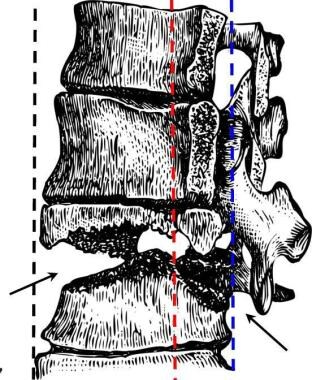 Drawing of Chance fracture of thoracolumbar junction. Defect follows irregular horizontal plane (arrows), which results in disruption of anterior (black dotted line), middle (red dotted line), and posterior columns (blue dotted line).
Pathophysiology
The thoracolumbar spinal junction represents a transitional area from the rigid thoracic spine to the more mobile lumbar region. The thoracic spine's intrinsic stability is a result of the ribs and their articulation with the spine, the smaller disk spaces, and the frontal orientation of its facet joints. As the lower two thoracic vertebrae (T11-12) lose the anterior rib articulations (floating ribs), the facet joints also change orientation to become more oblique or sagittal, allowing an increase in mobility. [14]
Flexion-distraction forces are responsible for the Chance fracture. Usually related to lap seatbelt wear, this mechanism can result in complete ligamentous injury or a combination of bony, ligament, and disk involvement. (See the image below.)
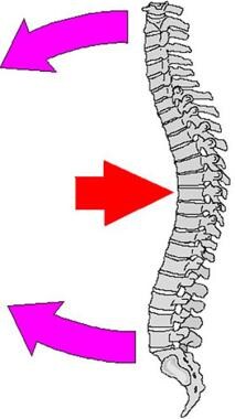 Chance fracture or modified compression fracture of upper lumbar spine may occur when weight of upper body moves forward (red arrow) while person's waist and upper body are fixed in position by seatbelt or steering wheel of automobile (pink arrows). Consequent fixed-position stress results in fracture.
Etiology
The most common history is that of a back-seat passenger who was involved in a motor vehicle accident while restrained by a lap seatbelt [4] or that of a person who fell from a height. Lumbar Chance fracture from lap seatbelt injury sustained during an airplane landing incident has been reported. [15]
Hu and Lieberman reported a case in which a 67-year-old woman with osteoporosis and thoracic kyphosis experienced a proximal vertebral body Chance fracture after pedicle screw instrumentation and fusion. [16] Pitta et al described a case in which a patient with ankylosing spondylitis experienced a lumbar Chance fracture after undergoing total hip arthroplasty via the direct anterior approach. [17]
Epidemiology
Fewer than 10% of fractures involving the lumbar spine are a result of flexion-distraction forces. These injuries tend to occur between T12 and L4, with the highest incidence at L2.
Prognosis
With proper recognition and early management of a Chance fracture, near-anatomic reduction and healing can be expected. After 3 months of immobilization in a cast or thoracolumbosacral orthosis (TLSO), a rehabilitation exercise program with emphasis on the extensor muscles of the thoracolumbar spine can assist the return to preinjury activity levels. The ultimate result may not be determined for a year after the injury, with long-term back pain being the major complaint. [18]
-
Chance fracture or modified compression fracture of upper lumbar spine may occur when weight of upper body moves forward (red arrow) while person's waist and upper body are fixed in position by seatbelt or steering wheel of automobile (pink arrows). Consequent fixed-position stress results in fracture.
-
Anterior view of Chance fracture of L2 vertebral body. Fracture line follows horizontal plane through L2 vertebral body and transverse processes (arrows).
-
Drawing of Chance fracture of thoracolumbar junction. Defect follows irregular horizontal plane (arrows), which results in disruption of anterior (black dotted line), middle (red dotted line), and posterior columns (blue dotted line).


