Background
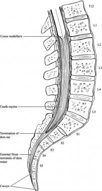
The spinal cord tapers and ends at the level between the first and second lumbar vertebrae in an average adult. The most distal bulbous part of the spinal cord is called the conus medullaris, and its tapering end continues as the filum terminale. Distal to this end of the spinal cord is a collection of nerve roots, which are horsetail-like in appearance and hence called the cauda equina (Latin for horse's tail). (See the image of cauda equina anatomy below.)
See Back Pain: Find the Cause, Watch for the Comeback, a Critical Images slideshow, to help diagnose and manage this common problem.
These nerve roots constitute the anatomic connection between the central nervous system (CNS) and the peripheral nervous system (PNS). They are arranged anatomically according to the spinal segments from which they originated and are within the cerebrospinal fluid (CSF) in the subarachnoid space with the dural sac ending at the level of second sacral vertebra.
Cauda equina syndrome refers to a characteristic pattern of neuromuscular and urogenital symptoms resulting from the simultaneous compression of multiple lumbosacral nerve roots below the level of the conus medullaris (see the image below). These symptoms include low back pain, sciatica (unilateral or, usually, bilateral), saddle sensory disturbances, bladder and bowel dysfunction, and variable lower extremity motor and sensory loss (see Clinical).
Although the lesion is technically involves nerve roots and represents a "peripheral" nerve injury, damage may be irreversible and cauda equina syndrome may be a surgical emergency (see Treatment). [1]
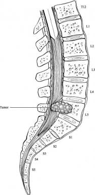 Illustration demonstrating an example of cauda equina syndrome secondary to a spinal neoplasm.
Lesions involving the termination of the spinal cord (conus medullaris) are not discussed in this article. Please see the article Spinal Cord Injuries.
Anatomy
The spinal cord, which is the downward continuation of medulla that starts just below the foramen magnum, serves as a conduit for the ascending and descending fiber tracts that connect the peripheral and spinal nerves to the brain. The cord projects 31 pairs of spinal nerves on either side (8 cervical, 12 thoracic, 5 lumbar, 5 sacral, 1 coccygeal) that are connected to the peripheral nerves.
A cross-section of the spinal cord reveals butterfly-shaped gray matter in the middle, surrounded by white matter. As in the cerebrum, the gray matter is composed of cell bodies. The white matter consists of various ascending and descending tracts of myelinated axon fibers, each with specific functions.
During development, the vertebral column grows more rapidly than the spinal cord. Spinal nerves exit the vertebral column at progressively more oblique angles because of the increasing distance between the spinal cord segments and the corresponding vertebrae. Lumbar and sacral nerves travel nearly vertically down the spinal canal to reach their exiting foramen.
The spinal cord ends at the intervertebral disc between the first and second lumbar vertebrae as a tapered structure called the conus medullaris, consisting of sacral spinal cord segments. The upper border of the conus medullaris is often not well defined. The fibrous extension of the cord, the filum terminale, is a nonneural element that extends down to the coccyx.
The cauda equina (CE) is a bundle of intradural nerve roots at the end of the spinal cord, in the subarachnoid space distal to the conus medullaris. Cauda is Latin for tail, and equina is Latin for horse (ie, the "horse's tail"). The CE provides sensory innervation to the saddle area, motor innervation to the sphincters, and parasympathetic innervation to the bladder and lower bowel (ie, from the left splenic flexure to the rectum). [100]
The nerves in the CE region include lower lumbar and all of the sacral nerve roots. The pelvic splanchnic nerves carry preganglionic parasympathetic fibers from S2-S4 to innervate the detrusor muscle of the urinary bladder. Conversely, somatic lower motor neurons from S2-S4 innervate the voluntary muscles of the external anal sphincter and the urethral sphincter via the inferior rectal and the perineal branches of the pudendal nerve, respectively.
Hence, the nerve roots in the CE region carry sensations from the lower extremities, perineal dermatomes, and outgoing motor fibers to the lower extremity myotomes.
The conus medullaris obtains its blood supply primarily from 3 spinal arterial vessels: the anterior median longitudinal arterial trunk and 2 posterolateral trunks. Less prominent sources of blood supply include radicular arterial branches from the aorta, lateral sacral arteries, and the fifth lumbar, iliolumbar, and middle sacral arteries. The latter contribute more to the vascular supply of the cauda equina, although not in a segmental fashion, unlike the blood supply to the peripheral nerves.
The nerve roots may also be supplied by diffusion from the surrounding CSF. Moreover, a proximal area of the nerve roots may have a zone of relative hypovascularity.
Pathophysiology
In understanding the pathological basis of any disease involving the conus medullaris, keep in mind that this structure constitutes part of the spinal cord (the distal part of the cord) and is in proximity to the nerve roots. Thus, injuries to this area often yield a combination of upper motor neuron (UMN) and lower motor neuron (LMN) symptoms and signs in the dermatomes and myotomes of the affected segments. On the other hand, a cauda equina lesion is an LMN lesion because the nerve roots are part of the PNS.
Cauda equina syndrome may result from any lesion that compresses CE nerve roots. These nerve roots are particularly susceptible to injury, since they have a poorly developed epineurium. A well-developed epineurium, as peripheral nerves have, protects against compressive and tensile stresses. [2]
The microvascular systems of nerve roots have a region of relative hypovascularity in their proximal third. Increased vascular permeability and subsequent diffusion from the surrounding cerebral spinal fluid supplement the nutritional supply. This property of increased permeability may be related to the tendency toward edema formation of the nerve roots, which may result in edema compounding initial and sometimes seemingly slight injury.
Several studies in different animal models have assessed the pathophysiology of cauda equina syndrome. [3, 4] Olmarker et al, using a graded balloon pressure method in a porcine model, reported that the venules in the CE region begin to compress at a pressure as low as 5 mm Hg and the arterioles begin to occlude as the balloon pressure surpasses the mean arterial pressure. [5, 6, 7, 3, 8] Despite this, even a pressure as high as 200 mm Hg failed to completely shut off nutritional supply to the CE.
These studies showed that not only the magnitude but also the length and the speed of obstruction were also important in damaging the CE region. [9] Similar results were reported in other studies. Takahashi et al reported a reduction in blood flow to the intermediate nerve segment when 2 pressure points were applied along the path of the nerve in the CE. [10]
Others have studied compound action potentials in afferent and efferent segments of nerves in the CE region after application of balloon compression. [11, 12, 13] These researchers reported that 0-50 mm Hg of pressure did not affect the action potentials (the threshold for disturbances in action potentials was 50-75 mm Hg), and significant deficits were observed when pressure rose to 100-200 mm Hg.
Etiology
Cauda equina syndrome is caused by any narrowing of the spinal canal that compresses the nerve roots below the level of the spinal cord. [14] Numerous causes of cauda equina syndrome have been reported, including disc herniation, intradural disc rupture, spinal stenosis secondary to other spinal conditions, traumatic injury, primary tumors such as ependymomas and schwannomas, metastatic tumors, infectious conditions, arteriovenous malformation or hemorrhage, and iatrogenic injury. [14, 15]
The most common causes of cauda equina and conus medullaris syndromes are the following:
-
Lumbar stenosis (multilevel)
-
Spinal trauma including fractures [16]
-
Neoplasm, including metastases, astrocytoma, neurofibroma, and meningioma; 20% of all spinal tumors affect this area
-
Spinal infection/abscess (eg, tuberculosis, herpes simplex virus, meningitis, meningovascular syphilis, cytomegalovirus, schistosomiasis) [20]
-
Idiopathic (eg, spinal anesthesia [21] ): these syndromes may occur as complications of the procedure or of the anesthetic agent (eg, hyperbaric lidocaine, tetracaine)
-
Spina bifida and subsequent tethered cord syndrome [22]
Other, rare causes include the following:
-
Spinal hemorrhage, especially subdural and epidural hemorrhage causing compression within the spinal canal
-
Intravascular lymphomatosis
-
Congenital anomalies of the spine/filum terminale, including tethered cord syndrome
-
Conus medullaris lipomas
-
Multiple sclerosis
-
Spinal arteriovenous malformations
-
Late-stage ankylosing spondylitis
-
Neurosarcoidosis
-
Deep venous thrombosis of the spinal veins (propagated)
-
Inferior vena cava thrombosis [23]
A retrospective study of 66 consecutive cases of patients admitted to a neurosurgical unit with suspected cauda equina syndrome found that almost half had no evidence of structural pathology on MRI. [24] These researchers suggested that the symptoms have a functional origin in such cases.
Trauma
Traumatic events leading to fracture or subluxation can lead to compression of the cauda equina. [16, 25, 26, 27, 28] Penetrating trauma can cause damage or compression of the cauda equina. Spinal manipulation resulting in subluxation has caused cauda equina syndrome. [29] Rare cases of sacral insufficiency fractures have been reported to cause cauda equina syndrome. [30] Acute and delayed presentations of CES due to hematomas and posttraumatic arachnoid cysts have also been reported. [31, 32, 33]
Herniated disk
The reported incidence of cauda equina syndrome resulting from herniated lumbar disk (see the image below) varies from 1-15%. [34, 35, 19] Ninety percent of lumbar disk herniations occur either at L4-L5 or L5-S1. [36, 37]
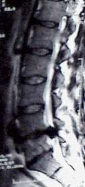 Sagittal MRI of a patient with cauda equina syndrome secondary to a large lumbar disk herniation.
Of cases of herniated disks leading to cauda equina syndrome, 70% occur in patients with a history of chronic low back pain; in 30%, cauda equina syndrome is the first symptom of lumbar disk herniation. [18] Men in the fourth and fifth decades of life are most prone to cauda equina syndrome secondary to disk herniation. [38]
Most cases of cauda equina syndrome secondary to disk herniation involve either a large central disc or an extruded disc fragment that compromises a significant amount of the spinal canal diameter. [39] The presentation may be acute or that of a more protracted course, with the latter bearing a better prognosis. [35] Individuals with congenital stenosis who sustain a disk herniation are more likely to develop cauda equina syndrome because even a small herniation can drastically limit the space available for the nerve roots.
Rare cases of intradural disk herniations have been reported to cause cauda equina syndrome. [40] Myelography in these instances typically demonstrates a complete block of the contrast material. If an intradural disc fragment is identified, transdural removal of the extruded disc fragment may be helpful to prevent further stretching of the already compromised nerve root.
Spinal stenosis
Narrowing of the spinal canal can be due to a developmental abnormality or degenerative process. Although unusual, cauda equina syndrome from spinal stenosis secondary to spinal disorders such as ankylosing spondylitis, spondylosis, and spondylolisthesis have all been reported. [41, 42, 43, 44, 45, 46, 47]
Neoplasms
Cauda equina syndrome can be caused by primary or metastatic spinal neoplasms. Among the primary tumors able to cause CES include myxopapillary ependymoma, schwannoma, and paraganglioma.
Myxopapillary ependymoma is the most common tumor of the filum. Recovery of the function after surgery depends on the duration of symptoms and the presence or absence of sphincter dysfunction [48] Paraganglioma of the filum, when present, needs to be differentiated from other tumors of this region. [49] Although rare, this entity may present as CES.
Schwannomas are benign encapsulated neoplasms that are structurally identical to a syncytium of Schwann cells. [50] These growths may arise from peripheral or sympathetic nerves. Schwannomas, whether solitary or as a part of a syndrome, may cause CES if present at the level of the conus or filum terminale. Primary tumors that affect the sacrum, such as chordoma and giant cell tumor of the bone, may produce similar symptoms as a result of bony destruction and collapse. [51]
Ependymomas are gliomas derived from relatively undifferentiated ependymal cells. They often originate from the central canal of the spinal cord and tend to be arranged radially around blood vessels. Ependymomas are found most commonly in patients aged approximately 35 years. They can lead to increased intracranial pressure (ICP), and cerebrospinal fluid (CSF) has an increased protein level.
Metastatic lesions of the spine are being reported with increasing frequency because of earlier diagnosis, better imaging, and more effective treatment modalities. Although metastasis accounts for most tumors in the spine in general, metastatic tumors in the cauda equina are relatively rare compared with primary tumors.
For the spine in general, sources of spinal metastases are as follows [52] :
-
Lung cancer (40-85%)
-
Breast cancer (11%)
-
Renal cell carcinoma (4%)
-
Lymphatic cancer (3%)
-
Colorectal cancer (3%)
Although lung cancer is the most common source of spine metastases, in one study, only 0.7% of the lung cancer metastases to the spine produced cauda equina syndrome; most of the metastatic lesions were not at the level of the cauda equina. [52] Up to 8% of patients with prostate cancer experience malignant spinal cord compression; however, the percentage of cases involving cauda equina syndrome is unknown. [53]
The CE region is also a favored site for drop metastases from intracranial ependymoma, germinoma, and other tumors. [54] Other unusual metastatic spread from genitourinary and gynecologic cancer have also been reported at the conus region, causing neurological compromise. [55, 56]
Inflammatory and infectious conditions
Long-lasting inflammatory conditions of the spine, including Paget disease and ankylosing spondylitis, can lead to cauda equina syndrome secondary to spinal stenosis or fracture.
Infectious conditions, including epidural abscess, can lead to deformity of the nerve roots and spinal cord. [57] Symptoms generally include severe back pain and a rapidly progressing motor weakness.
Infectious causes for cauda equina syndrome may be pyogenic or nonpyogenic. Pyogenic abscesses are generally found in an immunocompromised or poorly nourished host. Staphylococcus aureus causes epidural abscesses in 25-60% of cases, but, recently, an increasing incidence of infections with methicillin-resistant S aureus, Pseudomonas species, and Escherichia coli have been recorded. A high index of suspicion is helpful in correct diagnosis and management. [57]
Nonpyogenic causes for abscess are rare and include tuberculosis. Resurgence of tuberculosis secondary to immunocompromise in individuals with HIV infection requires a high index of suspicion, as the development of cauda equina syndrome may follow an indolent course. [58] Other uncommon organisms, such as Nocardia asteroides and Streptococcus milleri, have also been reported as a cause of abscess that leads to the development of CES. [59, 60]
Iatrogenic causes
Complications of spinal instrumentation have been reported to cause cases of cauda equina syndrome, including misplaced pedicle screws [61] and laminar hooks. [62, 63] Continuous spinal anesthesia also has been linked to cases of cauda equina syndrome. [64]
Rare cases of cauda equina syndrome caused by epidural steroid injections, fibrin glue injection, [65] and placement of a free-fat graft have been reported. [66]
Several cases have involved the use of hyperbaric 5% lidocaine for spinal anesthesia. Recommendations are that hyperbaric lidocaine not be administered in concentrations greater than 2%, with a total dose not to exceed 60 mg. [67, 68]
Medical and surgical situations such as bone screw fixation, fat grafts, lumbar arthrodesis for spondylolisthesis, lumbar discectomy, intradiscal therapy, lumbar puncture forming an epidural hematoma, chiropractic manipulation, and a bolus injection of anesthetic during spinal anesthesia have been related to the development of cauda equina syndrome–like syndromes. [35, 69, 70, 71, 72, 73]
Epidemiology
Cauda equina and conus medullaris syndromes are classified as clinical syndromes of the spinal cord; epidemiological data on the 2 syndromes are often not available separately from the general data on spinal cord injury.
Cauda equina syndrome is uncommon, both atraumatically as well as traumatically. It is often reported as a case report due to its rarity. Although infrequent, it is a diagnosis that must be considered in patients who complain of low back pain coupled with neurologic complaints, especially urinary symptoms.
Age-related differences in incidence
Traumatic cauda equina syndrome is not age specific. Atraumatic cauda equina syndrome occurs primarily in adults as a result of surgical morbidity, spinal disk disease, metastatic cancer, or epidural abscess.
Prognosis
Morbidity and especially mortality rates are determined by the underlying etiology. Multiple conditions can result in cauda equina or conus medullaris syndrome. The prognosis improves if a definitive cause is identified and appropriate treatment occurs early in the course. Surgical decompression may be performed emergently, or, in some patients, delayed, depending on the etiology. Residual weakness, incontinence, impotence, and/or sensory abnormalities are potential problems if therapy is delayed.
Investigators have attempted to identify specific criteria that can aid in predicting the prognosis of patients with cauda equina syndrome. Patients with bilateral sciatica have been reported to have a less favorable prognosis than patients with unilateral pain. Patients with complete perineal anesthesia are more likely to have permanent paralysis of the bladder. [39]
The extent of perineal or saddle sensory deficit has been reported to be the most important predictor of recovery. [74] Patients with unilateral deficits have a better prognosis than patients with bilateral deficits. Females and patients with bowel dysfunction have been reported to have worse outcomes postoperatively. [75]
Prognosis can be predicted with the American Spinal Injury Association (ASIA) impairment scale (see Physical Examination ), as follows:
-
ASIA A: 90% of patients remain incapable of functional ambulation (reciprocal gait of 200 feet or more)
-
ASIA B: 72% of patients are unable to attain functional ambulation
-
ASIA C/D: 13% are unable to attain functional ambulation 1 year after injury
Ambulatory motor index also is used to predict ambulatory capability. It is calculated by scoring hip flexion, hip abduction, hip extension, knee extension, and knee flexion on both sides, using a 4-point scale (0=absent, 1=trace/poor, 2=fair, 3=good or normal); the score is expressed as a percentage of the maximum score of 30. Prognostic significance is as follows:
-
A patient with a score of 60% or more has a good chance for community ambulation with no more than one knee-ankle-foot orthosis (KAFO)
-
A patient with a score of 79% or higher may not need an orthosis
-
A patient with a score of 40% or less may require 2 KAFOs for community ambulation
Patient Education
Patient education needs will vary with the type and severity of persistent deficits, and may include the following:
-
Training in self-catheterization and finger fecal disimpaction, if required
-
Use of measures to prevent pressure ulcers, such as skin inspection/care, positioning, turning and transferring tactics, use of skin protectors, or pressure-reducing support surfaces
-
Maintenance of endurance and strength-training exercises
-
Regular follow-up by the consulting teams who treated the patient in the hospital
-
Instructions on how and when medications should be taken and when follow-up laboratory tests should be performed
For patient education information, see the Erectile Dysfunction Center and Brain and Nervous System Center, as well as Impotence/Erectile Dysfunction, Erectile Dysfunction FAQs, and Cauda Equina Syndrome.
-
Muscle groups, surface anatomy, peripheral sensory innervation, and dermatomes of the anterior lower limb. This image should be correlated with Tables 1 and 2 in the text. Image courtesy of Nicholas Y. Lorenzo, MD.
-
Muscle groups, surface anatomy, peripheral sensory innervation, and dermatomes of the posterior lower limb. This image should be correlated with Tables 1 and 2 in the text. Image courtesy of Nicholas Y. Lorenzo, MD.
-
Conus/epiconus infarction in the setting of sickle cell crisis. Image courtesy of Matthew J. Baker, MD.
-
Conus/epiconus infarction in the setting of sickle cell crisis in the same patient shown in the above image. Image courtesy of Matthew J. Baker, MD.
-
Conus/epiconus infarction in the setting of sickle cell crisis in the same patient shown in the images above. Image courtesy of Matthew J. Baker, MD.
-
Illustration demonstrating the relevant anatomy of the cauda equina region.
-
Illustration demonstrating an example of cauda equina syndrome secondary to a spinal neoplasm.
-
Sagittal MRI of a patient with cauda equina syndrome secondary to a large lumbar disk herniation.
-
Epidural abscess with effacement of thecal sac in a 56-year-old man.
Tables
|
Conus Medullaris Syndrome |
Cauda Equina Syndrome |
Presentation |
Sudden and bilateral |
Gradual and unilateral |
Reflexes |
Knee jerks preserved but ankle jerks affected |
Both ankle and knee jerks affected |
Radicular pain |
Less severe |
More severe |
Low back pain |
More |
Less |
Sensory symptoms and signs |
Numbness tends to be more localized to perianal area; symmetrical and bilateral; sensory dissociation occurs |
Numbness tends to be more localized to saddle area; asymmetrical, may be unilateral; no sensory dissociation; loss of sensation in specific dermatomes in lower extremities with numbness and paresthesia; possible numbness in pubic area, including glans penis or clitoris |
Motor strength |
Typically symmetric, hyperreflexic distal paresis of lower limbs that is less marked; fasciculations may be present |
Asymmetric areflexic paraplegia that is more marked; fasciculations rare; atrophy more common |
Impotence |
Frequent |
Less frequent; erectile dysfunction that includes inability to have erection, inability to maintain erection, lack of sensation in pubic area (including glans penis or clitoris), and inability to ejaculate |
Sphincter dysfunction |
Urinary retention and atonic anal sphincter cause overflow urinary incontinence and fecal incontinence; tend to present early in course of disease |
Urinary retention; tends to present late in course of disease |
Nerve Root |
Pain |
Sensory Deficit |
Motor Deficit |
Reflex Deficit |
L2 |
Anterior medial thigh |
Upper thigh |
Slight quadriceps weakness; hip flexion; thigh adduction |
Slightly diminished suprapatellar |
L3 |
Anterior lateral thigh |
Lower thigh |
Quadriceps weakness; knee extension; thigh adduction |
Patellar or suprapatellar |
L4 |
Posterolateral thigh, anterior tibia |
Medial leg |
Knee and foot extension |
Patellar |
L5 |
Dorsum of foot |
Dorsum of foot |
Dorsiflexion of foot and toes |
Hamstrings |
S1-2 |
Lateral foot |
Lateral foot |
Plantar flexion of foot and toes |
Achilles |
S3-5 |
Perineum |
Saddle |
Sphincters |
Bulbocavernosus; anal |
Muscle |
Nerve |
Root |
Iliopsoas |
Femoral |
L2, 3, 4 |
Adductor longus |
Obturator |
L2, 3, 4 |
Gracilis |
Obturator |
L2, 3, 4 |
Quadriceps femoris |
Femoral |
L2, 3, 4 |
Anterior tibial |
Deep peroneal |
L4, 5 |
Extensor hallucis longus |
Deep peroneal |
L4, 5 |
Extensor digitorum longus |
Deep peroneal |
L4,5 |
Extensor digitorum brevis |
Deep peroneal |
L4, 5, S1 |
Peroneus longus |
Superficial peroneal |
L5, S1 |
Internal hamstrings |
Sciatic |
L4, 5, S1 |
External hamstrings |
Sciatic |
L5, S1 |
Gluteus medius |
Superior gluteal |
L4, 5, S1 |
Gluteus maximus |
Inferior gluteal |
L5, S1, 2 |
Posterior tibial |
Tibial |
L5, S1 |
Flexor digitorum longus |
Tibial |
L5, S1 |
Abductor hallucis brevis |
Tibial (medial plantar) |
L5, S1, 2 |
Abductor digiti quinti pedis |
Tibial (lateral plantar) |
S1, 2 |
Gastrocnemius lateral |
Tibial |
L5, S1, 2 |
Gastrocnemius medial |
Tibial |
S1, 2 |
Soleus |
Tibial |
S1, 2 |
Features |
Cauda Equina Syndrome |
Conus Medullaris |
Vertebral level |
L2-sacrum |
L1-L2 |
Spinal level |
Injury to the lumbosacral nerve roots |
Injury of the sacral cord segment (conus and epiconus) and roots |
Severity of symptoms and signs |
Usually severe |
Usually not severe |
Symmetry of symptoms and signs |
Usually asymmetric |
Usually symmetric |
Pain |
Prominent, asymmetric, and radicular |
Usually bilateral and in the perineal area |
Motor |
Weakness to flaccid paralysis |
Normal motor function to mild or moderate weakness |
Sensory |
Saddle anesthesia, may be asymmetric |
Symmetric saddle distribution, sensory loss of pin prick, and temperature sensations (Tactile sensation is spared.) |
Reflexes |
Areflexic lower extremities; bulbocavernosus reflex is absent in low CE (sacral) lesions |
Areflexic lower extremities (If the epiconus is involved, patellar reflex may be absent, whereas bulbocavernosus reflex may be spared.) |
Sphincter and sexual function |
Usually late and of lesser magnitude; lower sacral roots involvement can cause bladder, bowel, and sexual dysfunction |
Early and severe bowel, bladder, and sexual dysfunction that results in a reflexic bowel and bladder with impaired erection in males |
EMG |
Multiple root level involvement; sphincters may also be involved |
Mostly normal lower extremity with external anal sphincter involvement |
Outcome |
May be favorable compared with conus medullaris syndrome |
The outcome may be less favorable than in patients with CES |


