Practice Essentials
Calcium pyrophosphate deposition (CPPD) disease is a metabolic arthropathy caused by the deposition of calcium pyrophosphate dihydrate in and around joints, especially in articular cartilage and fibrocartilage (see the images below). [1] (See Etiology, Presentation, and Workup.)
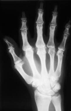 Calcium pyrophosphate deposition disease. Radiograph of the wrist and hand showing chondrocalcinosis of the articular disc of the wrist and atypical osteoarthritis involving the metacarpophalangeal joints in a patient with underlying hemochromatosis.
Almost any joint may be involved by CPPD, although the knees, wrists, and hips are most often affected. This condition is the most common cause of secondary metabolic osteoarthritis. (See Presentation.)
The European Alliance of Associations for Rheumatology (EULAR) recognizes at least four different clinical presentations of CPPD, as follows [2, 3] :
-
Asymptomatic (lanthanic) CPPD
-
Osteoarthritis (OA) with CPPD
-
Acute CPP crystal arthritis (pseudogout)
-
Chronic CPP inflammatory crystal arthritis (previously termed pseudo–rheumatoid arthritis)
Other forms of CPPD described in the literature include pseudo–polymyalgia rheumatica (pseudo-PMR), pseudo–neuropathic arthropathy, and tumoral (tophaceous) CPPD. [3]
Patients with CPPD can experience significant morbidity due to the pain of an acute attack of pseudogout or to symptoms of chronic arthropathy. Treatment of symptomatic CPPD is important to prevent further end-organ damage, but it cannot reverse the joint disease. (See Treatment and Medication).
For patient education information, see Arthritis and Pseudogout.
Etiology
Although the exact mechanism for the development of CPPD remains unknown, increased adenosine triphosphate breakdown with resultant increased inorganic pyrophosphate in the joints results from aging, genetic factors, or both. Changes in the cartilage matrix may play an important role in promoting CPPD deposition. Rare hereditary forms of CPPD occur, generally inherited in an autosomal dominant mode.
Overactivity of enzymes that break down triphosphates, such as nucleoside triphosphate pyrophosphohydrolase, has been observed in the cartilage of patients with CPPD disease. Therefore, inorganic pyrophosphate can bind calcium, leading to CPPD deposition in the cartilage and synovium. [4, 5] Hyaline cartilage is affected most commonly, but fibrocartilage, such as the meniscal cartilage of the knee, can also be involved. [6]
Hypotheses based on in vitro studies propose that pyrophosphohydrolase activity and inorganic phosphate content, as noted above, are generalized phenomena that occur in fibroblasts. [7] Although these phenomena are generalized, the reason they occur only in joints remains unknown.
Genetics
Genetic defects have been identified as specific gene mutations in a few kindred families. [8, 9] The mutations were in the genes ANKH and COL, which may be involved in crystal-induced inflammation. This is related to synovial tissue and direct cartilage activation, leading to the arthritis caused by CPPD. The ANKH protein is involved in transport of inorganic pyrophosphate (PPi), which regulates calcification, bone mineralization, and bone resorption. [10]
The gene TNFRSF11B encodes osteoprotegerin, which has a critical role in regulating osteoclast development. In a study of patients with familial osteoarthritis with chondrocalcinosis, Ramos et al identified a mutation in TNFRSF11B that results in a form of osteoprotegerin with enhanced capacity to inhibit osteoclastogenesis and bone resorption. [11]
Subsequent messenger RNA expression analysis of the relevant genes in this pathway, in articular cartilage of independent patients undergoing joint replacement surgery for osteoarthritis, showed that upregulation of TNFRSF11B is a general phenomenon in the pathophysiological process of osteoarthritis. [11]
Epidemiology
Occurrence in the United States
CPPD is a common condition that occurs with aging in all races. In a retrospective study of 1070 consecutive computed tomographic scans of the abdomen and pelvis in patients over 65 years of age, the prevalence of symphysis pubis chondrocalcinosis was 21.1%. [12] Nearly 50% of people older than 85 years have radiologic evidence of chondrocalcinosis.
Sex- and age-related demographics
CPPD is slightly more common in women than in men. The exact female-to-male ratio is unknown but is probably 1.4:1.
CPPD usually occurs in individuals who are in the fifth decade of life or older, with increasing prevalence as age increases. When it occurs early, before the fourth decade of life, it is usually associated with a secondary cause, such as an underlying metabolic disease, or with a familial cause.
Comorbidities
In a cross-sectional study in the national Veterans Affairs (VA) population that included 25,157 patients with CPPD, the strongest positive associations with CPPD were as follows [13] :
-
Hyperparathyroidism (odds ratio [OR] 3.35)
-
Gout (OR 2.82)
-
Osteoarthritis (OR 2.26)
-
Rheumatoid arthritis (OR 1.88)
-
Hemochromatosis (OR 1.87)
In addition, positive associations were seen with osteoporosis (OR 1.26), hypomagnesemia (OR 1.23), chronic kidney disease (OR 1.12), and calcium supplementation (OR 1.15). Negative associations were seen with use of proton-pump inhibitors or loop diuretics. [13]
-
Calcium pyrophosphate deposition disease. Radiograph of the knee showing chondrocalcinosis involving the meniscal cartilage, as well as evidence of osteoarthritis.
-
Calcium pyrophosphate deposition disease. Radiograph of the wrist and hand showing chondrocalcinosis of the articular disc of the wrist and atypical osteoarthritis involving the metacarpophalangeal joints in a patient with underlying hemochromatosis.
-
Calcium pyrophosphate deposition disease. Appearance of calcium pyrophosphate dihydrate crystals obtained from the knee of a patient with pseudogout. The crystals are rhomboid-shaped with weakly positive birefringence, as seen by compensated polarized microscopy. The black arrow indicates the direction of the compensator.
-
Calcium pyrophosphate deposition disease. High-powered view of calcium pyrophosphate dihydrate crystals with compensated polarized microscopy. The black arrow indicates the direction of the compensator. Crystals parallel to the compensator are blue, while those perpendicular to the compensator are yellow.
-
Calcium pyrophosphate deposition disease. High-powered view of calcium pyrophosphate dihydrate crystals with compensated polarized microscopy. The crystals parallel to the compensator were blue, while those perpendicular to the compensator were yellow. However, the crystals have been rotated 90%, resulting in a color change in both of them. The direction of the compensator was not changed and is indicated by the black arrow.
-
Calcium pyrophosphate deposition disease. Ultrasonography of the wrist demonstrates chondrocalcinosis.
-
Intraoperative photographs demonstrate extensive precipitate deposition of the calcium pyrophosphate crystals in the articular cartilage, meniscus, and synovium of a knee. Left images depict femoral and tibial surfaces. Right images depict anterior cruciate ligament.
-
Intraoperative photographs demonstrate extensive precipitate deposition of the calcium pyrophosphate crystals in the articular cartilage, meniscus, and synovium of a knee. Upper left image depicts anterior horn medial meniscus. Lower left image depicts undersurface of meniscus. Upper right image depicts medial femoral condyle. Lower right image depicts synovium.
-
Calcium pyrophosphate deposition disease. Ultrasound scan of the triangular fibrocartilage complex (TFCC) of the wrist shows thin hyperechoic bands parallel to the surface of the hyaline cartilage. Other findings include a punctate pattern consisting of several hyperechoic spots and homogeneous hyperechoic nodular or oval deposits in the articular surface.


