Practice Essentials
Tibial tubercle (tuberosity) fractures are infrequent fractures affecting physically active adolescents. [1, 2, 3, 4, 5, 6] Activities involving powerful contraction of the knee extensors, such as springing and jumping movements, can result in avulsion fractures of the tibial tuberosity apophysis. [7, 8, 9] This condition should be distinguished from Osgood-Schlatter disease, a chronic apophysitis of the tibial tuberosity due to recurrent traction injury.
Medical therapy typically involves analgesia for pain control and thromboprophylaxis. Nondisplaced type I injuries can be managed conservatively by means of cast immobilization in a long leg cast in full-knee extension. All other injuries are best treated by means of open reduction and internal fixation (ORIF) with cast immobilization for 6-8 weeks. (See Treatment.)
Anatomy
The extensor complex of the thigh exerts its force through the ligamentum patellae on the tibial tuberosity. During its histogenesis, the tibial tuberosity is an anterior extension of the proximal tibial epiphysis separated from the rest of the tibia by the growth plate. As the growth plate closes in late puberty, it is transiently replaced by fibrocartilaginous elements, which predispose it to traction injury as a result of its weaker tensile strength.
Pathophysiology
The proximal tibia has two ossification centers, the proximal tibial epiphysis and the tibial tuberosity, which are separated by a cartilage bridge (see the image below). Before ossification, the tibial tuberosity is composed of fibrocartilage that has good tensile strength. However, during ossification, columnated cartilaginous cells with poor tensile strength replace the fibrocartilage, and it is within this small window between fibrocartilage and ossified matrix that the tibial tuberosity is at risk of avulsion fractures.
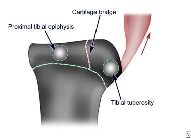 Ossification centers and epiphyseal cartilages of the proximal tibia and tibial tuberosity.
As a result of the direction of pull of the patella tendon, the tibial tuberosity along with the proximal tibial epiphysis can be avulsed upward in a fracture in one or more fragments (see the image below).
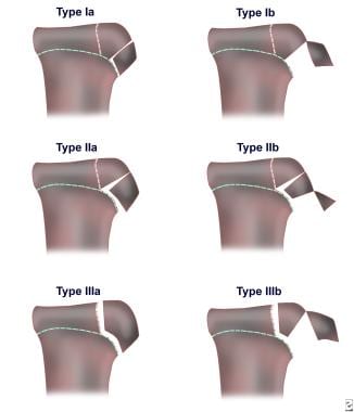
Watson-Jones classified the fractures into the following three types [10] :
-
Type I - The fracture is within the most distal portion of the tibial tuberosity ossification center and usually results in avulsion of the most distal portion
-
Type II - Extension of the fracture line occurs into the proximal end of the tibia through the cartilage bridge but does not involve the articular surface
-
Type III - This is an intra-articular fracture in which the fracture line has propagated into the joint
Ogden modified this classification by adding the subtypes A and B, [11] with A representing single fractures and B representing comminuted fractures.
Modifications to Ogden's classification were subsequently proposed. A modification in current use is as follows [12] :
-
Type I - Fracture of the secondary ossification center near the patella tendon insertion
-
Type II - Fracture between the primary and secondary ossification centers
-
Type III - Fracture that traverses the primary and secondary ossification centers (most common type)
-
Type IV - Fracture through the entire physis
-
Type V - Avulsion of the periosteal sleeve
The modifiers A (nondisplaced) and B (displaced) are also used.
Etiology
Tibial tubercle fracture is caused by injury from violent tensile forces on the tibial tuberosity. The force is delivered through eccentric contraction of the extensor mechanism of the knee from either of the following:
-
Violent contraction of the extensors without shortening (eg, springing off when jumping)
-
Forceful flexion of the knee against the powerful contraction of the quadriceps (eg, landing from a jump)
In other words, it occurs when sudden acceleration or deceleration of the extensor mechanism occurs.
Patients with Osgood-Schlatter disease may be predisposed to tibial tuberosity fractures. [13] Similarly, patients with these fractures may have a family history of Osgood-Schlatter disease or a history of fractures of the tibial tuberosity.
A few cases of tibial tubercle fracture occurring after bone–patellar tendon–bone (BPTB) autograft for anterior cruciate ligament (ACL) reconstruction have been reported. [14]
Epidemiology
In the United States, the frequency of tibial tubercle fracture has not been determined, though the injury is known to occur infrequently. At one major center, 15 cases of tibial tuberosity fracture were diagnosed in 5 years. Tibial tuberosity fractures typically occur in individuals aged 14-17 years. As the growth plate closes in late puberty, it is transiently replaced by fibrocartilaginous elements. These elements predispose the tibial tuberosity to traction injury as a result of its weakened tensile strength.
Internationally, the frequency is not known. As in the United States, the condition occurs infrequently.
Prognosis
Pretell-Mazzini et al systematically reviewed the English-language literature from 1970 through 2013 (23 studies; 336 fractures; mean follow-up, 33.56 mo [range, 5.7-115]) in order to determine the following with regard to tibial tubercle fractures in pediatric patients (mean age at surgery, 14.6 y) [15] :
-
Frequency and type of associated injuries
-
Frequency of concomitant Osgood-Schlatter disease
-
Methods of treatment
-
Functional and radiologic outcomes according to fracture type
-
Complications
The most common fracture reported was type III (50.6%). The rate of associated injury was 4.1% overall and was highest for type III fractures (4.7%). [15] Compartment syndrome occurred in 3.57% of cases. ORIF was performed in 98% of surgical cases. Regardless of the type of fracture, 98% of patients were able to regain their preinjury activity and knee range of motion (ROM), and 99.4% achieved fracture consolidation. The overall complication rate was 28.3%; removal of an implant because of bursitis (55.8%) was the most common complication, followed by tenderness/prominence (17.9%) and refracture (6.3%).
In a retrospective case series that included 228 subjects aged 18 years old or younger treated for 236 tibial tubercle fractures at a single institution, Haber et al found that most patients returned to sports (88%). [16] Compartment syndrome was identified in four patients (2%), three of whom had type IV fractures.
A single-institution retrospective review (N = 19; average age, 14.6 y; average body mass index [BMI], 25.8) by Yang et al reported outcomes after surgical treatment of displaced tibial tubercle fractures in male adolescents. [17] The fractures in this series occurred during athletic activity. Unicortical screws/pins were used with no loss of fixation; no patient was treated with bicortical screws. Routine use of advanced imaging was unnecessary. One patient presented with acute compartment syndrome and underwent fasciotomy. No growth arrest occurred. All patients returned to preinjury athletic activities at an average of 18.5 weeks.
-
Ossification centers and epiphyseal cartilages of the proximal tibia and tibial tuberosity.
-
Classification of tibial tuberosity fractures.


