Practice Essentials
An ankle sprain is usually that of an inversion-type twist of the foot, followed by pain and swelling. The most commonly injured site is the lateral ankle complex, which is composed of the anterior talofibular, calcaneofibular, and posterior talofibular ligaments. [1, 2]
Signs and symptoms
Signs and symptoms of an ankle sprain include the following:
-
Pain/tenderness
-
Swelling and/or bruising
-
Cold foot or paresthesia (possible neurovascular compromise) [1]
-
Muscle spasm
See Clinical Presentation for more detail.
Diagnosis
The physical examination confirms a diagnosis made on the basis of patient history and differentiates an ankle sprain from a fracture. Examination in patients may include the following tests:
-
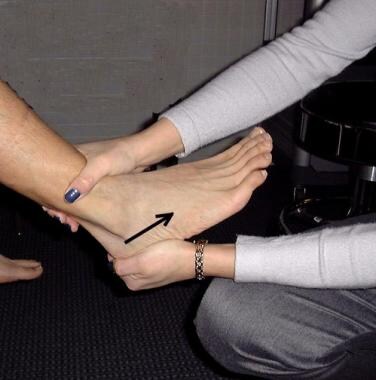
Anterior drawer test: To assess for ankle instability (see the image below)
-
Prone anterior drawer test: Also tests for ligamentous instability
-
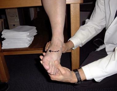
Talar tilt test (or inversion stress maneuver): To assess integrity of the calcaneofibular ligament (see the image below)
-
External rotation test: To evaluate the integrity of the syndesmotic ligaments
-
Kleiger test: Variation of the external rotation test; to assess the integrity of the deltoid ligament
-
Squeeze test (or fibular compression test): To evaluate for syndesmotic or fibular injury
-
Neurovascular evaluation: To assess neurovascular status of the affected limb
Imaging studies
The following radiologic studies may be used to evaluate ankle sprains:
-
Plain radiography: Guided by the Ottawa Ankle Rules to diagnose ankle or foot fractures
-
Stress-view radiography: May provide further assessment for ankle stability; accuracy of study increases with use of local anesthesia
-
Computed tomography scanning: May be indicated for imaging of soft tissues or for bone imaging beyond radiography; useful for evaluating osteochondritis dissecans and stress fractures
-
Ankle arthrography: May be useful for determining capsular damage and the number of ankle ligaments damaged
-
Bone scanning: To detect subtle bone abnormalities (eg, stress fracture, osteochondral defects) and syndesmotic disruptions
See Workup for more detail.
Management
Conservative therapy
Conservative therapy for acute ankle sprains may be described by the acronyms RICE (rest, ice, compression, and elevation) and PRICES (combination of protection, relative rest, ice, compression, elevation, and support). Protective devices include air splints or plastic and Velcro braces. Ankle taping can also increase ankle stability, but its effectiveness is highly dependent on the expertise of the individual who performs the taping.
Physical therapy during the recovery phase is aimed at the patient regaining full range of motion, strength, and proprioceptive abilities, and may include the following:
-
Strengthening exercises: Starts with isometric exercises, then advances to use of elastic bands or surgical tubing
-
Proprioception rehabilitation: Starts with single-leg-stance exercise in a single plane, then progresses to multiplanar exercises
-
Other exercises: Uses a balance or tilt board, then advances to functional drills, jogging, sprinting, and cutting, and then progresses to figure-of-eight and carioca drills [5]
Pharmacotherapy
The following medications are used in the management of ankle sprain:
-
Analgesics (eg, acetaminophen)
-
Nonsteroidal anti-inflammatory agents (eg, ibuprofen, naproxen)
Surgery
In most patients, there is no improved outcome with operative repair of third-degree anterior talofibular ligament tears and medial ankle ligament tears.
Indications for operative intervention in patients with an ankle sprain include the following:
-
Distal talofibular ligament third-degree sprain that causes widening of the ankle mortise
-
Deltoid sprain with the deltoid ligament caught intra-articularly and with widening of the medial ankle mortise
-
In selected young patients with high athletic demands who have both anterior talofibular and calcaneofibular complete ruptures
Surgical procedures for chronic ankle instability and sprains include the Watson-Jones procedure, the Evans procedure, and the Chrisman-Snook procedure.
See Treatment and Medication for more detail.
Background
The history of an ankle sprain is usually that of an inversion-type twist of the foot followed by pain and swelling. An individual with an ankle sprain can almost always walk on the foot, albeit carefully and with pain.
In an individual with normal local sensation and cerebral function, the ability to walk on the foot usually excludes a fracture. (See Clinical Presentation.) Suspect neurovascular compromise if the patient reports a cold foot or describes paresthesias. [1] Bone tenderness in the posterior half of the lower 6 cm of the fibula or tibia and the inability to bear weight immediately after the injury and in the emergency department are indications to obtain radiographic imaging. These Ottawa ankle rules have been validated for patients aged 5-55 years. [6, 7]
Ankle sprains are classified into the following 3 grades:
-
Grade 1 injuries involve a stretch of the ligament with microscopic tearing but not macroscopic tearing. Generally, little swelling is present, with little or no functional loss and no joint instability. The patient is able to fully or partially bear weight.
-
Grade 2 injuries stretch the ligament with partial tearing, moderate-to-severe swelling, ecchymosis, moderate functional loss, and mild-to-moderate joint instability. Patients usually have difficulty bearing weight.
-
Grade 3 injuries involve complete rupture of the ligament, with immediate and severe swelling, ecchymosis, an inability to bear weight, and moderate-to-severe instability of the joint. Typically, patients cannot bear weight without experiencing severe pain.
Drawer and talar tilt examination techniques are used to assess ankle instability; however, the use of these techniques in acute injuries is in question because of pain, edema, and muscle spasm.
Pain reduction is essential, but improvement of any loss of motion, strength, and/or proprioception is equally important. [8] Rest, ice, compression, and elevation (ie, RICE) are the mainstays of acute treatment; more comprehensively, the combination of protection, relative rest, ice, compression, elevation, and support (PRICES) is used. [1] (See Treatment.)
Physical therapy during the recovery phase is aimed at the patient regaining full range of motion (ROM), strength, and proprioceptive abilities.
For recurrent lateral ankle sprains, treatment should begin with a trial of conservative therapy for approximately 2-3 months. The recurrence rate for lateral ankle sprains has been reported to be as high as 80%. [9]
It is generally accepted that for most patients, operative repair of third-degree anterior talofibular ligament (ATFL) tears and medial ankle ligament tears does not contribute to an improved outcome. [10] One of the few absolute indications for surgery in patients with a sprained ankle is a distal talofibular ligament third-degree sprain that causes widening of the ankle mortise. A second indication is a deltoid sprain with the deltoid ligament caught intra-articularly and with widening of the medial ankle mortise.
Anatomy
The ankle joint is a hinged synovial joint with primarily up-and-down movement (plantar flexion and dorsiflexion). The other joints around the ankle are responsible for other movements, giving the ankle a total range of motion (ROM) comparable to that of a ball and socket. The combined movement in the dorsiflexion and plantarflexion directions is greater than 100°; bone-on-bone abutment beyond this range protects the anterior and posterior ankle capsular ligaments from injury. The anterior and posterior ankle capsular ligaments are relatively thin compared with the medial and lateral ankle ligaments.
Pathophysiology
The lateral ankle complex, which is composed of the anterior talofibular, calcaneofibular, and posterior talofibular ligaments, is the most commonly injured site. [1, 2] Approximately 85% of such sprains are inversion sprains of the lateral ligaments, 5% are eversion sprains of the deltoid or medial ligament, and 10% are syndesmotic injuries. The ATFL is the most likely component of the lateral ankle complex to be injured in a lateral ankle sprain. Osteochondral or chondral injuries of the talar dome should be considered when diagnosing an ankle injury.
During forced dorsiflexion, the PTFL can rupture. With forced internal rotation, ATFL rupture is followed by injury to the PTFL. Extreme external rotation disrupts the deep deltoid ligament on the medial side, and adduction in neutral and dorsiflexed positions can disrupt the CFL. In plantarflexion, the ATFL can be injured.
The strongest ankle capsule-ligament complex is the deltoid ligament, which has 2 parts: the superficial component and the deep component. The superficial component runs the farthest from the medial malleolus to the medial aspect of the calcaneus, posteriorly. The medial malleolus usually fractures before the deltoid ligament fails mechanically.
Ankle spurs may occur at any of the bony ligament attachments. On lateral radiographs, it is not uncommon to see an anterior spur at the neck of the talus, where the anterior ankle capsule attaches. This is caused by ossification of the hematoma organization associated with anterior ligament sprains.
Because of its great strength, the syndesmotic ligament, which has a deep portion between the bones and superficial, anterior, and posterior portions, is rarely sprained. This distal tibiofibular ligament holds the distal tibia and fibular bones together at the ankle joint and maintains the integrity of the ankle mortise. It takes a great amount of force to strain this ligament, which normally does not have much excursion. A significant tear of this ligament requires surgical treatment. Severe posttraumatic arthritis of the tibiotalar joint (ankle) can result quickly if a syndesmosis tear remains unrecognized and untreated. A syndesmotic ligament tear is usually a part of an ankle fracture that needs to be treated specifically. This is not generally true of the other ankle ligament tears.
Etiology
Mechanical forces exceeding the tensile limits of the ankle joint capsule and supportive ligaments cause ankle sprains.
There are a number of contributing factors, which can be classified as either predisposing or provocative, as follows:
-
Predisposing factors can result from a lack of physical conditioning; they include poor muscle tone and shortened and/or contracted joint capsule or tendons. Poor proprioception can also be a factor, as can inadequate training or experience with the physical activity being performed.
-
Provocative factors include accidents and other unforeseen circumstances that result in mechanical stresses that exceed the tensile limits of the ankle joint capsule and ligaments. Obesity can contribute to sprains by increasing kinetic energy to a point that exceeds joint-design stress limits.
A cohort study analyzed risk factors in ankle injuries from the Cadet Illness and Injury Tracking System (CIITS) database at the United States Military Academy (USMA) from 2005-2009. The results found higher risk of syndesmotic ankle sprains in males who performed at a higher level of athletic competition; male athletes were 3 times more likely to experience medial ankle sprains than female athletes. [11]
Recurrent sprains
The exact etiology of recurrent ankle sprains is unknown; however, many factors may play a role.
One possibility is that recurrent sprains result primarily from ligaments healing in a lengthened position due to scar tissue filling in the gap between the torn, separated ends. Furthermore, the weakness of the healed ligament may be due to the inherent weakness of the scar.
In a study by Bosien et al, 22% of patients with recurrent ankle sprains had persistent peroneal weakness. [12] The authors believed that this contributed to recurrent injury, especially in incompletely rehabilitated ankle sprains.
An unrecognized disruption of the distal tibiofibular ligament has been cited as a potential culprit. This condition is diagnosed based on tenderness over the anterior syndesmosis and pain when the fibula is squeezed against the tibia at midshaft, with dorsiflexion and external rotation or with excessive medial-lateral motion of the tibiotalar joint.
Freeman et al suspected that functional instability that resulted in recurrent sprains was secondary to loss of proprioception in the foot. [13] Mechanoreceptors and their afferent nerve fibers have been shown to exist in the ligaments and capsule of the ankle. Furthermore, disruption of the ligaments and joint capsule with a grade 3 sprain (a complete ligament tear; see Clinical Presentation) leads to impairment of the reflex stabilization of the foot, causing the foot to give way. [14] In addition, dysfunction of the peroneal nerve can result in delayed muscle response, causing a delay in the activation of the peroneal muscles and leading to functional instability.
Impingement by the distal fascicle of the AITF ligament and/or impingement of the capsular scar tissue in the talofibular joint is another cause, and hereditary hypermobility of joints is an additional suggested etiologic factor.
Epidemiology
Most ankle sprains are probably self-treated and are never reported to a health care provider; therefore, many ankle sprains are not documented. Sprained ankles have been estimated to constitute up to 30% of injuries seen in sports medicine clinics and are the most frequently seen musculoskeletal injury seen by primary care providers. [15] More than 23,000 people per day in the United States, including athletes and nonathletes, require medical care for ankle sprains. Stated another way, incident cases have been estimated at 1 case per 10,000 persons per day. [16] A US Army study found that ankle sprains are the most common foot and ankle injury in active-duty Army personnel with a rate of 103 sprains per 1000 persons per year. [17]
Female athletes are 25% more likely to sustain ankle injuries than male athletes. Female basketball players are at a higher risk of a first-time inversion injury than those participating in other sports. [18] Soccer and volleyball are other leading causes of ankle sprains in high school and college female athletes. [19, 20] Some studies attribute a higher incidence of ankle injuries in high school football, basketball, and soccer players. [19, 21, 22] Other studies conclude that in college men, the risk of suffering an ankle sprain appears to be similar with basketball, soccer, and football. [18]
A cohort study analyzed risk factors in ankle injuries from the Cadet Illness and Injury Tracking System (CIITS) database at the United States Military Academy (USMA) from 2005-2009. The results found higher risk of syndesmotic ankle sprains in males who performed at a higher level of athletic competition; male athletes were 3 times more likely to experience medial ankle sprains than female athletes. [11]
Ankle injuries are very common, with ankle sprain being the most common injury. Most individuals have complete recovery; however, a substantial number have chronic problems that usually lead to modification of physical activity. [23, 10]
Prognosis
Acute injuries
The prognosis for isolated and adequately treated ankle sprains is excellent. The prognosis for a patient with ankle sprains and other traumatic injuries is related to the prognosis for the other injuries.
In a systematic literature review, 36-85% of patients with acute ankle sprains reported full recovery at 2 weeks to 36 months, independent of the initial grade of sprain, [24] with most recovery occurring within the first 6 months. [25] After 12 months, the risk of recurrent ankle sprain returns to preinjury levels. [26] However, 3-34% of patients reported re-sprains at 2 weeks to 96 months after the initial injury. Furthermore, after 3 years, some patients still had residual pain and instability. One risk factor for residual symptoms seems to be participation in competitive sports. [24]
Recurrent sprains
If recurrent ankle sprains are treated early and appropriate rehabilitation is initiated, the prognosis is excellent with conservative treatment. [27, 28] The prognosis becomes even more important to consider for patients who require surgical correction. Reconstructive procedures can vary significantly in their ability to correct any persistent instability.
With respect to chronic syndesmotic sprains, long-term outcome studies are few in number. In a study conducted at West Point, all patients who were studied returned to full duty without further problems. One of these patients was surgically treated, and all had full ROM of the ankle. [29]
In 6 cases of frank diastasis over 4- to 60-month follow-up, 4 cases had good results, and 2 had fair results in that the patients had residual mild ankle pain and restricted ROM, according to Edwards and DeLee. There was 1 case of postoperative skin slough that healed uneventfully, and 1 fixation device failed. [30]
In 5 patients with subacute or chronic syndesmotic injury, all of the affected ankles achieved fusion with no complications within 10 weeks, in a study by Katznelson et al. Each patient was treated with operative stabilization and bone grafting to the tibiofibular joint, which formed a synostosis. One patient developed traumaticosteoporosis that resolved in 6 months; this patient had mild loss of dorsiflexion. [31]
Results are limited for subtalar instability, because this condition is mostly recognized during surgery for chronic lateral ligamentous instability. Most of the available results are intermingled with the results of lateral ankle procedures.
Chrisman and Snook had 3 patients with subtalar and ankle instability that were treated by their eponymous procedure. [32] One patient had a failed Watson-Jones procedure with persistent instability. The 3 patients with subtalar instability had a 20° limitation of inversion compared with the opposite, normal side. These patients had no symptoms of instability at 2- to 6 year follow-up. [33]
Posttraumatic osteoarthritis is common in patients with repetitive ankle injuries. One study estimates that approximately 12% of the overall prevalence of osteoarthritis (OA) is in fact posttraumatic OA of the hip, knee, or ankle. The financial burden is significant, estimated at $3.06 billion dollars annually. [34]
Patient Education
Review the self-care techniques of acute sprain with patients so that they can take an active part in their care. Teach them the mnemonic PRICES, as follows:
-
Protection
-
Relative rest
-
Ice
-
Compression
-
Elevation
-
Support
Provide patients with information on when to call for advice. Instruct them to call a doctor or nurse if one of the following conditions is observed:
-
The joint is wobbly or moves past its normal ROM
-
The bone is deformed or bends abnormally
-
Pain prevents putting weight on the injured area after 24 hours
-
Weight-bearing still is difficult after 4 days
-
Extreme pain, bruising, or severe swelling is present
-
The toes below the injury feel cold to the touch or become numb or blue
These instructions can be tailored to each practice and by no means should be construed as all-inclusive.
-
Anterior drawer test.
-
Talar tilt test.
-
Tilt board.
-
strengthening using an elastic band.
-
Ankle brace
-
Example of a lace-up ankle support. Courtesy of Swede-O, Inc.
-
Example of a brace for immobilization or functional purposes. Courtesy of Swede-O, Inc.
-
Example of a brace that can be used for functional purposes. Courtesy of Swede-O, Inc.
-
Example of a brace secured with Velcro straps. Courtesy of Swede-O, Inc.
-
Example of a lace-up ankle support brace with figure-8 straps. Courtesy of Swede-O, Inc.


