Practice Essentials
Fractures around joint replacement prostheses are commonly called periprosthetic fractures, whereas fractures around plates, rods, or prostheses can be more generally termed peri-implant fractures. Nonprosthetic peri-implant fractures may be considered to represent a separate entity. [1] Interprosthetic fractures are fractures that occur between two prostheses or implants (usually a femur fracture between a total knee replacement and a total hip replacement.)
More than 300,000 hip fractures occur yearly, and almost all are treated operatively with internal fixation or prosthetic replacement. The number of orthopedic implants placed in the femur is increasing. [2] Furthermore, the number of implants placed in other bones is increasing, with expanding indications for shoulder, elbow, and ankle replacement, and internal fixation continues to be used in all the long bones, especially the tibia and the humerus.
More than 123,000 total hip arthroplasties (THAs) and 150,000 total knee arthroplasties (TKAs) are completed each year in the United States, with the numbers expected to increase as the population ages. [3] Additionally, the indications for joint replacement have expanded significantly over the past few decades to include much older and younger patients, both of which have a higher risk of periprosthetic fracture. [4]
As the number of implants placed and life expectancy increases, it is inevitable that associated fractures also become more common. [5]
The major complications of total joint arthroplasty are loosening and osteolysis. The rate of osteolysis increases with time, and osteolytic bone defects are stress risers, which predispose the patient to fractures. [6] The use of computer-assisted navigation and robotic-assisted surgery in joint replacement requires the use of tracking pins placed in the femur and tibia. Periprosthetic fractures through pin sites have been reported infrequently, but as the frequency of computer-assisted techniques increases, the incidence of these fractures will likely also increase. [7]
Mayo Clinic and Swedish registry studies found that 94% and 70% of patients with periprosthetic fracture had a loose stem before fracture. [8] The rate of periprosthetic fracture around primary total hip replacement is about 1%, with a 20-year fracture probability of 3.5%. Rates of periprosthetic fracture after revision hip replacement range from 1.5% to 7.8%, with a 20-year fracture probability of 11%. The number of periprosthetic fractures is expected to rise by 4.6% per decade over the next 30 years. [9] Abdel et al reported a 12% incidence of intraoperative fracture in revision total hip replacement. [10]
Improvements in cancer treatment also have resulted in longer life spans with increased likelihood of metastatic bone lesions and impending or actual pathologic fractures that require internal fixation. The ability of tumor to "outgrow" a fixation device and the reduced ability of irradiated or tumor-replaced bone to heal fractures also result in an increasing frequency of peri-implant fractures.
Fractures around implants pose unique fixation challenges. The original placement of the implant may predispose to later fracture, the long-term presence of the device may change the structure of the bone and increase susceptibility to fracture, and the implant itself may interfere with healing or the placement of other fixation devices. [11, 12] Common problems include malalignment, stiffness, and nonunion. [12] If malalignment occurs after a periprosthetic fracture, the abnormal joint biomechanics may cause a high rate of revision secondary to loosening. [11]
The implant may impair fracture healing because of endosteal ischemia. [13] Rates of nonunion for supracondylar fractures proximal to total knee prostheses are higher than those for supracondylar fractures without the implant. [13] Stems, rods, screws, and methylmethacrylate may block the medullary canal, preventing intramedullary fixation of fractures. Stems and rods also block screw fixation through the medullary canal to hold plates on bone. The techniques for treating peri-implant fractures may be more difficult, with more limited options and more frequent complications than the techniques used in treating fractures without the presence of an implant.
Pathophysiology
Patients with inflammatory arthritis (including rheumatoid arthritis [RA]) have a higher incidence of periprosthetic fracture due to associated osteoporosis and to bone erosion and defects resulting from the simultaneous activity of osteoclasts and inflammatory cytokines. [14] Early periprosthetic femur fracture is also associated with an age of 75 years or greater as an independent risk factor. [15]
Treatment of periprosthetic fractures requires strict adherence to the basic principles of treating any fracture. The surgeon must restore the biomechanical integrity of the bone. This requires restoration of a biologic environment in which the bone can heal and a mechanically stable construct that supports the bone to give it a chance to heal.
Biology is maintained by strict soft-tissue and indirect reduction techniques, when possible, to preserve the periosteal or endosteal blood supply. The surgeon should minimize periosteal stripping, avoid dead space, and consider bone grafting if the biologic environment is compromised. The patient's medical condition should be optimized. A patient who smokes should be encouraged to stop.
Mechanical stability is obtained by restoring the anatomic integrity of the bone and by following Arbeitsgemeinschaft für Osteosynthesefragen (AO)/Association for the Study of Internal Fixation (ASIF) principles with adequate fixation distal and proximal to the fracture.
The rate of intraoperative fracture may be lessened through careful preoperative planning and templating. [16]
Etiology
Peri-implant fractures can result from the same forces that cause fractures without an implant present, but in addition, they can be caused by factors specifically related to the placement of the implant or the presence of the implant. Osteoporosis, medications, and medical comorbidities all contribute.
Of concern is the potential for low-energy subtrochanteric fractures related to long-term use of bisphosphonates to decrease the risk of fracture in postmenopausal women and others with decreased bone mass. Bisphosphonate-associated atypical femur fractures have been identified in periprosthetic hip fractures around THAs. [17, 18] Atypical femoral neck fractures have also occured in patients with RA and associated osteoporosis treated with bisphosphonates. [14]
Osteoporosis, inflammatory arthritis, corticosteroid use, increased age (>80 y), decreased age (< 60 y), female sex, and previous revision arthroplasty (especially if previously complicated by infection or fracture/nonunion) all increase the risk of periprosthetic fracture. [4, 15] Loosening of implants, RA, Paget disease, tumors, polyneuropathies, extruded cement, and varus stem position increase the risk as well. [19] A meta-analysis by Bissias et al found that female sex, RA, uncemented technique, and revision arthroplasty were risk factors, but obesity, advanced age, and poor general health were not. [14] Osteoarthritis was associated with a decreased incidence of periprosthetic fracture.
The most frequent mechanism of injury is a low-energy fall, causing 75% of primary total hip periprosthetic fractures and 56% of revision total hip periprosthetic fractures. [8]
Uncemented implants develop periprosthetic fracture three times more often than cemented implants, probably because of stabilization of weak bone by the increased mechanical strength of the cement. Additionally, failure of bone ingrowth or microfracture during insertion may predispose to later fracture. [14]
Peri-implant fractures can be caused by technical problems during their placement. Many studies have implicated notching of the anterior cortex of the femur during knee arthroplasty as the cause of supracondylar fracture, [13] with a 40% fracture rate even 8 years after surgery. [11] Other studies, however, have questioned the association. [20] Periprosthetic fractures occur significantly closer to the prosthesis (leaving less room for fracture fixation) in patients with anterior notching than in patients without notching. [21]
Different implants have different risk for peri-implant fracture. The Fakler classification of distal femur periprosthetic fractures attempted to classify and predict fracture characteristics according to implant type. [22] There is a significantly lower rate of peri-implant fracture for hip implants with collared stems and "fit-and-fill" designs. [23] There is no statistical difference between short and long cephalomedullary nails with regard to the rate of peri-implant fractures. [24]
The calcar may fracture during hip arthroplasty, [25, 26] the stem may penetrate the femoral shaft, or distal femoral fracture can occur with manipulation and preparation of the femur (see the image below). [27, 26]
An increased risk was reported by Hartford and Knowles in hip replacements using the direct anterior approach in female patients, those with morbid obesity (body mass index [BMI] >40), those with smaller implants, and those with an increased Dorr (calcar-to-canal) ratio. [10] Sershon et al confirmed the increased risk in females but also found that low BMI (< 20) and age greater than 65 years doubled the risk of periprosthetic fracture. In their review, surgical approach did not influence fracture rate. [23] Anatomic implants may decrease the risk of fracture during insertion of stems. [14]
Intraoperative fracture occurs 14 times more frequently in patients with uncemented stems, especially in female patients older than 65 years. [28] Increased use of cementless acetabular cups is predicted to increase the occurrence of future periprosthetic fractures of the acetabulum after THA. [29]
A study by Sappey-Marinier et al compared dual-mobility (DM) cups (n = 73) with single-mobility (SM) cups (n = 53) to assess their relative effects on the incidence of dislocation and periprosthetic fracture in the setting of revision THA. [30] They found that the use of DM cups increased hip stability and reduced dislocation as compared with SM cups but also led to a higher rate of periprosthetic fracture through load transfer on the femur. Further studies with a larger cohort and longer follow-up would be needed to confirm these findings.
Risk factors for cortical perforation during revision total hip replacement include "shorter [patient] stature, proximal location of the femoral isthmus, narrow femoral canal, and smaller radius of curvature" of the anterior bow of the femur. [16] In revision hip replacement, the rate of periprosthetic fracture was three times higher with uncemented stems. [31]
Fractures can occur during internal fixation when screws are placed too close or bone-holding devices crack the bone, especially in osteoporotic bone. Any drill hole up to 20% of the diameter of the bone weakens the bone by 40% of its original strength. Some 90% of fractures around fracture fixation implants occur through a drill hole (see the image below). [32]
Displacement of unrecognized femoral neck fracture or new fracture occurs in 3% of intramedullary nailings of femoral shaft fractures. [33, 34] With any implant, the end of the device becomes a stress riser in which the weaker osteoporotic bone tends to fracture first when excessive load is applied. [32]
Removal of devices is also associated with refracture. After plate removal, the cortical bone has been stress-shielded and needs to be protected. Zickel intramedullary hip nails have been associated with subtrochanteric fracture when removed, [32] and the more modern intramedullary hip screw systems may do the same. During prosthetic revisions, the rate of fracture is 17.6%, compared with 3.5% for primary procedures; osteoporotic bone or bone with osteolytic defects may fail while the prosthesis or its cement is being removed. [26]
Epidemiology
The incidence of supracondylar fracture after total knee replacement is in the range of 0.3-2.5%. [13, 11, 4] Fracture can occur more than 10 years after joint replacement [35] ; thus, as the number of patients with replacements accumulates, more fractures occur. In data from the Mayo Clinic Joint Registry, the incidence of periprosthetic fracture after primary total hip replacement was 1.1%, and it was 4% after revision total hip replacement. Periprosthetic fracture after total hip replacement may be the second leading cause of revision, after aseptic loosening. [36]
The exact incidence and frequency of other peri-implant fractures have not been established.
Prognosis
A good outcome and a favorable prognosis are expected if the surgeon restores the biomechanical function of the limb. Failure to do so results in a poor outcome. [37] However, even with improved techniques and implant designs, these fractures remain challenging.
For example, Hoffmann et al, in a review of periprosthetic femur fractures proximal to a total knee implant, showed that only 69.4% healed after the initial surgery, with 8% experiencing hardware failure. [21] Nonunion rates were lower with submuscular plate insertion than with an extensive lateral approach. Range of motion (ROM) was reduced in most patients, and 13.5% had a 5° extension lag. In 23% of patients, more than mild pain was reported. Similarly, Hou et al reported only a 75% rate of union after periprosthetic fractures around total knee implants. [38]
For periprosthetic femur fracture after THA, Holley et al reported that only 74% of patients healed after the initial surgical treatment (a 26% nonunion rate) and only 84% were healed even after additional surgical interventions (a 14% nonunion rate). [36] The complication rate was 29%. Brand et al summarized the literature, reporting that only 48% of patients regained their prior walking status. [19]
When treating periprosthetic fractures, the surgeon must evaluate the stability of the implant carefully. Loose implants used for fixation allow motion at the fracture site that hinders healing and physically interferes with the placement of more stable fracture fixation. Loose prostheses used for joint replacement are painful and interfere with adequate fracture fixation.
If the implant is loose or malaligned, the implant should be revised while the fracture is fixed at the same setting. If the implant is stable and sufficient bone stock is available for fracture stabilization, the implant should be retained while the fracture is fixed according to standard treatment principles. When treating peri-implant fractures of the femur, the surgeon should have a flexible approach, using the best-fitting device, following basic fracture principles of rigid internal fixation and restoration of the anatomy and preservation of soft-tissue attachments.
For optimal results in treating periprosthetic fractures, the following are essential [39] :
-
Assess the stability of the fracture
-
Restore mechanical stability
-
Respect the biologic environment
-
Maintain flexibility
-
Choose the device that fits
-
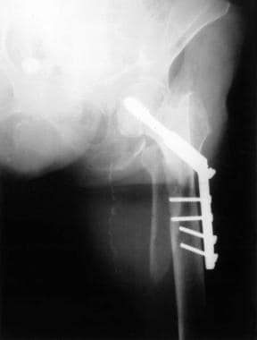
Distal femur fracture during hip arthroplasty.
-
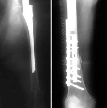
Failed fixation caused by fracture through screw holes.
-
Fracture around loose prosthesis treated with replacement.
-
Fracture at the end of implant treated with replacement.
-
Fracture around stable prosthesis treated with flexible rods.
-
Fracture around plate implant treated with rigid rod.
-
Fracture around stable prosthesis treated with rigid rod.
-
Fracture around stable prosthesis treated with standard plate.
-
Fracture around stable rod implant treated with plate.
-
Fracture around plate treated with a rod (pathologic).
-
Open reduction and internal fixation with 2 "combi" fixed-angle locking screw plates (anterior and lateral placement to help control both anterolateral and mediolateral forces).
-
Fracture around stable implant treated with less invasive stabilization system (LISS) plate.
-
Failed periprosthetic repair (total hip above, total knee below).
-
Failed periprosthetic fracture repair.
-
Vancouver B1 fracture treated with locked plate.
-
Vancouver B1 fracture at tip of well-fixed hip replacement stem.
-
Vancouver B1 fracture treated with locked plating technique.
-
Periprosthetic fracture at stem of hip replacement with insufficient proximal bone for internal fixation.
-
Periprosthetic fracture at stem of hip replacement with insufficient proximal bone for internal fixation; proximal femur replacement prosthesis with internal fixation.
-
Periprosthetic fracture.
-
Third periprosthetic fracture.
-
Third periprosthetic fracture.
-
Third periprosthetic fracture.
-
Healed third periprosthetic fracture after minimally invasive locked plating.
-
Healed third periprosthetic fracture after minimally invasive locked plating.
-
Healed third periprosthetic fracture after minimally invasive locked plating.
-
Healed third periprosthetic fracture after minimally invasive locked plating.
-
Healed third periprosthetic fracture after minimally invasive locked plating.
-
Example of periprosthetic fracture at tip of proximal stem of total knee replacement in elderly patient with osteoporotic bone that was treated with Ilizarov circular multiplanar thin wire external fixator with excellent outcome. Courtesy of Springer Nature [Nozaka K, Miyakoshi N, Hongo M, et al. Effectiveness of circular external fixator in periprosthetic fractures around the knee. BMC Musculoskelet Disord. 2020;21(317). Online at: https://bmcmusculoskeletdisord.biomedcentral.com/articles/10.1186/s12891-020-03352-9. Reused without alteration under Creative Commons Attribution 4.0 International License.]
-
Nonunion of distal femur periprosthetic fracture above total knee replacement. Preoperative AP radiograph for distal femur replacement. Courtesy of William J Hopkinson, MD, FACS, FAAOS.
-
Nonunion of distal femur periprosthetic fracture above a total knee replacement. Preoperative lateral radiograph for distal femur replacement. Courtesy of William J Hopkinson, MD, FACS, FAAOS.
-
Distal femur replacement after distal femur peri-implant fracture with prior plate fixation. Courtesy of William J Hopkinson, MD, FACS, FAAOS.


