Practice Essentials
Olecranon fractures are a diverse group of injuries, ranging from simple nondisplaced fractures to complex fracture-dislocations of the elbow joint. [1] The unique prehensile skill of human beings largely depends on the integrity of the bones, ligaments, and muscles around the elbow joint. The elbow not only bends the arm but also permits pronation and supination of the hand. Fractures of the olecranon are common and are usually detected easily but require careful treatment for an optimal result. [2, 3, 4]
Olecranon fractures can be complex injuries, presenting the physician with a wide array of surgical and nonsurgical therapeutic options. A successful functional outcome after olecranon fractures correlates directly with accuracy of anatomic joint reduction, restoration of mechanical stability that allows early motion, respect for the soft tissues, and maintenance of an intact extensor mechanism.
Nondisplaced olecranon fractures with intact extensor mechanisms are generally treated nonoperatively. Nonoperative treatment is often desirable in patients with significant associated medical conditions. Surgical treatment (see Treatment) is indicated for the following:
-
Fractures with significant displacement (>1-2 mm)
-
All patients lacking active extension of the elbow
-
Most fractures associated with elbow instability
-
Cases in which nonoperative treatment has failed
Controversy exists regarding the amount of acceptable articular displacement for closed treatment. The method chosen for open treatment of olecranon fractures is also controversial. Future treatment of olecranon fractures may very well involve percutaneous fixation accompanied by arthroscopic assistance.
For patient education resources, see Broken Elbow.
Anatomy
The elbow is a complex hinge joint. The major stabilizers to valgus stress (ie, bending away from the body) are the medial (ulnar) collateral ligament and the radial head. The major stabilizer to varus stress (ie, toward the body) is the lateral collateral ligament complex. The coronoid process stabilizes the humerus against the distal ulna.
The olecranon (the proximal bony projection of the ulna at the elbow; see the image below) also prevents anterior translation of the ulna with respect to the distal humerus. The anterior surface of the olecranon is covered with articular cartilage. Therefore, all fractures (except the rare tip fractures) are intra-articular fractures. The olecranon articulates with the trochlea of the humerus. The triceps inserts into the posterior third of the olecranon and proximal ulna. The periosteum of the olecranon blends with the triceps.
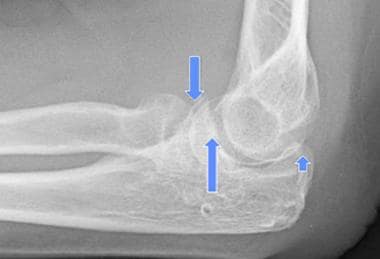 Olecranon process consists of bone of proximal ulna from base of coronoid process (down arrow) proximally. Trochlear notch (up arrows; also called semilunar notch) is articular surface shown between two arrows.
The ulnar nerve lies on the posterior aspect of the elbow, posterior to the medial collateral ligament. The ulnar nerve sweeps anteriorly to join the ulnar artery. The ulnar neurovascular bundle may be at risk during Kirschner wire (K-wire) fixation.
Fracture displacement is largely due to the pull of the triceps, which tends to pull a separated fragment upward but is resisted by the strong fibrous covering on the olecranon. This fibrous covering is formed by the blending of fibers in the lateral ligaments, the elbow capsule, and some triceps fibers that blend with the periosteum.
If the fracture force does not tear the fibrous sheath, little or no tendency toward displacement exists, even in the presence of comminution. Usually, wide separation of fragments indicates an extensive tearing of the fibrous sheath in which the unopposed triceps is contracted, drawing the separated fragment upward.
Etiology
The most common mechanism of an olecranon fracture is a fall on the semiflexed supinated forearm. As the hand strikes the ground, muscles are tensed to break the fall, and the powerful triceps snaps the olecranon over the lower end of the humerus, which acts as a fulcrum. The next most frequent cause of this injury is direct trauma, as in falls on or blows to the point of the elbow. [5]
Occasionally, the olecranon may be fractured by hyperextension injuries, such as those resulting in elbow dislocation in adults or supracondylar fractures in children. Very rarely is the olecranon broken by muscular violence, as in throwing. Stress fractures can occur in baseball players and other throwing athletes. [6]
Epidemiology
Even though the olecranon is a very heavy, strong process of bone, it is fractured frequently in adults. This is partly because of its exposed position on the point of the elbow, where most direct injuries to the elbow occur, and partly because of the tremendous cross-strain put on the olecranon during falls on the flexed forearm.
The olecranon process is rarely broken in children, because in early life, it is short, thick, and relatively much stronger than the lower end of the humerus. Usually, children sustain supracondylar fractures of the humerus instead. Nevertheless, the clinician should have a high index of suspicion in children because olecranon fractures do occur, often associated with radial head subluxation or dislocation; failure to diagnose the fracture can have long-term negative consequences.
Open fractures occur in 2-31% of cases. Neurologic injuries to median, radial, or ulnar nerves may occasionally occur. Ulnar neurapraxia has been reported in 2-5% of cases. Generally, symptoms resolve with conservative treatment, but late neurolysis or transposition may occasionally be required.
Prognosis
Approximately 95% of patients with olecranon fractures are expected to have near-normal function; 20-25% of patients will develop radiographic evidence of arthrosis at 15- to 20-year follow-up, but these patients are usually asymptomatic.
Age is the most important factor influencing outcome: Younger patients generally do better than older ones. [7]
One study retrospectively reviewed the outcome of 18 patients who underwent locking-plate osteosynthesis after open reduction for comminuted olecranon fractures. [8] In all cases, complete union was achieved. The findings indicated that whereas the risk of limited elbow motion is high in cases with concomitant injuries, locking plates are an additional and often successful option for olecranon fracture fixation.
Buijze et al compared stiffness and strength with contoured locking compression plate fixation (combined with an intramedullary screw) and one-third tubular plate fixation (combined with bicortical screws) in a cadaveric comminuted olecranon fracture model with a standardized osteotomy. [9] Stiffness was measured by subjecting the specimens to cyclic loading while measuring gapping at the osteotomy site, and strength was measured by subjecting specimens to ramp load until failure. The two fixation methods did not differ significantly with regard to construct stiffness and strength, and all failures consisted of failure of the bone, not of hardware.
In a study by Buijze and Kloen, 19 patients with an acute comminuted olecranon fracture were managed with a contoured locking compression plate and intramedullary screw fixation, 16 of whom were available for follow-up at a minimum of 12 months after fixation. [10] The authors noted that in patients managed with plate fixation for olecranon fractures, placement of an axial intramedullary screw may obstruct the placement of bicortical screws in the ulnar shaft. As a solution, they assessed the effectiveness of unicortical screws with a contoured locking compression plate.
In this study, all 19 fractures healed, and the mean time to fracture union was 4 months. [10] The mean Disabilities of the Arm, Shoulder and Hand (DASH) score was 13. According to the Mayo Elbow Performance Index and the Broberg and Morrey grading system, 15 of the 16 patients followed had a good or excellent outcome. In nine patients, hardware removal was necessary; after removal, the mean elbow extension deficit improved from 34º to 10º, and mean flexion improved from 118º to 138º.
According to Iannuzzi et al, in comminuted fractures of the olecranon (Mayo type IIB), it may be difficult or even impossible to preserve the olecranon's normal articulation with the trochlea of the humerus. [11] The authors therefore described a modified technique for reconstructing these fractures when stable anatomic reduction and fixation cannot be achieved. In this technique, the comminuted fragments are excised, and the proximal olecranon fragment is advanced past the resulting defect and fixed to the distal ulna. The authors presented two cases with clinical follow-up and noted that satisfactory preservation of range of motion (ROM) and elbow stability were achieved in each case.
-
Lateral radiograph of elbow in 78-year-old man who fell on his outstretched hand. Displaced fracture of olecranon was noted.
-
Drawing depicting radial bow of proximal third of ulna.
-
Anteroposterior radiograph following reduction and internal fixation of fracture with 7.3-mm cannulated screw and 1.6-mm cable.
-
Lateral radiograph demonstrating threads of screw engaging cortices of ulna.
-
Typical relatively transverse olecranon fracture.
-
Pediatric olecranon fracture.
-
Olecranon process consists of bone of proximal ulna from base of coronoid process (down arrow) proximally. Trochlear notch (up arrows; also called semilunar notch) is articular surface shown between two arrows.
-
Incorrect tension band technique: pins should be anchored in anterior cortex and not placed down medullary canal.
-
Transverse olecranon fracture without comminution.
-
Transverse olecranon fracture treated with tension band technique (ideally, both K-wires should have been anchored in anterior cortex).
-
Lateral plate position for olecranon fracture fixation.
-
Posterior plate position for olecranon fracture.
-
Example of distal olecranon fracture.
-
Plate fixation of distal olecranon fracture.
-
Comminuted olecranon fracture.
-
Plate fixation of comminuted olecranon fracture.
-
Example of specialized olecranon plate.
-
20-year-old man with comminuted olecranon fracture extending distally into ulnar shaft from gunshot injury.
-
Bridge plating of comminuted olecranon fracture extending into proximal ulna diaphysis after gunshot injury.
-
Monteggia-variant fracture-dislocation: olecranon and radial head fractures.
-
Monteggia-variant fracture-dislocation treated with fixation of both fractures.
-
Monteggia-variant fracture of proximal ulna and radial head.
-
Monteggia-variant fracture-dislocation treated with plate fixation of olecranon and proximal ulna and replacement of radial head.
-
Example of intramedullary screw fixation of olecranon fracture. In this case, screw diameter was too small and loss of fixation has occurred.
-
Intramedullary rod fixation of olecranon osteotomy used in repair of distal humerus fracture.
-
79-year-old man with olecranon fracture.
-
AP radiograph of tension band wiring of olecranon fracture in 79-year-old man.
-
Lateral radiograph of tension band wiring in 79-year-old man with olecranon fracture. Note excellent technique with anatomic reduction, K-wires anchored in anterior cortex, and tension band wire placed dorsally.
-
Nonunion of olecranon fracture in 79-year-old man despite excellent tension band wiring technique.
-
Excision of olecranon after nonunion of fracture in 79-year-old man.
-
Comminuted olecranon fracture.
-
Comminuted olecranon fracture plated with proximal edge off bone to allow two more screws in proximal segment and fixed angled intramedullary screw (bending plate distorts locking hole).
-
Healed comminuted olecranon fracture after removal of prominent plate fixation.
-
Olecranon nonunion after nonoperative treatment.
-
Repair of olecranon nonunion with double-plate technique (original fracture was treated nonoperatively).
-
AP radiograph of healed olecranon nonunion after original nonoperative treatment; repaired with double-plate technique.
-
Lateral radiograph of healed olecranon nonunion after original nonoperative treatment; repaired with double-plate technique.
-
MRI showing olecranon stress fracture.
-
Use of pins to raft joint surface of comminuted olecranon fracture with plate fixation of fracture as well.


