Practice Essentials
Osteosarcoma, the most common malignant bone tumor, [1, 2] is a deadly form of musculoskeletal cancer that most commonly causes patients to die of pulmonary metastatic disease (see the image below). [3, 4] It is an ancient disease that is still incompletely understood.
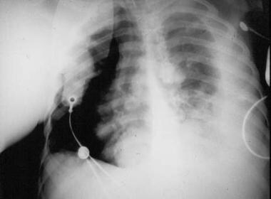 Chest radiograph of patient with osteosarcoma who died from pulmonary metastatic disease. Note the presence of a pneumothorax as well as radiodense (bone-forming) metastatic lesions.
Osteosarcoma is thought to arise from primitive mesenchymal bone-forming cells, and its histologic hallmark is the production of malignant osteoid. Other cell populations may also be present, as these types of cells may also arise from pluripotential mesenchymal cells, but any area of malignant bone in the lesion establishes the diagnosis as osteosarcoma.
Most osteosarcomas arise as solitary lesions within the fastest growing areas of the long bones of children. The top three affected areas are the distal femur, the proximal tibia, and the proximal humerus, but virtually any bone can be affected.
Not all osteosarcomas arise in a solitary fashion. Multiple sites may become apparent within a period of about 6 months (synchronous osteosarcoma), or they may be noted over a period longer than 6 months (metachronous osteosarcoma). [5] Such multifocal osteosarcoma is decidedly rare, but when it occurs, it tends to be in patients younger than 10 years.
The most common presenting symptom is pain, particularly with activity. Pathologic fractures are not particularly common, except with the telangiectatic type of osteosarcoma. Extremity pain may result in a limp. There may or may not be a history of swelling. Systemic symptoms are rare. Tumor spread to the lungs only rarely results in respiratory symptoms. Metastases to other sites are extremely rare; thus, other symptoms are unusual.
Physical examination findings are usually limited to the site of the primary tumor and may include the following:
-
Mass
-
Decreased range of motion (ROM)
-
Lymphadenopathy
-
Respiratory findings
The mainstay of therapy is surgical removal of the malignant lesion. Most often, limb-sparing (limb-preserving) procedures can be used to treat patients with this disease and, thus, preserve function. Amputation may be warranted in some circumstances. Chemotherapy is also required to treat micrometastatic disease, which is present but often not detectable in most patients (~80%) at the time of diagnosis. [6]
Guidelines for the management of osteosarcoma have been published (see Guidelines).
Pathophysiology
Osteosarcoma is a bone tumor and can occur in any bone, usually in the extremities of long bones near metaphyseal growth plates. The most common sites are as follows:
-
Femur (42%, 75% of which are in the distal femur)
-
Tibia (19%, 80% of which are in the proximal tibia)
-
Humerus (10%, 90% of which are in the proximal humerus)
-
Skull and jaw (8%)
-
Pelvis (8%)
A number of variants of osteosarcoma exist, including conventional types (osteoblastic, chondroblastic, and fibroblastic), telangiectatic, multifocal, parosteal, and periosteal. This article only addresses conventional osteosarcoma (often referred to simply as osteosarcoma).
Etiology
The exact cause of osteosarcoma is unknown. However, a number of risk factors have been identified. [3, 4, 7, 8, 9, 10, 11, 12, 13, 14]
Rapid bone growth appears to predispose persons to osteosarcoma, as suggested by the increased incidence during the adolescent growth spurt, the high incidence among large-breed dogs (eg, Great Dane, St Bernard, German shepherd), and osteosarcoma's typical location in the metaphyseal area adjacent to the growth plate (physis) of long bones.
Genetic predisposition plays a role. Bone dysplasias, including Paget disease, fibrous dysplasia, enchondromatosis, and hereditary multiple exostoses and retinoblastoma (germline form) are risk factors. The combination of constitutional mutation of the RB gene (germline retinoblastoma) and radiation therapy is linked with a particularly high risk of developing osteosarcoma, Li-Fraumeni syndrome (germline p53 mutation), and Rothmund-Thomson syndrome (autosomal recessive association of congenital bone defects, hair and skin dysplasias, hypogonadism, and cataracts).
The only known environmental risk factor is exposure to radiation. Radiation-induced osteosarcoma is a form of secondary osteosarcoma and is not discussed further in this article.
Epidemiology
In the United States, the incidence of osteosarcoma has been cited at 3.1 per million (4.4 per million population < 25 years). [15] The National Cancer Institute (NCI) Surveillance, Epidemiology, and End Results (SEER) Pediatric Monograph 1973-2004 found the incidence to be slightly higher in Blacks than in Whites, as follows [15] :
-
Blacks – 3.4 cases per million per year (5.0 per million < 25 years)
-
Whites – 3.0 cases per million per year (4.2 per million < 25 years)
The incidence of osteosarcoma is slightly higher in males than in females. According to SEER data, the average male-to-female ratio is 1.22:1. [15]
Osteosarcoma is very rare in young children (0.5 cases per million per year in children < 5 years). However, the incidence increases steadily with age, rising more dramatically in adolescence in correspondence with the adolescent growth spurt, as follows [16] :
-
Age 5-9 years – 2.6 (Black) or 2.1 (White) cases per million per year
-
Age 10-14 years – 8.3 (Black) or 7 (White) cases per million per year
-
Age 15-19 years – 8.9 (Black) or 8.2 (White) cases per million per year
A second peak of incidence exists in individuals older than 60 years. [15]
Prognosis
The present understanding of outcome and prognosis for osteosarcoma is driven by certain serum markers, clinical staging, and histologic response to chemotherapeutic agents. [17]
According to SEER data for 1973-2004, the overall relative 5-year survival rates were as follows [15] :
-
Age < 25 years - 61.6% (females, 65.8%; males, 58.4%)
-
Age 25-59 years - 58.7% (females, 64%; males, 54.6%)
-
Age 60-85+ years - 24.2% (females, 27.0%; males, 19.9%), with a drastic drop with advancing age (from ~50% for patients in their 50s to 17% for those in their middle-to-late 60s and to 10.8% for those aged 80 years or older)
Patients with an elevated ALP at diagnosis are more likely to have pulmonary metastases. In patients without metastases, those with an elevated LDH are less likely to do well than are those with a normal LDH.
Bu et al conducted a meta-analysis of eight published studies to determine whether p16(INK4a) is a prognostic factor for patients with osteosarcoma. [18] The meta-analysis showed that a high level of expression of p16(INK4a) was significantly associated with favorable overall survival. The investigators concluded that p16(INK4a) is an effective biomarker of survival for patients with osteosarcoma.
Ma et al conducted a study to determine the diagnostic and prognostic value of circulating miR-148a in the peripheral blood of patients with osteosarcoma. [19] Expression of miR-148a was significantly associated with tumor size and distant metastasis. High expression of miR-148a was associated with poor overall survival and poor disease-specific survival. The investigators concluded that detection of circulating miR-148a expression in the peripheral blood is useful in identifying patients with osteosarcoma who have a poor prognosis.
A study by Zhao et al found that high expression of the oncoprotein transient receptor potential melastatin member 8 (TRPM8) was predictive of a poor prognosis in patients with osteosarcoma, in that it was associated with higher clinical stage and distant metastasis, as well as with shorter overall survival and disease-free survival. [20] TRPM8 may prove to be a useful molecular target for therapy in osteosarcoma patients.
Clinical staging as it relates to prognosis is discussed elsewhere (see Staging).
In a retrospective study by Kim et al, the records of 331 patients with stage II osteosarcoma who had undergone surgery and chemotherapy were reviewed. [21] The authors found that initial tumor size appears to be associated with histologic response and is an important prognostic factor in osteosarcoma. Other studies have shown that patients in whom a good histopathologic response to neoadjuvant chemotherapy has been achieved (>95% tumor cell kill or necrosis) have a better prognosis than those whose tumors do not respond as favorably.
-
Chest radiograph of patient with osteosarcoma who died from pulmonary metastatic disease. Note the presence of a pneumothorax as well as radiodense (bone-forming) metastatic lesions.
-
Clinical appearance of a teenager who presented with osteosarcoma of the proximal humerus (same patient as in the following images). Note the impressive swelling throughout the deltoid region, as well as the disuse atrophy of the pectoral musculature.
-
Radiographic appearance (plain radiograph) of a proximal humeral osteosarcoma (same patient as previous image). Note the radiodense matrix of the intramedullary portion of the lesion, as well as the soft-tissue extension and aggressive periosteal reaction.
-
Intense radionuclide uptake of the proximal humerus is noted on a bone scan (same patient as previous 2 images).
-
A comparison bone scan of the involved shoulder (right image) with the uninvolved shoulder (left image) (same patient as previous 3 images).
-
Magnetic resonance image appearance (T1-weighted image) of osteosarcoma of the proximal humerus (same patient as previous 4 images). Note the dramatic tumor extension into the adjacent soft-tissue regions.
-
Core needle biopsy instruments commonly used for bony specimens. Craig needle set.
-
Close-up view of Craig needle biopsy instruments. Cutting cannula with T-handle attached (top) and sheath through which the cutting cannula passes (bottom).
-
Resected specimen of a proximal tibia osteosarcoma. The primary lesion was such that the knee joint was resected with the primary lesion. Note that the previous longitudinal biopsy tract was completely excised with the specimen.
-
Intraoperative consultation with the pathologist, in which the surgeon and pathologist view the microscopic appearance of the biopsy specimen.
-
Intraoperative consultation with the pathologist. A frozen section of the biopsy specimen is being performed.
-
Intraoperative photograph of a Van Ness rotationplasty procedure. Osteosynthesis of the tibia to the residual femur is being performed. Courtesy of Alvin H. Crawford MD, FACS.
-
Clinical photograph taken at the conclusion of a Van Ness rotationplasty procedure (same patient as previous image). Note that the new "knee" of the operative side (left side) is purposely reconstructed distal to the normal right knee. This is in anticipation of the future growth potential of the unoperated limb. Courtesy of Alvin H. Crawford MD, FACS.