Background
Macular corneal dystrophy (MCD) is a rare but severe stromal corneal dystrophy. Macular corneal dystrophy is an IC3D category 1 dystrophy and is an autosomal-recessive condition. It is characterized by multiple irregular gray-white opacities in the corneal stroma that extend out into the peripheral cornea and down to the Descemet membrane.
Corneal dystrophy is defined as a bilateral noninflammatory clouding of the cornea, the clear outer layer of the front of the eye. Corneal dystrophies can be placed into categories based on their location within the cornea and their etiologies, as follows: [1]
-
Anterior corneal dystrophies affect the corneal epithelium and subepithelium.
-
Transforming growth factor beta–induced (TGFBI) dystrophies affect the epithelium and stroma.
-
Stromal corneal dystrophies affect the stroma (the central layer of the cornea).
-
Posterior corneal dystrophies involve the Descemet membrane and endothelium.
Whereas macular corneal dystrophy is now classified as a stromal corneal dystrophy, granular and lattice corneal dystrophies are now classified as epithelial-stromal TGFBI dystrophies owing to their involvement in multiple layers of the cornea. [1] The age of onset for most corneal dystrophies is less than 20 years (exceptions include map-dot-fingerprint dystrophy and Fuchs corneal dystrophy). Patients with macular corneal dystrophy tend to have more severe vision loss earlier than patients with lattice or granular dystrophy; however, macular corneal dystrophy is less common than both lattice and granular dystrophies.
Most corneal dystrophies are inherited in an autosomal-dominant pattern. Exceptions that are autosomal recessive include macular corneal dystrophy and congenital hereditary endothelial dystrophy (CHED) (previously known as autosomal recessive CHED2, differentiated from autosomal dominant CHED1, which has been eliminated owing to its similarity to posterior polymorphous corneal dystrophy). [1]
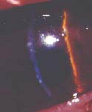
Macular corneal dystrophy, unlike granular corneal dystrophy, has no clear areas between opacities. [2] Opacities usually first appear in adolescence but may become apparent anytime from early infancy to the sixth decade of life. Affected individuals usually experience severe visual impairment before the fifth decade of life once opacities have coalesced and the entire stroma becomes cloudy. [3] Examples of macular dystrophy are shown in the images below.
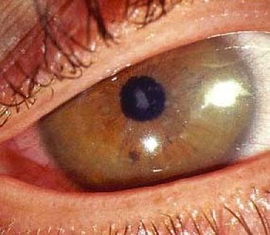 Macular dystrophy. Image courtesy of James J. Reidy, MD, FACS, Associate Professor of Ophthalmology, State University of New York, School of Medicine & Biomedical Sciences, Buffalo, New York.
Pathophysiology
The metabolic defect for macular corneal dystrophy appears to be an error in the synthesis of keratan sulfate, which leads to accumulation of glycosaminoglycans in the cornea. Subgroups of macular dystrophy can be identified by immunohistochemical methods. Keratan sulfate was not detected in the serum of patients with histopathologically confirmed macular corneal dystrophy. Because keratan sulfate in the serum appears to be predominantly derived from the normal turnover of cartilage, [4] these studies strongly suggest that the defect in keratan sulfate synthesis in macular corneal dystrophy is not restricted to corneal cells and that this condition is one manifestation of a systemic disorder of keratan sulfate.
Epidemiology
Frequency
United States
Macular corneal dystrophy is uncommon, but areas with the highest prevalence include parts of the United States.
International
Although relatively uncommon, macular corneal dystrophy is most prevalent in India, Saudi Arabia, Iceland, and parts of the United States. [3]
Mortality/Morbidity
Corneal changes become visible in the first decade of life. A significant reduction in vision usually occurs by age 20-40 years. Patients can develop decreased corneal sensitivity. Eye pain due to recurrent corneal erosions is rare is much less common than in patients with lattice or granular corneal dystrophies.
Sex
No sexual predilection has been reported.
Age
Corneal changes become visible in the first decade of life; vision may be significantly reduced by age 20-40 years.
Prognosis
Of all the stromal corneal dystrophies, macular corneal dystrophy results in the earliest visual loss. This visual loss is due to the lack of clear spaces between the denser gray-white macular opacities.
Symptomatic patients are eligible for either excimer laser phototherapeutic keratectomy (PTK) when the bulk of the opacity is superficial or, more commonly, corneal transplantation.
-
Macular dystrophy. Image courtesy of James J. Reidy, MD, FACS, Associate Professor of Ophthalmology, State University of New York, School of Medicine & Biomedical Sciences, Buffalo, New York.
-
Macular dystrophy.

