Continually Updated Clinical Reference
 
 
  All Sources     eMedicine     Medscape     Drug Reference     MEDLINE
 
eMedicine - Posterior Polymorphous Corneal Dystrophy : Article by

Quick Find
Authors & Editors
Introduction
Clinical
Differentials
Workup
Treatment
Medication
Follow-up
Miscellaneous
Multimedia
References

Related Articles
Dystrophy, Fuchs Endothelial

Glaucoma, Primary Congenital


Patient Education
Click here for patient education.


Author: Dustin J Coupal, MD, FRCSC, Eye Specialist and Surgeon, Private Practice

Dustin J Coupal is a member of the following medical societies: American Academy of Ophthalmology, Canadian Medical Association, Canadian Medical Protective Association, and Canadian Ophthalmological Society

Coauthor(s): W Keith Hamilton, MD, Clinical Assistant Professor, Department of Ophthalmology, Saskatoon City Hospital Eye Centre

Editors: Fernando H Murillo-Lopez, MD, Senior Surgeon, Unidad Privada de Oftalmologia CEMES; Francisco Talavera, PharmD, PhD, Senior Pharmacy Editor, eMedicine; Christopher J Rapuano, MD, Professor, Department of Ophthalmology, Jefferson Medical College; Co-Chairman of the Cornea Service, Co-Chairman of Refractive Surgery Department, Wills Eye Hospital; Ralph Garzia, OD, Assistant Dean for Clinical Programs, Associate Professor, School of Optometry, University of Missouri at St Louis; Hampton Roy Sr, MD, Associate Clinical Professor, Department of Ophthalmology, University of Arkansas for Medical Sciences

Author and Editor Disclosure

Synonyms and related keywords: PPMD, posterior polymorphous dystrophy, posterior endothelial dystrophy, hereditary deep dystrophy, PPCD

Background

First described by Koeppe in 1892, posterior polymorphous corneal dystrophy (PPMD) is a dominantly inherited condition characterized by particular alterations of the Descemet membrane and the corneal endothelium. Typically, the corneal changes are either slowly progressive or nonprogressive. In severe cases, corneal decompensation and edema can occur. Although PPMD is most often a bilateral condition, marked asymmetry in the degree of involvement may be seen. Most persons with PPMD are asymptomatic.

Pathophysiology

Three main abnormalities are described: vesicular changes, endothelial band lesions, and irregular opacities of the posterior corneal surface. The corneal endothelium undergoes a transformation and demonstrates many epithelial characteristics on examination with electron microscopy and immunohistochemical analysis. Often, the endothelium is found to be multilayered.

Frequency

United States

The frequency of PPMD is not well documented. Although it is considered to be uncommon, PPMD may be recognized and diagnosed more often in recent years than in the past.

Mortality/Morbidity

The effect of PPMD on patients is highly variable, with a broad clinical spectrum of findings, ranging from nonprogressive asymptomatic disease to progressive or advanced debilitating corneal disease with corneal decompensation and glaucoma.

Race

No racial predilection exists.

Sex

No sexual predilection exists.

Age

Although PPMD is an inherited corneal dystrophy, the age at diagnosis is highly variable because of the broad spectrum of disease severity. Findings may be present at birth. Most patients are first identified at age 30-50 years; however, this is likely only indicative of a more common age for ocular examinations.

  • Some patients may present at birth with congenital disease, exhibiting advanced disease with corneal edema.
  • Presentation in adulthood is indicative of a more stable disease state with a decreased probability of progression to corneal decompensation.
  • Most patients with significant symptoms present at age 25-50 years.


History

  • The clinical findings in patients with PPMD are highly variable, with a broad clinical spectrum of findings, ranging from only occasional Descemet membrane vesicles to progressive debilitating corneal disease with corneal decompensation and glaucoma.
  • A family history of PPMD should be assessed.
  • Although most patients with PPMD are asymptomatic, the most common symptoms in those with more significant involvement are as follows:
    • Photophobia
    • Decreased visual acuity
    • Foreign body sensation

Physical

  • The most characteristic finding on slit lamp biomicroscopy is multiple vesicles or blisters, either isolated or grouped in clusters, on the posterior corneal surface. The vesicles often have identifiable surrounding halos.
  • Slit lamp biomicroscopy may also identify all or some of the following characteristics depending on the extent of the disease:
    • Posterior corneal opacities occurring in linear bands or other polymorphous configurations with irregular scalloped edges
    • Areas of more diffuse posterior corneal opacity
    • Focal excrescents of the Descemet membrane
    • Iridocorneal adhesions
    • Pupillary ectropion
    • Corectopia
    • Stromal and epithelial edema (advanced cases)

Causes

  • The precise etiology of PPMD remains unknown. PPMD is a congenital inherited dystrophy involving abnormalities of the corneal endothelium and the Descemet membrane.
    • Most cases of PPMD are transmitted in an autosomal dominant fashion with variable expression, although autosomal recessive transmission has also been reported.
    • A number of autosomal dominant cases of PPMD have been linked to a mutation in an unidentified gene located at band 20q11, whereas other cases have reported a mutation in a gene encoding for collagen VIII located on chromosome 1 (COL8A2) as the cause of PPMD.
    • In other cases, the genetic locus remains unknown.
  • PPMD has been associated with other conditions, including the following:
    • Alport syndrome
    • Keratoconus


Dystrophy, Fuchs Endothelial
Glaucoma, Primary Congenital

Other Problems to be Considered

Congenital hereditary endothelial dystrophy (CHED)
Iridocorneal endothelial (ICE) syndrome


Imaging Studies

  • Specular microscopy has proven to be valuable in identifying the characteristic vesicular changes of the Descemet membrane in patients with PPMD. Typically, the endothelial cell count is only mildly diminished with varying amounts of polymegethism and pleomorphism.
  • Confocal microscopy has also been used to identify the typical features of PPMD.
  • On most occasions, PPMD can be clinically diagnosed using slit lamp examination; however, mild cases may require further imaging studies to confirm the diagnosis.

Other Tests

  • Tonometry is used to measure intraocular pressure, which may be elevated in up to 40% of patients with PPMD.
  • The secondary open-angle glaucoma that occurs in some patients with PPMD is believed to be due to an abnormality of the trabecular endothelium.
  • Examination of family members may also be helpful in making the diagnosis.

Histologic Findings

Light microscopy typically demonstrates 4 types of cells: normal endothelial cells, degenerated endothelial cells, fibroblast-like cells, and epithelial-like cells. In 60% of cases, a multilayered endothelium can be identified that has many characteristics similar to that of the epithelium. Immunohistochemical studies have also shown positive staining of the endothelium using epithelial cell markers.

Scanning electron microscopy and transmission electron microscopy of corneal tissue show lamination of the Descemet membrane, fibroblast-like endothelial cells, and microvilli characteristic of epithelial transformation of the corneal endothelium.

Staging

No formal staging system has been described for PPMD.


Medical Care

Management of PPMD varies widely based on the differences in severity of corneal decompensation. Many patients who are asymptomatic and show minimal signs of PPMD can be treated conservatively and do not require therapy.

  • Ruptured corneal bullae should be treated similar to a corneal abrasion.
  • Hyperosmotic saline drops and ointments may be used in cases of corneal failure with corneal edema.
  • Secondary glaucoma may require medical or surgical management.
  • A bandage soft contact lens may be used as a temporary measure to treat bullous keratopathy.

Surgical Care

  • Corneal transplantation is usually reserved for patients with substantially decreased visual acuity or when the disease is advanced and painful due to ruptured epithelial bullae. Penetrating keratoplasty (full thickness corneal transplantation) has been the transplant procedure of choice in the past. The newer techniques of posterior endothelial keratoplasty may have some advantages for some patients.
  • Rare cases of corneal graft failure due to recurrence of PPMD in the donor graft have been reported, but the most common problem following a corneal transplant is related to uncontrolled glaucoma.
  • To treat pain in eyes without good visual potential, other procedures, such as anterior stromal micropuncture, excimer laser phototherapeutic keratectomy, amniotic membrane transplantation, and conjunctival flap surgery, can also be considered.
  • When glaucoma medications no longer adequately control the intraocular pressure, glaucoma laser or incisional surgery may be required to prevent glaucomatous optic nerve damage.

Consultations

Depending on the severity of the condition, consultation with corneal and glaucoma subspecialists may be warranted.


The goals of pharmacotherapy are to reduce morbidity and to prevent complications.

Drug Category: Hypertonic agents

Sodium chloride hypertonic ophthalmic solutions are used to dehydrate the cornea.

Drug NameSodium chloride (Muro 128 Ophthalmic)
DescriptionUsed for temporary relief of corneal edema. Available as 2% and 5% ophthalmic solution concentrations and 5% ointment.
Adult DoseSolution: 1-2 gtt into affected eye(s) q3-4h
Ointment: Apply 0.5-inch ribbon into affected eye(s) q3-4h
Pediatric DoseAdminister as in adults
ContraindicationsDocumented hypersensitivity
InteractionsNone reported
PregnancyC - Safety for use during pregnancy has not been established.
PrecautionsMay cause temporary burning and irritation upon use; if pain, change in vision, continued redness, or irritation of the eye occurs or if the initial condition/problem worsens or persists, reevaluate therapy; do not use product if it changes color or becomes cloudy


Further Outpatient Care

  • The frequency of follow-up care for patients with PPMD depends on the severity of disease.

Complications

  • Corneal erosion, secondary corneal ulcers, corneal scarring, graft rejection, endothelial failure, and other complications can occur and lead to corneal graft failure.
  • Glaucoma can be present prior to corneal transplantation or may result from corneal transplantation.

Prognosis

  • The prognosis is generally dependent on the severity of disease. In most cases, PPMD is a slowly progressive or nonprogressive disease. Patients with mild disease that is identified in adulthood have the best prognosis and are unlikely to require penetrating keratoplasty during their lifetime.


Medical/Legal Pitfalls

  • Failure to have patients who have recently been diagnosed with PPMD undergo renal evaluation and hearing tests to assess for Alport syndrome, which has been associated with PPMD.
  • Patients need to be monitored for the development of glaucoma.

Special Concerns

  • Family members should be examined for evidence of asymptomatic corneal disease, which may help establish a diagnosis of PPMD.


Media file 1:  
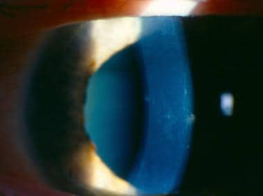
Slit lamp image demonstrates posterior corneal vesicles and opacities in linear bands and other polymorphous configurations typical of posterior polymorphous corneal dystrophy.
Click to see larger pictureClick to see detailView Full Size Image
Media type:  Photo


  • Anderson NJ, Badawi DY, Grossniklaus HE, Stulting RD. Posterior polymorphous membranous dystrophy with overlapping features of iridocorneal endothelial syndrome. Arch Ophthalmol. Apr 2001;119(4):624-5. [Medline].
  • Bechara SJ, Grossniklaus HE, Waring GO, Wells JA 3rd. Keratoconus associated with posterior polymorphous dystrophy. Am J Ophthalmol. Dec 15 1991;112(6):729-31. [Medline].
  • Boruchoff SA, Weiner MJ, Albert DM. Recurrence of posterior polymorphous corneal dystrophy after penetrating keratoplasty. Am J Ophthalmol. Mar 15 1990;109(3):323-8. [Medline].
  • Brooks AM, Grant G, Gillies WE. Differentiation of posterior polymorphous dystrophy from other posterior corneal opacities by specular microscopy. Ophthalmology. Nov 1989;96(11):1639-45. [Medline].
  • Chiou AG, Kaufman SC, Beuerman RW, et al. Confocal microscopy in posterior polymorphous corneal dystrophy. Ophthalmologica. 1999;213(4):211-3. [Medline].
  • Colville DJ, Savige J. Alport syndrome. A review of the ocular manifestations. Ophthalmic Genet. Dec 1997;18(4):161-73. [Medline].
  • Grupcheva CN, Chew GS, Edwards M, et al. Imaging posterior polymorphous corneal dystrophy by in vivo confocal microscopy. Clin Experiment Ophthalmol. Aug 2001;29(4):256-9. [Medline].
  • Henriquez AS, Kenyon KR, Dohlman CH, et al. Morphologic characteristics of posterior polymorphous dystrophy. A study of nine corneas and review of the literature. Surv Ophthalmol. Sep-Oct 1984;29(2):139-47. [Medline].
  • Klintworth GK. The molecular genetics of the corneal dystrophies--current status. Front Biosci. May 1 2003;8:d687-713. [Medline].
  • Koeppe L. Klinische Beobachtungen mit der Nerstspaltlampe und dem Hornhautmikroskop. Albrecht von Graefe's Arch Klin Exp Ophthalmol. 1916;91:375-379.
  • Krachmer JH. Posterior polymorphous corneal dystrophy: a disease characterized by epithelial-like endothelial cells which influence management and prognosis. Trans Am Ophthalmol Soc. 1985;83:413-75. [Medline].
  • Laganowski HC, Sherrard ES, Muir MG, Buckley RJ. Distinguishing features of the iridocorneal endothelial syndrome and posterior polymorphous dystrophy: value of endothelial specular microscopy. Br J Ophthalmol. Apr 1991;75(4):212-6. [Medline].
  • Moroi SE, Gokhale PA, Schteingart MT, et al. Clinicopathologic correlation and genetic analysis in a case of posterior polymorphous corneal dystrophy. Am J Ophthalmol. Apr 2003;135(4):461-70. [Medline].
  • Presberg SE, Quigley HA, Forster RK, Green WR. Posterior polymorphous corneal dystrophy. Cornea. 1985-86;4(4):239-48. [Medline].
  • Teekhasaenee C, Nimmanit S, Wutthiphan S, et al. Posterior polymorphous dystrophy and Alport syndrome. Ophthalmology. Aug 1991;98(8):1207-15. [Medline].
  • Weissman BA, Ehrlich M, Levenson JE, Pettit TH. Four cases of keratoconus and posterior polymorphous corneal dystrophy. Optom Vis Sci. Apr 1989;66(4):243-6. [Medline].

Posterior Polymorphous Corneal Dystrophy excerpt

Article Last Updated: Feb 1, 2007