Background
Trochlear nerve palsy is mentioned in ophthalmology texts dating to the mid nineteenth century. However, it received little more than a brief mention and was no doubt an underrecognized entity. In 1935, Bielschowsky correctly noted that trochlear nerve palsy was the most common cause of vertical diplopia and introduced his classic head-tilt test. With greater clinical interest, the number of identified fourth nerve palsies has increased.
Key Considerations
A fourth nerve palsy is a common cause of binocular vertical oblique diplopia in isolation.
The fourth cranial nerve exits dorsally and has the longest intracranial course.
An isolated fourth cranial nerve palsy usually can be diagnosed using the 3-step test.
The primary action of the superior oblique muscle is intorsion.
History of the Procedure
The introduction of the Harada-Ito procedure in the 1960s and Knapp's surgical approach in the 1970s enhanced the ability to successfully treat this challenging clinical entity. [1]
Problem
The fourth cranial nerve innervates the superior oblique muscle, which intorts, depresses, and abducts the globe. [2, 3] Fourth nerve palsy can be congenital or acquired, unilateral or bilateral; each of these presents with a distinct clinical picture. [2, 4] Clinicians must carefully assess the patient to determine both the etiology and extent of disease. Acquired weakness of this muscle usually leads to complaints of binocular vertical or oblique diplopia, sometimes with a torsional component. Surgery may be required to treat these patients. Thorough assessment and careful preoperative planning maximize the chances of a successful surgical outcome. [2]
Epidemiology
Frequency
Most cases of isolated fourth nerve palsy are believed to be congenital. [5] However, estimating the true frequency of congenital fourth nerve palsy is difficult. Many patients compensate with use of head-tilt or large fusional amplitudes; therefore, it may not present to an ophthalmologist until adulthood, when their fusional control begins to deteriorate. [2]
Some of the best information regarding the incidence of acquired fourth nerve palsy can be found in the Mayo Clinic series. Several studies reported the incidence and etiology of acquired cranial nerve palsies in adult and pediatric patients. Trochlear nerve palsy was less common than abducens or oculomotor palsies. Of 4,373 acquired cases of extraocular muscle palsy in adults, there were only 657 cases of isolated fourth nerve disease. [6] Fourth nerve palsy also was the least frequent in a pediatric population. In a similar Mayo Clinic study of 160 children, 19 of them had isolated fourth nerve palsy. [7, 8]
Etiology
The most common cause of congenital trochlear nerve palsies is congenital cranial dysinnervation syndrome, followed by an abnormal superior oblique tendon. [9, 10, 11, 2]
The most common cause of acquired isolated fourth nerve palsy, after idiopathic, is head trauma. [12, 13, 14, 2]
One must consider the possibility of underlying structural abnormalities (eg, skull based tumor) if fourth nerve palsy results after only minor trauma.
Microvasculopathy secondary to diabetes, atherosclerosis, or hypertension also may cause isolated fourth nerve palsy. [15]
There are rare reports of thyroid ophthalmopathy and myasthenia gravis mimicking an isolated fourth nerve palsy. These patients eventually develop other findings, unmasking the underlying diagnosis.
Tumor, aneurysm, multiple sclerosis, or iatrogenic injury may present with isolated fourth nerve palsy that may evolve over time to include other cranial nerve palsies or neurologic symptoms. [16]
Fourth nerve palsy may become manifest after cataract surgery. Patients with underlying, well-controlled, and asymptomatic fourth nerve palsy may decompensate gradually as they lose binocular function resulting from cataract. After restoration of good vision, these patients become aware of diplopia.
Pathophysiology
Congenital Trochlear Nerve Palsy
A series of high-definition magnetic resonance imaging (MRI) studies by Yang et al have identified 2 etiologies of congenital trochlear nerve palsies, with the most common being congenital cranial dysinnervation syndrome. This syndrome was present in 73% of congenital trochlear nerve palsy cases and is characterized by absence of the trochlear nerve and secondary atrophy of the superior oblique muscle. The remaining 27% had a normal trochlear nerve and superior oblique muscle size, but an abnormal superior oblique tendon, which may explain the variations in superior oblique tendon laxity encountered surgically. [9, 10, 11]
Helveston, in a series of 36 patients with congenital superior oblique palsy, found 33 abnormal superior oblique tendons. [17] The tendon may be abnormally lax, have an abnormal insertion, or be absent altogether.
Acquired Trochlear Nerve Palsy
The long course of the trochlear nerve makes it especially susceptible to injury in association with severe head trauma. Contrecoup forces can compress the nerve against the rigid tentorium, which lies adjacent to the nerve for much of its course. Injury to nerve can occur anywhere along its course from midbrain to orbit. Lesions at the nucleus cause contralateral superior oblique palsy, since the nerve decussates at the anterior medullary velum, caudal to the inferior colliculus. Midbrain trauma can produce bilateral superior oblique palsy by contusive injury of decussation of nerves. Compression or ischemia at this site also can produce bilateral palsy. [2]
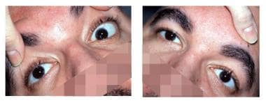 Patient with traumatic bilateral superior oblique palsy; note right hypertropia on right head tilt and left hypertropia on left head tilt.
One should suspect a lesion to the trochlear nucleus or fascicle when palsy is associated with a contralateral Horner syndrome or an ipsilateral relative afferent pupillary defect (RAPD, especially without concomitant visual loss [ie, tectal RAPD]). This is due to the close proximity of the sympathetic pathways in the dorsolateral tegmentum of the midbrain and the pretectal afferent pupillary fibers that run through the superior colliculus.
Tumors or aneurysms causing compressive injury in the subarachnoid space generally damage adjacent structures and produce associated neurologic signs. The same is true of lesions in the area of cavernous sinus and orbital apex, which generally produce multiple cranial neuropathies. In rare cases, fourth nerve palsy may result from any cause of increased intracranial pressure such as pseudotumor cerebri or meningitis. Direct orbital injury can result in a clinical picture that resembles fourth nerve palsy, but superior oblique weakness in this setting most likely is due to direct damage to muscle or tendon.
Presentation
The superior oblique muscle intorts, depresses, and abducts the globe.
In acquired lesions of the fourth nerve, patients report vertical, torsional, or oblique diplopia. Diplopia is usually worse on downgaze and gaze away from side of affected muscle.
In case of trauma, patients usually report symptoms immediately after regaining consciousness.
Torsional diplopia and downgaze horizontal diplopia may be predominant complaints in bilateral palsies. [18]
Patients may adopt a characteristic head tilt, away from the affected side, to reduce their diplopia. Interestingly, some patients develop head tilt toward the side of the lesion. This so-called paradoxic head tilt is used to create a wider separation of images, which allows the patient to suppress or ignore one image. [18] Old photographs may provide clear documentation of a head tilt in congenital fourth nerve palsy.
Congenital fourth nerve palsies may present with several unique findings, as follows:
-
Patients with long-standing head tilt present during early childhood may develop facial asymmetry. Characteristically, there is shallowing of the face between the lateral canthus and the side of the mouth on the side of the head tilt. Any condition that leads to torticollis in early life may result in similar facial asymmetry.
-
Patients with congenital palsies also tend to develop large, vertical fusional amplitudes, and they may have lack of subjective torsion even when large amounts of fundus torsion are present.
Patients with congenital superior oblique palsy who are lacking a trochlear nerve develop a head tilt at an earlier age. Patients with congenital superior oblique palsy who have a normal trochlear nerve demonstrate more overelevation in adduction and frequent dissociated vertical deviation. [9]
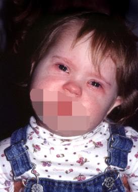 A 2-year-old girl with compensatory left head tilt due to congenital right superior oblique palsy.
Indications
For patients with decompensating congenital fourth nerve palsy, indications for intervention include cosmetically or functionally unacceptable head position, and onset of increasing frequency of diplopia.
Patients with acquired disease from tumors or compressive lesions usually are significantly disturbed by symptoms and are likely to require prism or, in some cases, surgical intervention.
Relevant Anatomy
The trochlear nucleus is located in the tegmentum of the midbrain, at the level of the inferior colliculus. [12, 3] The trochlear nerves decussate at the anterior medullary velum in the roof of the aqueduct before exiting from the dorsal aspect of the midbrain. The fourth nerve courses between the posterior cerebral and superior cerebellar arteries before entering the cavernous sinus. The fourth nerve then enters the orbit through the superior orbital fissure and the outside annulus of Zinn. From here, the nerve crosses medially over the levator palpebrae superioris and superior rectus muscles before entering the belly of the superior oblique muscle.
The superior oblique muscle originates from the orbital apex, above the annulus, and runs along the superonasal aspect of orbit before becoming a tendinous cord. The superior oblique tendon then passes through the trochlea and abruptly turns laterally and posteriorly to insert on the globe. The tendon is cordlike as it passes beneath the nasal border of the superior rectus, but fans out to form a broad insertion.
When performing a superior oblique tenotomy, the superior rectus muscle insertion may be used as a landmark. The portion of tendon that is cut during the tenotomy may be isolated by dissecting to a point approximately 8-12 mm posterior to nasal aspect of superior rectus insertion. Broad superior oblique insertion, which is 10-18 mm in length, has great functional importance. Anterior fibers act mainly to intort the globe and do little to abduct or depress the eye. Conversely, more posterior fibers are responsible for abduction and depression but have little torsional action. Surgical procedures designed to alleviate torsional diplopia, such as the Harada-Ito procedure, consist of advancing only anterior fibers of tendon insertion.
Contraindications
Patients with microvascular disease have a high likelihood of resolution. These patients may be observed and advised to patch 1 eye or use monovision lenses to minimize their symptoms.
Similarly, patients who have traumatic fourth nerve palsy may be observed for 6 months prior to surgical intervention because of the possibility of spontaneous resolution; however, some traumatic palsies may recover as late as 1 year after injury. [19]
Prognosis
The prognosis of a fourth nerve palsy depends on the underlying etiology. Congenital palsies are long standing and often remain static. Acquired, demyelinating (rare), traumatic, ischemic (microvascular), and idiopathic palsies usually resolve over time. The prognosis of fourth nerve palsies due to a structural lesion depends on the treatment of the underlying lesion. Most patients with symptoms that do not recover spontaneously can improve with prism or surgery.
Patient Education
Patients should be advised on the etiology and prognosis of the fourth nerve palsy. Prism or surgical therapy can be considered in patients who have stable and unresolved ocular deviations.
-
A 2-year-old girl with compensatory left head tilt due to congenital right superior oblique palsy.
-
Postoperative photo of same girl; note marked improvement of head tilt.
-
Patient with traumatic bilateral superior oblique palsy; note right hypertropia on right head tilt and left hypertropia on left head tilt.


