Overview
This article is not an exhaustive treatise of neuro-ophthalmic signs, but it emphasizes some practical aspects and common pitfalls of the physical examination.
Incomplete assessment of patients during routine examinations and the failure to correlate symptoms or neuroimaging findings with signs are probably more common reasons for missed neuro-ophthalmic diagnoses than the potential subtlety of neuro-ophthalmic signs. Suggestions for incorporating a more thorough, efficient eye examination in routine practice are included in the Examination efficiency, Bedside examination, and Functional visual loss sections below.
Combining the history, physical examination, and/or neuroimaging into a logical package is essential. A patient claiming bilateral, profound visual loss should not be dressed immaculately, unless groomed by a sighted caregiver. In patients with good binocular acuity complaining of constant binocular diplopia, examination should show a deviation on alternate-cover testing.
Reviewing the patient's old photographs helps to determine the chronicity of many lid, pupil, dysmotility, and orbital problems. The value of photo review should not be underestimated.
Indications
Indications for neuro-ophthalmic examination include the following:
-
Routine neuro-ophthalmic examination
-
Examination of patients who present with neuro-ophthalmic signs
Technique
Much information can be garnered if the physician escorts the patient to the examining lane. The patient's behavior in the waiting room (eg, use of cell phones), interaction with family or caregivers, and gait and navigational ability can be ascertained. The examining lane should be brightly illuminated to allow perusal of the patient's face and eyes. Anomalous head posture, such as head tilt, which may suggest contralateral palsy of cranial nerve (CN) IV, should be noted. The patient's dress and cleanliness often are clues to visual function. Post COVID, if one shakes hands with the patient, observe the patient's fingers, skin, and grip. Mental status, speech, and affect can be assessed during history taking.
Sensory testing
Stereovision testing and Worth 4-dot testing are not routine parts of the neuro-ophthalmic examination, but they are useful in certain situations.
Sensory testing is best performed before fusion is disrupted by ocular occlusion. The author's preferred stereovision test is the Titmus stereofly test.
Stereovision testing is used commonly in pediatric ophthalmologic examinations, and it is useful in patients with suspected functional visual loss.
A normal result on stereovision testing suggests 20/20 acuity in both eyes and is not compatible with poor visual acuity.
Worth 4-dot testing is useful if patients have difficulty expressing their diplopia. In patients with strabismus and good acuity in both eyes, central suppression suggests an element of amblyopia rather than an acquired motility deficit. The green filter should be placed before the better seeing of the 2 eyes. Red-green glasses can also be used for duochrome tests in patients suspected of malingering. The duochrome test should be checked by the physician personally prior to the patient being brought to the examination room in order to ensure that the colored filters are properly matched.
Refraction
The patient's refractive error should be documented. Although measuring pinhole acuity is useful, the pinhole corrects for only about 3 diopters (D) of refractive error. Patients with high refractive error, macular disease, or hand tremor may not improve their vision with the pinhole. Autorefractors are usually helpful.
Some patients with poor mentation or poor head control cannot use the phoroptor (ie, a machine containing a rotating bank of lenses). In such patients, complete the refraction with a trial frame.
It is not uncommon for patients with keratoconus (warped corneas) to present to the neuro-ophthalmologist with diplopia or blurry vision. A record of any scissoring of the retinoscopic reflex (an abnormal light reflex during objective refraction with a retinoscope) or irregular keratometry mires (a keratometer that measures corneal curvature) may be helpful if corneal topography is not readily available.
Cycloplegic refraction (refraction with cycloplegic drops instilled to relax accommodation) should be performed in all children with abnormal vision examination and should be considered in patients with comitant esotropia, unexplained headache, or unexplained visual complaints.
Visual acuity
Documentation of the patient's best-corrected visual acuity is essential. Carefully observe the patient during acuity testing to ensure that the chart is being read from only 1 eye at a time. In children, the author routinely tapes a tissue over the eye not being tested; alternately, the author asks the parent to occlude the child's eye not being tested.
Patients with homonymous hemianopsia may cut, or ignore, relevant portions of the eye chart and/or color-vision chart, or they may turn their face to compensate for the field loss.
Correlation of the distance vision with the near acuity is valuable. If near vision is better than distance vision or vice versa, ensure that the proper refraction was used and that the near card was held at the appropriate distance from the eyes.
In young patients, patients who are illiterate, or patients with decreased mentation, picture charts (eg, Allen figures), HOTV chart, a Sheridan Gardner chart, illiterate E charts, or Landolt C charts are useful. These charts allow the patient to indicate their acuity without having to read or verbalize. An optokinetic response suggests that the patient has at least 20/400 vision.
In patients with decreased Snellen acuity who have no visual complaints, exclude latent nystagmus. In patients with latent nystagmus, document binocular visual acuity and check their vision monocularly while fogging the other eye with an overplussed refraction (a high plus lens).
Color-vision testing
Ishihara color plates are a commonly used, effective means of screening the patient's color vision. Patients with vision as poor as 20/200 still may be able to see the control plate of the Ishihara.
In patients who have a congenital red-green defect, the Hardy-Rand-Rittler plates may be useful.
Asymmetry in the speed of recognition of the color plates may be significant.
The Farnsworth Panel D-15 and the Farnsworth-Munsell 100-hue test are more involved color-vision tests that allow determination of the axis of dyschromatopsia. The Farnsworth-Munsell 100-hue test also indicates the severity of dyschromatopsia.
Other tests
The author does not routinely perform spatial contrast sensitivity testing or photostress recovery. Some patients with optic neuropathy may have normal Snellen acuity but elevated contrast thresholds.
Visually evoked cortical potentials (VEPs) may be useful in patients who are nonverbal or malingering. Electroretinograms (ERGs) may be useful when retinal dystrophy cannot be distinguished from optic neuropathy. The author uses VEPs and ERGs in only a select minority of patients with neuro-ophthalmic complaints.
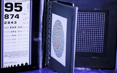
Amsler grid
The Amsler grid (see image below) is a valuable part of the neuro-ophthalmic examination and has utility beyond the examination of macular degeneration. With the Amsler grid, patients are asked if they can see a dot in the center of the grid. If they can see the dot, they are asked to fixate on the dot and then note if any areas of the square grid appear desaturated, if any areas of the grid have missing lines, or if the lines do not appear straight.
 Time can be saved if the near card and the Amsler grid are pasted into the color-vision test book. A self-adhesive note can be used to direct the patient to the appropriate section of the near card.
Amsler testing is performed monocularly with the near correction in place, if applicable. The Amsler grid may detect small central or paracentral scotomas missed between the 6° spacing of standard threshold automated perimetry.
The small homonymous paracentral scotomas of occipital stroke are screened more efficiently with the Amsler grid than with testing of central 10° automated perimetry. Confrontation fields at 4 m may also reveal a small homonymous scotoma.
Some patients with unexplained visual blurring may be referred for neuro-ophthalmic examination, which then reveals metamorphopsia (ie, the straight lines of the Amsler grid are distorted).
Non-ophthalmologists may mistake central serous retinopathy for optic neuritis. Central serous retinopathy often results in metamorphopsia or a scotoma.
Confrontation fields
Field defects respecting the vertical midline usually indicate chiasmal or retrochiasmal pathology. Field defects respecting the horizontal midline usually indicate ocular disease due to involvement of the nerve fiber layer or occlusion of a branch retinal vessel. A rare exception to this rule is occipital stroke above or below the calcarine fissure.
Confrontation fields are an essential part of the eye examination. It is not uncommon for patients to mistake homonymous visual loss for ipsilateral monocular visual loss. Furthermore, cognitively impaired patients may be incapable of performing formal perimetry.
Fields should be examined monocularly with the patient fixating on the examiner's nose. By convention, confrontation field defects are recorded from the perspective of the patient rather than the examiner.
Some examiners test confrontation fields by moving a finger, pen, or red-headed pin inward from the periphery while the patient is fixating centrally. The patient is asked to indicate if and when he or she sees the moving test object. However, this method of kinetic testing has some potential drawbacks. For instance, the speed at which the test object is moved may cause considerable variation in the results. In addition, moving objects are seen more readily than static ones (ie, statokinetic dissociation), rendering the kinetic test potentially less sensitive. Finally, the "yes" and "no" responses are somewhat nebulous compared to the exact quantitative response required by finger counting.
The author prefers to test confrontation fields by simultaneous finger presentations across the vertical midline in the upper quadrants and then in the lower quadrants (see image below). If the patient is unable to count fingers, then hand motions and light perception can be tested in each quadrant. If a subtle chiasmal lesion is suspected, the palms of the examiner's hands or identical red bottle tops can be placed on either side of the vertical midline to see if desaturation (fading) is present on 1 side. Clinical suspicions can be confirmed with formal perimetry testing.
 Confrontation visual fields are tested monocularly with the patient fixating on the examiner's eye or nose. The patient can be asked to add up the number of fingers, which are simultaneously presented on either side of the midline.
In some patients who are cognitively impaired, confrontation fields may provide a more accurate indication of the patient's problem than automated perimetry. If a patient does not fixate well for confrontation field testing, tangent screening, Goldmann perimetry, and automated perimetry are likely to be inaccurate.
Central 30° threshold automated perimetry is the preferred visual field test in neuro-ophthalmology and usually detects visual field defects of neuro-ophthalmic importance with the exception of the temporal crescent.
In patients with neuro-ophthalmic conditions, some clinicians feel that carefully performed Goldmann perimetry yields results comparable to those of automated perimetry.
The tangent screen is a useful screening device, but the results are difficult to reproduce. In most ophthalmic practices, the tangent screen is reserved for patients with suspected malingering and tunnel vision. The tangent screen allows fields to be checked easily at multiple distances. In functional patients complaining of tunnel vision, the field isopters do not expand geometrically despite increasing the test distance and the target size proportionately.
The tangent screen is occasionally useful in patients with small central scotomas because the 1-m test distance of the tangent screen reveals scotomas 3 times larger than those found on standard perimetry testing based on a 33-cm test distance.
Pupils
The pupils of any patient with unexplained visual loss, ptosis, or dysmotility should not be dilated until reviewed by the senior examiner, for the following reasons: (1) pupil examination is one of the few objective indicators of potential visual function, as the pupils are an important clue to patients with suspected functional visual loss; (2) subtle relative afferent pupillary defects are often difficult to detect; (3) Horner syndrome is easily missed unless the combination of anisocoria that is worse in the dark, pupil dilation lag, and ptosis can be correlated; (4) the presence or absence of pupil involvement may change the management of new-onset third nerve palsy; and (5) the pupils are clinically normal in myasthenia gravis.
To measure pupil size, use a pupil gauge or millimeter rule with the patient fixating in the distance, in both dark and illuminated environments. Dark irides make pupil examination more difficult than light irides; the view screen of the infrared autorefractor is often helpful. [1] In uncooperative children, the examiner can assess relative pupil size in the dark from a distance with the retinoscope or the direct ophthalmoscope.
If a difference in the size of the pupils (anisocoria) is present, determine if the anisocoria is greater in the dark or in the presence of light. If the anisocoria is less than 1 mm and remains the same in both the light and the dark and if the pupils are round and reactive, the patient may have physiologic anisocoria. Anisocoria greater in the dark suggests sympathetic disease, such as Horner syndrome. Anisocoria greater in the light suggests parasympathetic disease, such as Adie tonic pupil. Note that even if 1 eye is completely blind, the pupil sizes should be equal in the absence of iris trauma or synechiae.
Note the briskness of the pupil response. If the pupils do not react briskly, vermiform constriction, iris notches, and the response to near stimulus should be recorded. The near reaction is best tested with an accommodative target, such as a reading card. When testing the near response, do not use an illuminated light source as the accommodative target, since the pupillary light response will be added to the near response. If the pupil reacts better to near than to light, the patient has light-near dissociation. A common cause of afferent light-near dissociation is optic neuropathy. Well-known eponymous causes of light-near dissociation include Adie tonic pupil, Parinaud syndrome (ie, dorsal midbrain syndrome), and Argyll Robertson pupil (ie, tertiary syphilis).
In children, paradoxical pupillary constriction in darkness has been reported in patients with congenital stationary night blindness, congenital achromatopsia, cone disorders and Lebers congenital amaurosis, dominant optic atrophy, and amblyopia. In young children with nystagmus and paradoxical pupils, electroretinogram testing should be considered.
Testing for a relative afferent pupillary defect (RAPD) (or Marcus Gunn pupil) is an essential part of the eye examination. The designation of a "relative" afferent pupillary defect is important. Bilateral afferent pupillary defects are common (eg, patients with optic neuritis), but a bilateral RAPD cannot exist. In a dimly illuminated room, the patient fixates in the distance and a bright light is flashed for 2-3 seconds in 1 eye and then in the other eye. If the light is directed toward 1 eye and if the ipsilateral pupil appears to dilate "paradoxically", an RAPD is present. If the light is flashed on 1 eye and the pupil does not appear to constrict or increased pupillary escape from constriction occurs, an RAPD may also be present.
The RAPD still can be tested in patients in whom 1 pupil is dilated or unreactive. The physician examines the pupillary reaction of the undilated pupil while the light is flashed back and forth between the 2 eyes. In patients with strabismus, direct the light along the pupillary axis of each eye to avoid inducing an RAPD in the deviated eye. Although an RAPD may correspond with ipsilateral poor vision, this is not always the case. For example, a macular hole will result in poor acuity but not in an RAPD. In patients who have marked unilateral field loss or unilateral dyschromatopsia, an ipsilateral RAPD is usually present. A lesion confined to the optic tract may cause an incongruous homonymous field defect and RAPD contralateral to the optic tract lesion. In patients with lesions of the brachium of the superior colliculus, a contralateral RAPD without field loss (ie, midbrain RAPD) may be noted.
The differential diagnosis of heterochromia iridium (more commonly known as heterochromia iridis) is vast. Patients with heterochromia iridium may present to the neuro-ophthalmologist with congenital Horner syndrome, Sturge-Weber syndrome, or unilateral glaucoma treated with an antiprostaglandin, such as latanoprost.
Slit-lamp examination is of great utility when an abnormal pupil is suspected. Slight irregularities in the shape of the pupil are best seen with the slit lamp. Irregularities in pupil shape may be congenital, as with iris coloboma, or acquired, as with synechiae (iritis), iris notches (trauma), and iridocorneal endothelial syndrome (glaucoma).
In patients with type 1 neurofibromatosis, Lisch nodules closely resembling iris nevi may be seen by using the slit lamp.
Purse-stringing of the pupil may be seen with Adie tonic pupil; it is best elicited by quickly turning the slit-lamp beam on and off while the pupil is examined.
Pupil gaze synkinesis is a sign of aberrant regeneration of CN III. Most commonly, the pupil constricts on adduction. While the patient is fixating in the distance, the size of the pupils in primary gaze is compared with the pupil size in adduction.
Anterior segment
In addition to the slit-lamp findings of abnormal pupillary function, several other anterior segment findings may be of neuro-ophthalmic importance. Cells and flare, keratic precipitates, and posterior synechiae are typical manifestations of iritis. Iritis is seen with various neuro-ophthalmic conditions, including sarcoidosis, occasionally granulomatosis with polyangiitis, and orbital inflammatory syndrome. A low-grade iritis is seen in some cases of ocular ischemic syndrome caused by carotid occlusive disease. The phakomatoses have particular anterior segment findings. Patients with type 1 neurofibromatosis may have Lisch nodules, as mentioned above. Type 2 neurofibromatosis may be associated with posterior subcapsular cataracts. Conjunctival telangiectasias are seen in patients with Louis-Bar syndrome (ie, ataxia telangiectasia).
Corkscrew, arterialized conjunctival vessels and ipsilateral glaucoma suggest a dural arteriovenous malformation or carotid-cavernous fistula.
Acute angle-closure glaucoma is sometimes mistaken for cerebral aneurysm due to the dilated pupil, cephalalgia, and vomiting. However, a misty cornea, a narrow angle in the ipsilateral and usually the contralateral eye, and elevated intraocular pressure differentiate the 2 entities. Patients receiving topiramate may have narrow angles.
Examination of the fornices in a patient with proptosis occasionally reveals the salmon patch lesions of orbital lymphoma.
Fundi
Dilated fundus examination with stereo slit-lamp illumination and indirect ophthalmoscopy enables visualization of fundus details that may not be visualized with direct ophthalmoscopy. Notwithstanding, the use of the direct ophthalmoscope should not be overlooked. The direct ophthalmoscope provides high magnification of details of the fundus. Venous pulsations and venous sheathing and some epiretinal membranes may be seen through the direct ophthalmoscope. Use of the red-free light may show nerve fiber layer defects.
Numerous anomalies of the optic disc have neuro-ophthalmic significance, but only a few are reviewed here.
Optic nerve hypoplasia is sometimes overlooked if the examiner does not appreciate the true nerve diameter in patients with the double-ring sign. Hypoplastic discs appear as abnormally small heads of the optic nerve. The nerve may be gray or pale, and it is often surrounded by a mottled yellowish peripapillary halo and bordered by a ring of hyperpigmentation or hypopigmentation (the double ring). CNS midline defects (eg, abnormal hypothalamic-pituitary axis) should be excluded, especially if optic nerve hypoplasia is associated with increased venous tortuosity.
Findings suggestive of papilledema include blurring of the fine peripapillary vessels (due to edema of the nerve fiber layer), peripapillary hemorrhages, venous congestion, and disc leakage on fluorescein angiography. Spontaneous venous pulsations, disc drusen, or myelinated nerve fibers suggest pseudopapilledema. The differentiation between papilledema and pseudopapilledema is discussed in separate articles (see Papilledema and Pseudopapilledema).
Chalky white, pale, swollen discs are classically described in elderly patients presenting with giant cell arteritis.
Painless visual loss in a patient older than 50 years with sectoral disc edema, altitudinal field loss, and small cup-to-disc ratios suggests nonarteritic ischemic optic neuropathy. After about a month, the disc edema resolves, and sectoral disc pallor is seen. The author finds the red-free light most useful in detecting sectoral disc pallor in patients with suspected, past, or nonarteritic ischemic optic neuropathy.
A swollen optic nerve with peripapillary telangiectasias in the setting of painless visual loss, especially in a young male patient, suggests Leber hereditary optic neuropathy.
Optic atrophy with optociliary shunt vessels (ie, optochoroidal collaterals) is classically associated with meningioma of the optic nerve sheath, but it is also seen in other conditions, including central retinal vein occlusion, optic nerve glioma, and chronic papilledema.
Thinning of the neural rim, especially vertical notching, may be seen with glaucoma. Peripapillary splinter hemorrhages (ie, Drance hemorrhages) have been described with normal-tension glaucoma. Disc pallor disproportionate to cupping is not expected with glaucoma.
Many retinal findings are of potential neuro-ophthalmic importance. Central retinal artery occlusion may be seen with embolic disease and giant cell arteritis. Multiple occlusions of the branch retinal artery in a patient with deafness and cerebrovascular accident may suggest Susac syndrome. Retinochoroidal striae may indicate an orbital mass lesion.
Some of the phakomatoses have classic fundus findings. Astrocytic hamartomas (ie, mulberry-like tumors) may be seen with tuberous sclerosis. Retinal angiomatosis may be seen in patients with von Hippel disease. Retinal arteriovenous malformation may be seen with Wyburn-Mason syndrome. Diffuse choroidal hemangiomas of Sturge-Weber syndrome (ie, tomato-ketchup fundus) are best noted by comparing the 2 fundi during indirect ophthalmoscopy. Increased cupping may be seen in the involved eye of patients with Sturge-Weber syndrome.
Ocular motility
Versions (ie, movement of both eyes) should be tested in each of the cardinal positions of gaze. Asking the patient to indicate if he or she sees double as the target is being moved may be useful. Check the downgaze positions twice: first, without lifting the lids so that lid-inferior gaze synkinesis is not missed, and, second, with the lids lifted so that the lids do not obscure the eyes.
Smooth pursuit is tested by having the patient follow a slowly moving target. Saccades can be tested by asking the patient to move his or her eyes in a particular direction. Separating saccades from smooth pursuit may be clinically important. Smooth pursuit may be irregular in brainstem-cerebellar disease, and catch-up saccades may be required. In Parinaud syndrome, upgaze saccades may be affected more than upward smooth pursuit early on in the disease. In patients with small pontine strokes involving purely the paramedian pontine reticular formation (PPRF), saccades may be affected and smooth pursuit relatively preserved. This is because the fibers of the latter eye movement system do not synapse in the PPRF.
The author screens for ocular alignment by examining the corneal light reflexes and then by performing alternate-cover tests at distance and near. The alternate-cover test is used to measure both tropia and phoria. If an abnormality is found, lateral gaze positions and upgaze and downgaze prism measurements are also recorded. If a vertical deviation is noted, head tilts are performed. The versions should correspond with the prism measurements. Ensure that the patient is not fixating with the palsied eye.
Ocular misalignments may be comitant or incomitant. Incomitant misalignments indicate an innervational problem or a mechanical restriction. Ductions (ie, movement of a single eye with the other eye covered) may be better than versions in patients with innervational problems, with no difference between ductions and versions noted in patients with mechanical restrictions. Forced ductions are restricted with mechanical disease and normal with paralytic disease, unless the paralysis has caused chronic contracture of the muscle.
A thorough discussion of third, fourth, and sixth nerve palsies is presented in Oculomotor Nerve Palsy, Trochlear Nerve Palsy, and Abducens Nerve Palsy, respectively. Only the infranuclear motility deficits are discussed herein. If a patient is fixating with the palsied eye, the motility deficit may be more difficult to recognize.
In patients with CN III palsy, the presence or absence of afferent pupillary involvement must be documented. Pupil-involving lesions may indicate an underlying posterior communicating artery aneurysm, and neuroimaging studies are required.
Although computed tomography angiography and magnetic resonance angiography are now usually performed in patients with new onset third nerve palsy, classic dictum was that patients with new-onset, pupil-sparing CN III palsy were observed daily for 5 days to exclude pupillary involvement. In CN III palsy, the involved eyelid is often ptotic, and the globe is hypotropic and exotropic, because both the superior oblique and the lateral rectus are still functioning. Because the multiple innervations of CN III may mask other findings, check CN IV function (ie, intorsion of the conjunctival vessels), CN V function (ie, corneal sensation and cutaneous facial sensation), and CN VI function (ie, abduction) to exclude possible cavernous sinus involvement. In patients with CN III palsy, search for aberrant regeneration, as this suggests a compressive lesion.
Pupil gaze synkinesis is discussed in the Pupils, Anterior segment, and Fundi sections above.
With lid-gaze synkinesis, the ptotic lid may elevate in downgaze (ie, pseudo–von Graefe sign) or adduction (ie, inverse Duane sign). Superior division palsies and inferior division palsies were traditionally thought to be due to cavernous sinus disease, but divisional somatotopy begins in the midbrain. The various fascicular syndromes of CN III (eg, Nothnagel syndrome, Benedikt syndrome, Weber syndrome, Claude syndrome) may aid in further localization of the lesion.
In patients with suspected CN IV palsy, document the versions, prism measurements on the 3-step test, and fusional amplitude; review an old photograph of the patient to look for a head tilt; and consider performing double Maddox rod testing for torsion. If strabismus surgery is being contemplated, overelevation in adduction (marked overaction of the antagonist inferior oblique) on version testing is useful to note because an inferior oblique weakening procedure can be performed.
Fusional amplitude can be estimated by placing a vertical prism bar in front of one eye and determining the maximum diopter of prism through which the patient can maintain single binocular vision. Fusional amplitudes greater than 4 prism diopters and long-standing head tilt are strongly suggestive of congenital CN IV palsy. In trauma patients with CN IV palsy, recording the amount of excyclotorsion is useful. It is tested with double Maddox rods; excyclotorsion greater than 10° suggests bilateral superior oblique palsies. Prism measurements in the supine position may help distinguish skew deviation from CN IV palsy. Vertical prism measurements that decrease by more than 50% from the upright to the supine position are suggestive of skew deviation. [2]
Not all abduction deficits are CN VI palsies. The imitators of CN VI palsy include Duane syndrome, myasthenia gravis, spasm of the near reflex, decompensated esophoria, and thyroid-associated ophthalmopathy with medial rectus contracture. In all patients with CN VI palsy, document the presence or absence of papilledema. When one documents prism measurements for CN VI palsy, ensure that esotropia (rather than exotropia) is recorded. "XT" is commonly mistakenly noted on the medical charts of patients with CN VI palsy.
In patients with a vertical gaze deficit, perform a doll's-head maneuver. If the globes elevate on this maneuver, a supranuclear palsy is the likely diagnosis. If an upgaze deficit is noted, then consider performing downgoing optokinetic nystagmus (OKN) to better elicit the convergence-retraction nystagmus of a possible Parinaud syndrome.
Internuclear ophthalmoplegia (INO) is a lesion of the medial longitudinal fasciculus and should not be mistaken for medial rectus palsies or partial CN III palsy. An ipsilateral adduction deficit with contralateral abducting nystagmus is usually present in INO; however, INO may manifest clinically as an adduction lag (even though no latency on adduction is observed on eye movement recordings). The author finds that the easiest way to elicit INO is by asking the patient to make large horizontal saccades from side to side.
Myasthenia gravis should be considered in the differential diagnosis of all patients with ocular dysmotility, especially if they have ptosis. The hallmark of myasthenia gravis is variability and fatigability. Myasthenia gravis can imitate any pupil-sparing ocular dysmotility pattern, including INO. In those with myasthenia who perform sustained lateral gaze, there may be an eventual drift in the eyes back toward the primary position. In patients with Graves disease and an exotropia, concomitant myasthenia is a possibility (with Graves disease esotropia and hypotropia are much more common).
Optokinetic asymmetry in the setting of homonymous hemianopsia suggests a deep parietal lobe lesion. OKN response is less when the OKN stimulus is moved toward the diseased parietal lobe than when the stimulus is moved in the opposite direction. A simplistic explanation for this is that the smooth pursuit is controlled by the ipsilateral parieto-occipito-temporal junction. In the case of a right-sided parietal lobe lesion, for example, movement of the OKN stimulus to the right will elicit a poor smooth pursuit movement to follow the OKN stripes, resulting in a smaller amplitude corrective saccade.
The King-Devick test, which requires saccadic eye movements while reading test cards, has been suggested as a quick assessment of visual performance in patients with concussion and patients undergoing multiple sclerosis trials. [3]
Anomalous head positions
An anomalous head position may be due to ophthalmic or nonophthalmic causes. Nonocular causes of abnormal head position include cervical torticollis, unilateral deafness, and habit posture. Ophthalmic causes for a head posture may be due to nystagmus or non-nystagmus-related problems. Patients with nystagmus may adopt a head posture to place the eyes in the null point.
CN palsies may result in anomalous head posture. Patients with CN IV palsy usually tilt their heads to the side opposite the palsy. Patients with CN VI palsy usually turn their heads toward the side ipsilateral to the palsy to avoid diplopia.
Chin-up positions may be seen with ptosis, A -pattern esodeviations, or V -pattern exodeviations. Abnormal head postures may be a clue to field loss.
Eyelids
The normal upper eyelid margin is located 1-1.5 mm below the superior limbus. If the eyelid margin is too high, lid retraction occurs; if it is too low, blepharoptosis or ptosis occurs. Whenever lid retraction is suspected, exclude contralateral ptosis.
Lid retraction may result from prior lid surgery or lid trauma. Neuro-ophthalmic causes of true lid retraction include thyroid-associated orbitopathy and dorsal midbrain syndrome. Lid retraction may be unilateral or bilateral in thyroid-associated orbitopathy. When the patient is asked to look down slowly from upgaze, the lid may be seen to lag behind the movement of the globe. Other signs of thyroid-associated orbitopathy may be present, including injected recti and proptosis. If the inferior recti are involved, upgaze may be restricted to forced ductions and lack of upward movement may be observed with the doll's-head maneuver. If lid retraction is seen after sustained upgaze, the facilitation of Lambert-Eaton syndrome may be a possibility.
Parinaud syndrome or dorsal midbrain syndrome may be associated with bilateral upper lid retraction (ie, Collier sign) when the nucleus of the posterior commissure is involved. The eyes usually do not appear injected or proptotic, and though upward versions may be affected, the eyes elevate on the doll's-head maneuver.
Ptosis most commonly results from weakening of the levator aponeurosis due to age or trauma. Hypotropia ipsilateral to the droopy eyelid is an important cause for pseudoptosis that can be excluded by performing an alternate-cover test. Neuro-ophthalmic causes of true ptosis include Horner syndrome, CN III palsy, and myasthenia gravis. The upper-lid ptosis of Horner syndrome is at the most 2 mm because this is the degree of function of the sympathetically innervated Müller muscle. The lower lid may appear elevated in Horner syndrome (ie, inverse ptosis) because the inferior tarsal muscle is innervated sympathetically.
Ptosis in CN III palsy may be absent, partial, or complete depending on the degree of involvement of the superior division of the third nerve. Bilateral ptosis may be seen with nuclear third nerve palsies if the central caudal nucleus is damaged. Lid-gaze synkinesis is important to note (see the Ocular motility and Head posture sections). Lid-gaze synkinesis, when acquired, suggests a compressive lesion or trauma.
The hallmark of myasthenia gravis is variability in both ptosis and ocular alignment. In patients with myasthenia gravis, the upper lid height may vary from second to second on slit-lamp examination, as if the eyelid was being wafted by a gentle breeze. More marked variability in the lid height may be seen after the patient is well rested, with transient decrease or abolition of the ptosis; this is the basis of the sleep test. Ice may be applied to the ptotic lid for several minutes to see if ptosis improves; cold stimulation is believed to ameliorate muscle function in patients with myasthenia gravis.
The often-touted curtain sign is the elevation of 1 lid that causes the contralateral lid to droop; however, this sign is not specific for myasthenia gravis and is explained by the Hering law. If a patient with myasthenia gravis is asked to quickly look upward from a downward position, the lid may overelevate and then droop (ie, Cogan lid twitch). Just as patients may have difficulties in eyelid opening, they often have weakness of eyelid closure, as manifested by an inability to bury the eyelashes on forced lid closure or an inability to keep the eyes shut against resistance applied by the examiner. Analogous to this is the peek sign of myasthenia, where on attempted sustained gentle lid closure, the lids slowly open over 30 seconds.
The eyelids may show hyperactive movement (eg, blepharospasm) or too little movement (eg, Bell palsy). Essential blepharospasm due to basal ganglia disease ultimately becomes bilateral. It sometimes is accompanied by apraxia of lid opening. Hemifacial spasm usually due to a dolichoectatic vessel impinging on the entry zone of the facial nerve root is unilateral. In any patient with hemifacial spasm, closely examine the other cranial nerves. The eyelids may not close with CN VII palsy, or the spontaneous blink may be lost in patients who are recovering from Bell palsy.
An S -shaped lid may be seen in neurofibromatosis. Lid neurofibromas may contribute to ptosis or ectropion. Ecchymosis of the eyelid skin may be seen with neuroblastoma (children), leukemia, and amyloidosis.
Floppy eyelid syndrome is occasionally observed in heavy-set patients with pseudotumor cerebri. In such patients, the eyelid easily everts, and the palpebral conjunctiva may appear beefy red. Lash ptosis is often present, and a loss of eyelash parallelism may also occur.
Orbit
Pseudoproptosis may be seen with lid retraction, unilateral high myopia, facial asymmetry, and contralateral enophthalmos.
Proptosis can be screened from above by gazing at the patient's forehead and then quantified with an exophthalmometer. When one performs exophthalmometry, record the base measurement. Orbital retropulsion is decreased with orbital mass-occupying lesions, and it is important to document in patients with or without proptosis. If orbital compliance is markedly decreased, have an increased index of suspicion for compressive optic neuropathy or an orbital cause for strabismus.
The differential diagnosis for proptosis is vast. In adults with proptosis, look closely for lid lag and lid retraction because thyroid-associated orbitopathy is the most common cause in this age group. In children with proptosis, exclude orbital cellulitis and rhabdomyosarcoma. Acquired enophthalmos may be seen after orbital fractures or neurosurgical decompression of the orbital walls, and with scirrhous breast carcinoma.
Pulsatile proptosis may occur with carotid-cavernous fistula, sinus mucoceles, and neurofibromatosis with dysplasia of the sphenoid wing, and after removal of the bony orbital rim. If spontaneous orbital pulsations are not readily visible, they can be detected by placing long-handled cotton-tip applicators over the closed eyelids and forehead and watching for oscillations of the cotton tip applicators.
Orbital masses usually displace the globe in the direction opposite the tumor. For example, frontal sinus mucocele may cause inferior globe displacement; intraconal tumors cause axial proptosis.
CNs V, VII, and VIII
Numbness of the ophthalmic (V1), maxillary (V2), or mandibular (V3) divisions of the trigeminal nerve (CN V) in a patient with a motility deficit may indicate a cavernous sinus process. For screening purposes, the author simultaneously touches both sides of the forehead (V1), upper cheek (V2), and chin (V3) to elicit asymmetry in sensation. Traditionally, corneal sensation (V1) is tested with dental floss or tissue in 4 different quadrants of the cornea. Occasionally, the author instills artificial tears in the eyes instead and asks the patient to compare corneal sensation.
Bell palsy (ie, idiopathic lower motor neuron CN VII palsy) is discussed in a separate article and will be mentioned only briefly here. In acute lower motor neuron facial nerve palsies, the forehead, eyelid, and lower face will droop. Orbicularis oculi function should be tested by asking the patient to gently close the eyelid as if sleeping. The amount of lagophthalmos can be measured with a millimeter ruler.
The presence or absence of the Bell phenomenon should be documented, as it indicates how well the cornea is protected when the patient is asleep. Corneal epithelial defects and corneal sensation should be noted. Patients with CN VII palsy and corneal anesthesia may require early tarsorrhaphy. Loss of spontaneous blink may occur in patients with apparently normal voluntary lid closure and is a helpful clue to suggest prior CN VII palsy. Examination for otic vesicles (possible Ramsay-Hunt syndrome), palpation of the parotid gland with a gloved finger (eg, epithelial tumors or sarcoidosis), and examination of the tongue for furrows (ie, Melkersson-Rosenthal syndrome) should be performed in patients with CN VII palsy.
Testing hearing in patients with nystagmus, hemifacial spasm, facial nerve weakness, or Cogan syndrome is important. To test hearing, the author whispers a number in one ear while scratching a prescription pad over the patient's other ear (masking).
Extraocular examination
Although vital signs are usually not recorded in patients presenting with ophthalmologic complaints, they may be important to note. Blood pressure should be measured in patients with papilledema and fundus hemorrhages. In patients with retinal emboli, checking the pulse may confirm suspicion of atrial fibrillation.
Intracranial bruits often are better appreciated by the patient than by the examiner. The bell of the stethoscope can be applied to the affected orbit or temple to detect the bruit. Carotid bruits may be heard in patients with retinal emboli or stroke; however, patients with high-grade or complete occlusion may not have a bruit.
A prominent temple may be seen ipsilateral to a meningioma of the sphenoid wing. Patients with giant cell arteritis may have ropey temporal arteries, as well as asymmetry in the pulses of the superficial temporal arteries. Point tenderness of the artery may help guide temporal artery biopsy. Before temporal artery biopsy, both of the proximal superficial temporal arteries can be compressed for 1 minute to exclude the remote possibility of a critical collateral to cerebral circulation provided by the external carotid.
The differential diagnosis for saddle nose includes congenital syphilis, granulomatosis with polyangiitis, relapsing polychondritis, lethal midline granuloma, and intranasal cocaine abuse.
Oral examination may be an important part of the neuro-ophthalmic examination. [4] A palatal eschar should be excluded in any patient with suspected mucormycosis. Patients with vertical pendular nystagmus or, in some cases, torsional nystagmus may have palatal undulations indicative of oculopalatal myoclonus. Oral ulcers or tongue ulcers are occasionally seen in patients with giant cell arteritis and jaw claudication. Tongue atrophy may be seen with Möbius syndrome. The relevance of the oral examination in patients with CN VII palsy has been discussed already.
Although a full neurologic examination is useful in a neuro-ophthalmic workup, it is usually not feasible in the ophthalmologist's office and is better reserved for the neurologist. Notwithstanding, the patient's gait, speech, and mental status can be ascertained during the interview. In patients with homonymous hemianopsia or retinal emboli, the presence or absence of hemiparesis should be noted. Neurologic examination may help localize the various fascicular syndromes of CN III and VI palsies. For example, CN III palsy with a contralateral tremor may indicate involvement of the red nucleus and Benedikt syndrome.
Examination efficiency
Noting the best-corrected visual acuity is important. Having a paddle occluder with attached pinhole may save time (see image below). Autorefractors are usually accurate and help the clinician to quickly obtain the patient's optimal refraction.
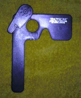 A paddle occluder with an attached pinhole facilitates the determination of visual acuity.
Time can be saved if the distance acuity, near acuity, color vision, Amsler grid, and confrontation field are all tested while one eye remains occluded, rather than alternately occluding each eye for every test. The author pastes a near card and Amsler grid into an abridged version of the Ishihara color plates. Near-vision checks are quicker if a self-adhesive note is used to direct the patient to the lower portions of the near card (see image below).
Time can be saved if the near card and the Amsler grid are pasted into the color-vision test book. A self-adhesive note can be used to direct the patient to the appropriate section of the near card.
After these tests, pupil testing and alternate-cover testing at distance and near can be performed while the patient fixates in the distance. Versions are documented, and Hirschberg testing and pupil testing are then performed. The pupils are tested at this time so that the bright lights do not obscure the visual function needed for proper performance of the tests already mentioned.
Much time, effort, and money can be saved if old photographs are reviewed to determine the chronicity of neuro-ophthalmic signs, such as abnormal head posture, anisocoria, strabismus, and lid retraction. At the time the appointment is made, having the receptionist ask the patient to bring in old photographs may be useful. If old photographs are not available, identification cards, such as a driver's license, may be of help. Small photographs can be viewed better with the indirect condensing lens, the direct ophthalmoscope, or the slit lamp.
A well-trained ophthalmic assistant will do much to increase office efficiency. However, the author's bias is that technicians working in the neuro-ophthalmology clinic not administer any dilating drops until the physician has reviewed the visual acuity and examined the pupils, lids, and motility. Even dilation with phenylephrine may obscure small amounts of ptosis.
Bedside examination
Eye examinations at the bedside are often more challenging than office visits for a variety of reasons. Compared with outpatients, hospitalized patients tend to be sicker, less reliable historians, and less cooperative or even unconscious during the eye examination. Lighting conditions may hamper examination of the pupils. Customary examining equipment may not be available. If neurologic vital signs are being followed up, pharmacologic dilation of the pupils and thorough fundus examination may not be possible.
When one checks near acuity, make patients sit up whenever possible so that they can take advantage of the near segment of their bifocals and of overhead lighting. A pair of reading glasses should be kept in the examining kit for presbyopic patients. In conscious patients who are intubated, vision can be checked by asking the patient to imitate numbers or movements of fingers at increasing distances.
Dyschromatopsia may be subjectively assessed by asking the patient to determine the perceived percentage of desaturation of a red eyedrop bottle top.
Examination of the anterior segment may be difficult if a portable slit lamp is not available. Shining a penlight or the vertical slit of the ophthalmoscope light through the 20-D- or 28-D indirect ophthalmoscopy condensing lens is a useful maneuver. Media opacities can be screened for by examining the red reflex through the direct ophthalmoscope.
Motility may change dramatically in patients who are not fully awake or who are sedated with medications. Dependency may also alter extraocular motility. The 3-step test does not work well in patients who are supine.
Corneal exposure should be excluded in all unconscious patients.
Carrying an OKN tape in the examination kit may be useful. If one is not available, removing alternate strips from a long row of brightly colored sticker labels (eg, allergy stickers) works well.
Functional visual loss
The neuro-ophthalmic examination of patients with suspected functional visual loss merits an article in itself. Numerous techniques may be used for detecting suspected functional visual loss, and each clinician should have a repertoire of them. Interested readers are referred to the excellent chapter by Miller and Keane in Walsh and Hoyt's Clinical Neuro-ophthalmology. [5]
In general, in functional patients, monocular visual loss is easier to diagnose than binocular visual loss. Likewise, feigned profound visual loss is easier to identify than moderate visual loss. In patients claiming profound visual loss in 1 or both eyes, mirror testing and OKN testing can be performed. The presence of OKN suggests vision of at least 20/400.
In patients claiming profound bilateral visual loss, observation of navigational skills, pupil reflexes, and prism-diplopia responses may be helpful. Patients claiming bilateral blindness can sometimes be loaded up with tissue in an examination room with a small garbage can adjacent to the examination chair. If the "blind patient" can discard the tissue into the garbage by himself it indicates functional vision.
The absence of an RAPD in patients with monocular visual loss and a normal fundus examination should be viewed with great suspicion. A diplopia response to 4-prism diopter base-out testing over the affected eye may provide further objective evidence of functional visual loss. When performing A-O vectography, cylinder fogging tests, duochrome tests, stereo acuity tests, and binocular visual field testing for suspected functional visual loss, ensure that the patient is not closing the affected eye. Ensure that the colored filters on the duochrome test are properly matched. [6]
In patients with functional field loss, confrontation fields, tangent screen testing, and Goldmann perimetry correlated with Amsler grid findings are often superior to automated perimetry. In patients with visual field constriction, tangent screen testing at increasing distances (with proportionate increase in target size) is typically performed. If a tangent screen is unavailable, performing confrontation testing at increasing distances will provide useful information if the patient is tunneling.
Patients with suspected functional visual loss often benefit from repeat examination or recheck in a different examination room. On the second examination, observe the patient's behavior as he or she enters and leaves the office and in the waiting room. The physician should recheck acuity by walking the patient up from the 20/10 line. Correlation of the distance and near acuities should be repeated. When testing near vision, ensure that the proper refraction and card-test distances are used. Carefully reexamine the pupillary responses, and then consider redilating the pupils, with possible cycloplegic refraction.
Organic pathology not uncommonly accompanies functional disease; the clinician should be wary of this association. If patients with functional visual loss do not improve, the author usually performs neuroimaging studies and, in select cases, initiates ERG and VEP testing. When possible, review the CT scans or MRIs with careful attention to the orbital apex and occipital lobe, as lesions in these locations might be overlooked.
Pediatric Neuro-Ophthalmic Examination
Toys, colorful objects, and stories should be on hand when examining young patients. Also, the examiner should consider removing his or her lab coat.
A complete examination may not be possible in patients who are young, autistic, or developmentally disabled, so the examination objectives should be prioritized.
Young children often feel more secure if they sit on their parent's lap in the examination chair. The parent can help with monocular occlusion.
In preverbal patients with horizontal nystagmus, a vertical optokinetic response is reassuring. However, the absence of an opticokinetic response does not necessarily indicate absent visual function.
Infants who are aged 1-3 months, with visual acuity of 20/400 or better, may manifest lid retraction when the room lights are suddenly diminished. If visual function is poor, the lid retraction may not be present.
Briskly reactive pupils in a child with profound visual dysfunction suggests cortical visual impairment. Paradoxical pupil constriction to darkness was discussed in the pupil section above.
Congenital motility disturbances such as Duanes syndrome not uncommonly go unnoticed until the child is older than 1 year. Review of old photographs and videos may be useful. In young children, the Doll's head maneuver can be used to assess limitations in motility. The examiner can also use the vestibulo-ocular reflex to advantage to test horizontal eye movements by rotating the infant in a circle.
Confrontation fields in children can be challenging. Young children can be directed to fixate centrally on a large toy while a smaller object is moved in from the periphery. The child's eye movement in the direction of the moving object suggests the presence of peripheral vision in that quadrant. When evaluating the temporal field, rotating the face to position the child's eye in full abduction may allow more accurate field testing, with less eye movement artefact.
To encourage compliance with the slit lamp, young children are often told that the slit lamp is a motorcycle. The chin rest and forehead rest are the "helmet" that the child must wear before the motorcycle ride starts. The child is told that it is important to look straight ahead while driving and that he will be given a headlight (slit light) because he is driving in the dark. Often, the 78D or 90D lens can be used to advantage in undilated children. If not, this author initially also uses indirect ophthalmoscopy to view the macula undilated, prior to the possible crying following dilating drop instillation.
-
A paddle occluder with an attached pinhole facilitates the determination of visual acuity.
-
Confrontation visual fields are tested monocularly with the patient fixating on the examiner's eye or nose. The patient can be asked to add up the number of fingers, which are simultaneously presented on either side of the midline.
-
Time can be saved if the near card and the Amsler grid are pasted into the color-vision test book. A self-adhesive note can be used to direct the patient to the appropriate section of the near card.


