Dunn EN, Gregori NZ, Goldhardt R. Phakic cystoid macular edema secondary to idiopathic macular telangiectasia type 1 responsive to topical anti-inflammatory agents. Semin Ophthalmol. 2013 Mar. 28 (2):84-7. [QxMD MEDLINE Link].
Spaide RF. RETINAL VASCULAR CYSTOID MACULAR EDEMA: Review and New Theory. Retina. 2016 Oct. 36 (10):1823-42. [QxMD MEDLINE Link].
Duplechain A, Conrady CD, Patel BC, Baker S. Uveitis. StatPearls. 2023 Jan. [QxMD MEDLINE Link]. [Full Text].
Guymer RH, Campbell TG. Age-related macular degeneration. Lancet. 2023 Apr 29. 401 (10386):1459-1472. [QxMD MEDLINE Link].
de Jong S, Tang J, Clark SJ. Age-related macular degeneration: A disease of extracellular complement amplification. Immunol Rev. 2023 Jan. 313 (1):279-297. [QxMD MEDLINE Link].
Romano F, Lamanna F, Gabrielle PH, Teo KYC, Battaglia Parodi M, Iacono P, et al. Update on Retinal Vein Occlusion. Asia Pac J Ophthalmol (Phila). 2023 Mar-Apr 01. 12 (2):196-210. [QxMD MEDLINE Link].
Moroi SE, Gottfredsdottir MS, Schteingart MT, Elner SG, Lee CM, Schertzer RM, et al. Cystoid macular edema associated with latanoprost therapy in a case series of patients with glaucoma and ocular hypertension. Ophthalmology. 1999 May. 106(5):1024-9. [QxMD MEDLINE Link].
Breazzano MP, Grewal MR, Tsang SH, Chen RWS. Etiology of Retinitis Pigmentosa. Methods Mol Biol. 2023. 2560:15-30. [QxMD MEDLINE Link].
O'Neal TB, Luther EE. Retinitis Pigmentosa. StatPearls. 2023 Jan. [QxMD MEDLINE Link]. [Full Text].
Kanukollu VM, Agarwal P. Epiretinal Membrane. StatPearls. 2023 Jan. [QxMD MEDLINE Link]. [Full Text].
Sivaprasad S, Bunce C, Crosby-Nwaobi R. Non-steroidal anti-inflammatory agents for treating cystoid macular oedema following cataract surgery. Cochrane Database Syst Rev. 2012 Feb 15. 2:CD004239. [QxMD MEDLINE Link].
Ciulla TA, Rosenfeld PJ. Anti-vascular endothelial growth factor therapy for neovascular ocular diseases other than age-related macular degeneration. Curr Opin Ophthalmol. 2009 May. 20(3):166-74. [QxMD MEDLINE Link].
Boyer DS, Yoon YH, Belfort R Jr, Bandello F, Maturi RK, Augustin AJ, et al. Three-year, randomized, sham-controlled trial of dexamethasone intravitreal implant in patients with diabetic macular edema. Ophthalmology. 2014 Oct. 121 (10):1904-14. [QxMD MEDLINE Link].
Bressler SB, Glassman AR, Almukhtar T, Bressler NM, Ferris FL, Googe JM Jr, et al. Five-Year Outcomes of Ranibizumab With Prompt or Deferred Laser Versus Laser or Triamcinolone Plus Deferred Ranibizumab for Diabetic Macular Edema. Am J Ophthalmol. 2016 Apr. 164:57-68. [QxMD MEDLINE Link].
Ali BM, Azmeh AM, Alhalabi NM. Suprachoroidal triamcinolone acetonide for the treatment of macular edema associated with retinal vein occlusion: a pilot study. BMC Ophthalmol. 2023 Feb 10. 23 (1):60. [QxMD MEDLINE Link].
Liu L, Wu X, Geng J, Yuan Z, Chen L. IVTA as Adjunctive Treatment to PRP and MPC for PDR and Macular Edema: A Meta-Analysis. PLoS One. 2012. 7(9):e44683. [QxMD MEDLINE Link]. [Full Text].
Parke DW 3rd, Sisk RA, Murray TG. Intraoperative intravitreal triamcinolone decreases macular edema after vitrectomy with phacoemulsification. Clin Ophthalmol. 2012. 6:1347-53. [QxMD MEDLINE Link]. [Full Text].
Bressler SB, Qin H, Beck RW, Chalam KV, Kim JE, Melia M, et al. Factors Associated With Changes in Visual Acuity and Central Subfield Thickness at 1 Year After Treatment for Diabetic Macular Edema With Ranibizumab. Arch Ophthalmol. 2012 Sep 1. 130(9):1153-1161. [QxMD MEDLINE Link].
Gregori NZ, Gaitan J, Rosenfeld PJ, Puliafito CA, Feuer W, Flynn HW Jr, et al. Long-term safety and efficacy of intravitreal bevacizumab (Avastin) for the management of central retinal vein occlusion. Retina. 2008 Oct. 28(9):1325-37. [QxMD MEDLINE Link].
Arevalo JF, Garcia-Amaris RA. Intravitreal bevacizumab for diabetic retinopathy. Curr Diabetes Rev. 2009 Feb. 5(1):39-46. [QxMD MEDLINE Link].
Altintas O, Yuksel N, Karabas VL, Demirci G. Cystoid macular edema associated with latanoprost after uncomplicated cataract surgery. Eur J Ophthalmol. 2005 Jan-Feb. 15(1):158-61. [QxMD MEDLINE Link].
Antcliff RJ, Spalton DJ, Stanford MR, Graham EM, ffytche TJ, Marshall J. Intravitreal triamcinolone for uveitic cystoid macular edema: an optical coherence tomography study. Ophthalmology. 2001 Apr. 108(4):765-72. [QxMD MEDLINE Link].
Boscia F, Furino C, Dammacco R. Intravitreal triamcinolone acetonide in refractory pseudophakic cystoid macular edema: functional and anatomic results. Eur J Ophthalmol. 2005 Jan-Feb. 15(1):89-95. [QxMD MEDLINE Link].
Callanan D, Fellman RL, Savage JA. Latanoprost-associated cystoid macular edema. Am J Ophthalmol. 1998 Jul. 126(1):134-5. [QxMD MEDLINE Link].
Conway MD, Canakis C, Livir-Rallatos C. Intravitreal triamcinolone acetonide for refractory chronic pseudophakic cystoid macular edema. J Cataract Refract Surg. 2003 Jan. 29(1):27-33. [QxMD MEDLINE Link].
Falcone PM. Vitreomacular traction syndrome confused with pseudophakic cystoid macular edema. Ophthalmic Surg Lasers. 1996 May. 27(5):392-4. [QxMD MEDLINE Link].
Feldman RB, Mayo SS, Robertson DM, Jones JD, Rostvold JA. Epiretinal membranes and cystoid macular edema in gyrate atrophy of the choroid and retina. Retina. 1989. 9(2):139-42. [QxMD MEDLINE Link].
Fung WE. Retina. Other causes of cystoid macular edema and retinal trauma after surgery. 1994. 2(2): 1811-7.
Gregori NZ, Rosenfeld PJ, Puliafito CA, Flynn HW Jr, Lee JE, Mavrofrides EC, et al. One-year safety and efficacy of intravitreal triamcinolone acetonide for the management of macular edema secondary to central retinal vein occlusion. Retina. 2006 Oct. 26(8):889-95. [QxMD MEDLINE Link].
Halperin LS, Goldman HB. Cystoid macular edema associated with topical echothiophate iodide. Ann Ophthalmol. 1993 Dec. 25(12):457-8. [QxMD MEDLINE Link].
Heckenlively JR, Jordan BL, Aptsiauri N. Association of antiretinal antibodies and cystoid macular edema in patients with retinitis pigmentosa. Am J Ophthalmol. 1999 May. 127(5):565-73. [QxMD MEDLINE Link].
Heier JS, Steinert RF, Frederick AR Jr. Cystoid macular edema associated with latanoprost use. Arch Ophthalmol. 1998 May. 116(5):680-2. [QxMD MEDLINE Link].
Hirakawa H, Iijima H, Gohdo T, Tsukahara S. Optical coherence tomography of cystoid macular edema associated with retinitis pigmentosa. Am J Ophthalmol. 1999 Aug. 128(2):185-91. [QxMD MEDLINE Link].
Igarashi H, Igarashi S, Ishiko S, Fukui K, Yoshida A. Cystoid macular edema as an initial symptom of inflammatory orbital pseudotumor. Ophthalmologica. 1997. 211(4):236-41. [QxMD MEDLINE Link].
Ikeda T, Sato K, Katano T, Hayashi Y. Attached posterior hyaloid membrane and the pathogenesis of honeycombed cystoid macular edema in patients with diabetes. Am J Ophthalmol. 1999 Apr. 127(4):478-9. [QxMD MEDLINE Link].
Ip MS, Gottlieb JL, Kahana A, Scott IU, Altaweel MM, Blodi BA, et al. Intravitreal triamcinolone for the treatment of macular edema associated with central retinal vein occlusion. Arch Ophthalmol. 2004 Aug. 122(8):1131-6. [QxMD MEDLINE Link].
Jonas JB, Akkoyun I, Kamppeter B, Kreissig I, Degenring RF. Branch retinal vein occlusion treated by intravitreal triamcinolone acetonide. Eye. 2005 Jan. 19(1):65-71. [QxMD MEDLINE Link].
Kersten AJ, Althaus C, Best J, Sundmacher R. Cystoid macular edema following immune recovery and treatment with cidofovir for cytomegalovirus retinitis. Graefes Arch Clin Exp Ophthalmol. 1999 Nov. 237(11):893-6. [QxMD MEDLINE Link].
Kim BY, Smith SD, Kaiser PK. Optical coherence tomographic patterns of diabetic macular edema. Am J Ophthalmol. 2006 Sep. 142(3):405-12. [QxMD MEDLINE Link].
Lee ST, Friedman SM, Rubin ML. Cystoid macular edema secondary to juxtafoveolar telangiectasis in Coats' disease. Ophthalmic Surg. 1991 Apr. 22(4):218-21. [QxMD MEDLINE Link].
Loeffler KU, Li ZL, Fishman GA, Tso MO. Dominantly inherited cystoid macular edema. A histopathologic study. Ophthalmology. 1992 Sep. 99(9):1385-92. [QxMD MEDLINE Link].
Maguire AM, Nichols CW, Crooks GW. Visual loss in cytomegalovirus retinitis caused by cystoid macular edema in patients without the acquired immune deficiency syndrome. Ophthalmology. 1996 Apr. 103(4):601-5. [QxMD MEDLINE Link].
McDonnell PJ, Ryan SJ, Walonker AF, Miller-Scholte A. Prediction of visual acuity recovery in cystoid macular edema. Ophthalmic Surg. 1992 May. 23(5):354-8. [QxMD MEDLINE Link].
Meyer CH. Current treatment approaches in diabetic macular edema. Ophthalmologica. 2007. 221(2):118-31. [QxMD MEDLINE Link].
Michael JC, De Venecia G. Retinal trypsin digest study of cystoid macular edema associated with peripheral choroidal melanoma. Am J Ophthalmol. 1995 Feb. 119(2):152-6. [QxMD MEDLINE Link].
Mishima H, Masuda K, Miyake K. The putative role of prostaglandins in cystoid macular edema. Prog Clin Biol Res. 1989. 312:251-64. [QxMD MEDLINE Link].
Park CH, Jaffe GJ, Fekrat S. Intravitreal triamcinolone acetonide in eyes with cystoid macular edema associated with central retinal vein occlusion. Am J Ophthalmol. 2003 Sep. 136(3):419-25. [QxMD MEDLINE Link].
Park DW, Folk JC, Whitcup SM, Polk TD, Kansupada K, Fountain C, et al. Phakic patients with cystoid macular edema, retinal periphlebitis, and vitreous inflammation. Arch Ophthalmol. 1998 Aug. 116(8):1025-9. [QxMD MEDLINE Link].
Rabena MD, Pieramici DJ, Castellarin AA, Nasir MA, Avery RL. Intravitreal bevacizumab (Avastin) in the treatment of macular edema secondary to branch retinal vein occlusion. Retina. 2007 Apr-May. 27(4):419-25. [QxMD MEDLINE Link].
Remky A, Beausencourt E, Hartnett ME, Trempe CL, Arend O, Elsner AE. Infrared imaging of cystoid macular edema. Graefes Arch Clin Exp Ophthalmol. 1999 Nov. 237(11):897-901. [QxMD MEDLINE Link].
Rocha G, Deschênes J. Pathophysiology and treatment of cystoid macular edema. Can J Ophthalmol. 1996 Oct. 31(6):282-8. [QxMD MEDLINE Link].
Rumelt S, Kraus E, Rehany U. Retinal neovascularization and cystoid macular edema in punctata albescens retinopathy. Am J Ophthalmol. 1992 Oct 15. 114(4):507-8. [QxMD MEDLINE Link].
Russo V, Barone A, Conte E, Prascina F, Stella A, Noci ND. Bevacizumab compared with macular laser grid photocoagulation for cystoid macular edema in branch retinal vein occlusion. Retina. 2009 Apr. 29(4):511-5. [QxMD MEDLINE Link].
Schubert HD. Cystoid macular edema: the apparent role of mechanical factors. Prog Clin Biol Res. 1989. 312:277-91. [QxMD MEDLINE Link].
Silverstein BE, Smith JH, Sykes SO, Jones MR, Schwartz D, Cunningham ET Jr. Cystoid macular edema associated with cytomegalovirus retinitis in patients with the acquired immunodeficiency syndrome. Am J Ophthalmol. 1998 Mar. 125(3):411-5. [QxMD MEDLINE Link].
Thach AB, Dugel PU, Flindall RJ, Sipperley JO, Sneed SR. A comparison of retrobulbar versus sub-Tenon's corticosteroid therapy for cystoid macular edema refractory to topical medications. Ophthalmology. 1997 Dec. 104(12):2003-8. [QxMD MEDLINE Link].
van Kooij B, Rothova A, de Vries P. The pros and cons of intravitreal triamcinolone injections for uveitis and inflammatory cystoid macular edema. Ocul Immunol Inflamm. 2006 Apr. 14(2):73-85. [QxMD MEDLINE Link].
Weene LE. Cystoid macular edema after scleral buckling responsive to acetazolamide. Ann Ophthalmol. 1992 Nov. 24(11):423-4. [QxMD MEDLINE Link].
Welzl-Hinterkorner E, Tholen H, Sturmer J, Opravil M, Bernauer W. [Bilateral cystoid macular edema after successful treatment of AIDS-associated cytomegalovirus retinitis]. Ophthalmologe. 1999 Feb. 96(2):87-91. [QxMD MEDLINE Link].
Wen JC, McCannel TA. Treatment of radiation retinopathy following plaque brachytherapy for choroidal melanoma. Curr Opin Ophthalmol. 2009 May. 20(3):200-4. [QxMD MEDLINE Link].
Whitcup SM, Csaky KG, Podgor MJ, Chew EY, Perry CH, Nussenblatt RB. A randomized, masked, cross-over trial of acetazolamide for cystoid macular edema in patients with uveitis. Ophthalmology. 1996 Jul. 103(7):1054-62; discussion 1062-3. [QxMD MEDLINE Link].
Yepremyan M, Wertz FD, Tivnan T, Eversman L, Marx JL. Early treatment of cystoid macular edema secondary to branch retinal vein occlusion with intravitreal triamcinolone acetonide. Ophthalmic Surg Lasers Imaging. 2005 Jan-Feb. 36(1):30-6. [QxMD MEDLINE Link].
Yang J, Cai L, Sun Z, Ye H, Fan Q, Zhang K, et al. Risk factors for and diagnosis of pseudophakic cystoid macular edema after cataract surgery in diabetic patients. J Cataract Refract Surg. 2017 Feb. 43 (2):207-214. [QxMD MEDLINE Link].
Iftikhar M, Dun C, Schein OD, Lum F, Woreta F. Cystoid Macular Edema after Cataract Surgery in the United States: IRIS® Registry (Intelligent Research in Sight) Analysis. Ophthalmology. 2023 Oct. 130 (10):1005-1014. [QxMD MEDLINE Link].
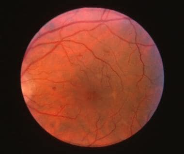 Fundus photograph of nonproliferative diabetic retinopathy with clinically significant macular edema and cystoid macular edema.


