Continually Updated Clinical Reference
 
 
  All Sources     eMedicine     Medscape     Drug Reference     MEDLINE
 
eMedicine - Marcus Gunn Jaw-winking Syndrome : Article by

Quick Find
Authors & Editors
Introduction
Clinical
Differentials
Workup
Treatment
Follow-up
Miscellaneous
Multimedia
References

Related Articles
Amblyopia

Extraocular Muscles, Actions

Extraocular Muscles, Anatomy

Oculomotor Nerve Palsy

Ptosis, Congenital


Patient Education
Click here for patient education.


Author: Sean M Blaydon, MD, FACS, Ophthalmic Plastic Surgery, Texas Oculoplastic Consultants, Austin, Texas

Sean M Blaydon is a member of the following medical societies: American Academy of Ophthalmology, American Medical Association, American Society of Ophthalmic Plastic and Reconstructive Surgery, Association of Military Surgeons of the US, International College of Surgeons US Section, Pan-Pacific Surgical Association, and Texas Medical Association

Editors: Michael J Bartiss, OD, MD, Medical Director, Ophthalmology, Family Eye Care of the Carolinas; Francisco Talavera, PharmD, PhD, Senior Pharmacy Editor, eMedicine; J James Rowsey, MD, Director of Corneal Services, St Luke's Cataract and Laser Institute, Florida; Lance L Brown, OD, MD, Ophthalmologist, Affiliated With Freeman Hospital and St John's Hospital, Regional Eye Center, Joplin, Missouri; Hampton Roy Sr, MD, Associate Clinical Professor, Department of Ophthalmology, University of Arkansas for Medical Sciences

Author and Editor Disclosure

Synonyms and related keywords: trigemino-oculomotor synkinesis, synkinetic jaw-winking phenomenon, wink reflex, jaw-winking ptosis

Background

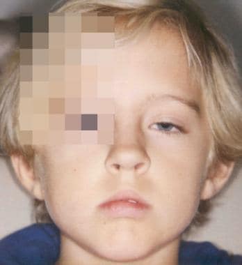
In 1883, Marcus Gunn described a 15-year-old girl with a peculiar type of congenital ptosis that included an associated winking motion of the affected eyelid on movement of the jaw. This synkinetic jaw-winking phenomenon now bears his name.

Patients with this condition have variable degrees of blepharoptosis in the resting, primary position. Although it usually is unilateral, it can present bilaterally in rare cases.

The wink reflex consists of a momentary upper eyelid retraction or elevation to an equal or higher level than the normal fellow eyelid upon stimulation of the ipsilateral pterygoid muscle. This response is followed by a rapid return to a lower position. The amplitude of the wink tends to be worse in downgaze. This rapid, abnormal motion of the eyelid can be the most disturbing aspect of the jaw-winking syndrome.

The wink phenomenon may be elicited by opening the mouth, thrusting the jaw to the contralateral side, jaw protrusion, chewing, or sucking. It often is discovered early, as the infant is bottle-feeding or breastfeeding.

Jaw-winking ptosis is almost always sporadic, but familial cases with an irregular autosomal dominant inheritance pattern have been reported.

Pathophysiology

Marcus Gunn jaw-winking is thought to be a form of synkinetic ptosis. An aberrant connection appears to exist between the motor branches of the trigeminal nerve (CN V3) innervating the external pterygoid muscle and the fibers of the superior division of the oculomotor nerve (CN III) that innervate the levator superioris muscle of the upper eyelid.

Electromyographic studies demonstrate this synkinetic innervation by showing simultaneous contraction of the external pterygoid and levator muscle. In rare cases, synkinesis may be present between the internal pterygoid and levator muscles. In these cases, the eyelid elevates on closing the mouth and clenching the teeth.

A few authors have speculated that the jaw-winking is not due to a new aberrant pathway, but rather the disinhibition of preexisting phylogenetically more primitive mechanisms. This is thought to explain why individuals who are not affected will often open their mouths while attempting to widely open their eyes to place eye drops or to apply makeup.

Since jaw-winking ptosis is believed to be due to abnormal innervation of the levator muscle and not secondary to myopathic changes, it is not surprising that most histopathologic studies have revealed normal striated muscle.

One recent study found variable degrees of fibrosis within the affected levator muscle, and to a lesser degree in the muscle of the normal, nonptotic eyelid.

Frequency

United States

Approximately 50% of blepharoptosis cases are congenital. Incidence of Marcus Gunn syndrome among this population is approximately 5%.

Mortality/Morbidity

  • Marcus Gunn syndrome is associated with strabismus in 50-60% of cases. Superior rectus palsy is found in 25% of cases and double elevator palsy in another 25% of cases. In double elevator palsy, a deficiency in elevation of the globe occurs in all positions of gaze, secondary to an apparent weakness of both the superior rectus and inferior oblique muscles. On rare occasions, horizontal strabismus in the absence of a vertical motility disturbance may occur.
  • Incidence of anisometropia among these patients is reported to be 5-25%. Anisometropia exists when a refractive difference between the 2 eyes of 1.25 diopters of sphere or 1 diopter of cylinder is present.
  • Amblyopia occurs in 30-50% of these patients and is almost always secondary to strabismus or anisometropia, and, only rarely, is due to occlusion by a ptotic eyelid. Amblyopia usually is defined as a decrease in vision of 2 or more lines on the Snellen chart.

Race

No known racial predilection exists.

Sex

Early reports showed jaw-winking ptosis to be more prevalent in females; however, larger case series have shown an equal prevalence among males and females.

Age

  • Marcus Gunn jaw-winking usually is evident at birth. The winking phenomenon often is first noted by the parents when the infant is feeding.
  • It has been suggested, and older patients often claim, that the jaw winking improves over time; however, it has not proven to be true on objective evaluation. More likely, patients stop seeking care as they get older, or they learn to compensate for and mask the wink response.
  • Likewise, the degree of ptosis may be underestimated with the patient able to adjust the height with varying jaw positions. This is referred to by some as "habitual" ptosis.


History

  • Signs and symptoms
    • Mild-to-moderate blepharoptosis, usually unilateral
    • Synkinetic upper eyelid movement with jaw winking after one of the following:

      • Mouth opening
      • Jaw movement toward contralateral side
      • Chewing
      • Sucking
      • Jaw protrusion
      • Clenching teeth together
      • Swallowing
    • Usually, parents first notice the phenomenon while the baby is breastfeeding or bottle-feeding.
    • Jaw winking worse in downgaze
    • Decreased vision secondary to amblyopia
    • Strabismus
      • Vertical deviation, usually a hypotropia on involved side
      • Horizontal deviation (rare cases)
  • Past ocular history
    • Occlusion or patching therapy for amblyopia
    • Strabismus surgery
    • Periorbital trauma (suggests an aberrant third nerve regeneration if accompanied by bizarre extraocular movements and diplopia.)
    • Previous eyelid surgery
    • Dry eyes (important if contemplating ptosis repair)
  • Past medical history
    • Details of birth history
    • Previous reactions to anesthesia
    • Previous diagnosis of a bleeding diathesis
    • Medication history to include anticoagulants or any nonsteroidal anti-inflammatory drugs (NSAIDs)

Physical

  • Complete ophthalmic examination
    • Pupillary examination
    • Visual acuity (rule out amblyopia in infants and children)
    • Cycloplegic refraction (rule out anisometropia)

  • External examination
    • Extraocular motility
    • Cover test (rule out a superior rectus or double elevator palsy)
    • Bell phenomenon (can be decreased with a superior rectus or double elevator palsy)
    • Head position

      • May elevate chin to see.
      • If a child does not elevate chin in the presence of moderate-to-severe ptosis, then consider amblyopia.
    • Ptosis evaluation

      • The degree of ptosis should be assessed with the jaw immobilized in a central position and after fusion is disrupted with brief ocular occlusion.
      • Vertical palpebral fissure (widest distance between the upper and lower eyelid)
      • Marginal reflex distance (MRD) - Distance from upper eyelid margin to corneal light reflex in primary position
      • Ptosis can be quantified - Mild (less than or equal to 2 mm), moderate (3 mm), or severe (greater than or equal to 4 mm)
      • Upper eyelid crease position (distance from crease to eyelid margin)
      • Levator function - Good (greater than or equal to 8 mm), fair (5-7 mm), or poor (less than or equal to 4 mm)
      • Measurement of eyelid position in downgaze
      • Presence of lid lag
    • Attempt to elicit synkinesis of eyelid movement.

      • Have the infant bottle-feed.
      • An older child can chew gum.
      • Have the patient open mouth, move jaw from side to side, or protrude jaw forward.
      • Jaw wink can be quantified - Mild (less than or equal to 2 mm), moderate (3-6 mm), or severe (greater than or equal to 7 mm)

Causes

See Pathophysiology.


Amblyopia
Extraocular Muscles, Actions
Extraocular Muscles, Anatomy
Oculomotor Nerve Palsy
Ptosis, Congenital

Other Problems to be Considered

Aberrant third nerve regeneration
Double elevator palsy


Lab Studies

  • Creatine phosphokinase level: If a past medical or family history of any reactions to anesthesia exists, rule out the possibility of malignant hyperthermia prior to any surgery.

Other Tests

  • Basic tear secretion test: Normal measurement is more than 5-10 mm of Schirmer filter paper wetting after 5 minutes following application of topical anesthetic, such as proparacaine.

Histologic Findings

Light microscopy of surgical specimens usually reveals normal striated muscle. Fibrosis within the muscle has been reported.


Medical Care

  • If amblyopia is encountered, treat aggressively with occlusion therapy and/or correction of anisometropia prior to any consideration of ptosis surgery.

Surgical Care

As with any patient who requires eyelid surgery, first address associated strabismus.

  • Superior rectus palsy
    • Superior rectus palsy can be corrected by resecting the superior rectus muscle but only in the absence of inferior rectus restriction.
    • Since the superior rectus is loosely bound to the overlying levator, the upper eyelid will be pulled inferiorly during resection, exacerbating any ptosis already present. This can be addressed during the subsequent ptosis repair.
  • Double elevator palsy
    • Double elevator palsy manifests as a deficit in elevation of the globe in all fields of gaze.
    • It may be the result of superior rectus and inferior oblique palsy and/or inferior rectus restriction.
    • Inferior rectus restriction may be suggested by the following:
      • Positive forced ductions in elevation
      • Normal force generations in upgaze indicating absence of superior rectus or inferior oblique palsy
      • Poor or absent Bell phenomenon on the affected side
    • Inferior rectus restriction is treated by recession of the inferior rectus muscle.
    • A combined superior rectus and inferior oblique (double elevator) palsy requires a transposition procedure to displace the medial and lateral recti muscles superiorly (Knapp procedure).
  • Consider eyelid surgery only when the parents (or the patient) and the surgeon agree about whether the most cosmetically objectionable condition is the ptosis or the jaw winking or whether it is a combination of both.
  • It is important to note that there are many techniques described for the correction of jaw-winking ptosis reflecting the ongoing controversy regarding the surgical management of this condition.
  • If the jaw winking is cosmetically insignificant, it can be ignored in the treatment of the ptosis.
    • If the ptosis is mild, the patient may elect not to proceed with surgery. If correction is desired, perform a conjunctivo-Müller muscle resection, a Fasanella-Servat procedure, or standard external levator resection.
    • If the ptosis is moderate to severe, a levator resection is indicated. Beard advocated performing more resection than normal to avoid undercorrection.1
    • In severe ptosis, a supermaximum (>30 mm) levator resection or frontalis suspension is necessary.
  • Although the amount of ptosis and synkinetic eyelid movement is variable, those patients with more severe ptosis tend to have the worse aberrant upper eyelid movement.
  • The jaw wink is considered cosmetically significant if it is 2 mm or more.
  • Any attempt to repair the ptosis without addressing the jaw winking would result in an exaggeration of the aberrant eyelid movement to a level well above the superior corneal limbus, which would be unacceptable to the patient.
  • If the jaw wink is significant, ablation of the levator and resuspension of the eyelid to the brow are necessary. Several techniques have been suggested to obliterate levator function, which effectively dampens the aberrant eyelid movement.
    • Bullock advocated complete excision of the levator aponeurosis and muscle all the way to the orbital apex.3
    • Dillman and Anderson argued that removal of a portion of the levator muscle above the Whitnall ligament (ie, myectomy) is adequate to obliterate its function without extensive dissection and damage to eyelid structures.4
    • Bowyer and Sullivan describe the removal of a portion of levator muscle above the Whitnall ligament through a posterior conjunctival approach.2
    • Dryden et al proposed suturing the transected levator aponeurosis to the arcus marginalis of the superior orbital rim.6 This technique not only effectively deactivates the muscle, but also allows the procedure to be reversed, if necessary.
  • Beard and others have advocated bilateral excision of the levator muscle and bilateral frontalis suspension.1 While this approach almost completely eliminates the wink and arguably results in better symmetry, it is often difficult to persuade the parents and the patient to perform surgery on and effectively damage the normal contralateral levator muscle.
  • Satisfactory and predictable results also can be obtained after only unilateral levator excision on the affected side, combined with bilateral frontalis suspension. This leaves the normal functioning levator muscle to elevate the nonptotic eyelid in primary position but produces a lag in downgaze for improved symmetry.
  • Kersten et al advocate unilateral levator muscle excision and frontalis sling only on the affected side.8 If the postoperative result is judged to be unsatisfactory, the parents or the patient can opt for further surgery to the contralateral side. Any amblyopia and strabismus should first be addressed, as there may be insufficient drive to lift the disinserted eyelid.
  • Islam et al described a technique of dissecting a frontalis flap hinged superiorly through a suprabrow incision that is then brought down into an eyelid crease incision.7 The frontalis flap is used to suspend the ptotic eyelid after extirpation of the levator muscle.
  • Lemagne and Neuhaus described techniques that involve transection of the involved levator followed by transposition of the distal segment to the brow, which effectively suspends the eyelid to the frontalis muscle.10, 12 Their techniques maintain normal eyelid contour, as the levator aponeurotic attachments are left undisturbed.

Consultations

  • Consult an oculoplastic surgeon or a strabismologist if the referring physician is uncomfortable with these procedures.
  • Forewarn the anesthesiologist that patients with Marcus Gunn jaw-winking ptosis are at a greater risk of developing arrhythmias during eyelid surgery.


Further Inpatient Care

  • Marcus Gunn jaw-winking ptosis usually is treated on an outpatient basis.

Further Outpatient Care

  • While the patient is awake, apply cold compresses to the eyelids for 20 minutes every 1-2 hours for 2-3 days to decrease swelling and bruising.
  • See the patient postoperatively in 5-7 days for suture removal.
  • Schedule regular follow-up appointments with a general or pediatric ophthalmologist for any child with preexisting amblyopia.

In/Out Patient Meds

  • Antibiotic ophthalmic ointment, such as erythromycin, is prescribed postoperatively, and it should be applied 2-4 times a day along sutures and in the eye for 1 week.

Complications

  • Complications of surgery to repair jaw-winking ptosis include the following:
    • Unacceptable eyelid height or contour
    • Exaggerated postoperative wink
    • Asymmetry
    • Poor wound healing
    • Wound infection
    • Bleeding or hematoma formation
    • Inflammation and granuloma formation around frontalis sling material

Prognosis

  • Satisfactory results usually are achieved by a medical and surgical approach to managing Marcus Gunn jaw-winking ptosis.

Patient Education

  • Discuss the following with the patient or the parents:
    • Connection between jaw movement and abnormal movement of the eyelid
    • Importance of treating amblyopia or strabismus prior to any surgical repair of ptosis
    • Various surgical approaches, to include possible benefits of bilateral eyelid surgery


Medical/Legal Pitfalls

  • Marcus Gunn jaw-winking syndrome is a rare disease, and early detection and treatment can prevent amblyopia.


Media file 1:  Congenital left upper eyelid ptosis.
Click to see larger pictureClick to see detailView Full Size Image
Media type:  Photo

Media file 2:  
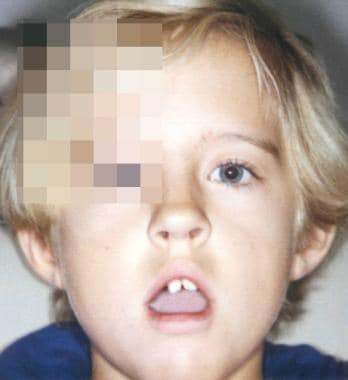
Marcus Gunn jaw-winking with left upper eyelid retraction on opening of the mouth.
Click to see larger pictureClick to see detailView Full Size Image
Media type:  Photo


  1. Beard C. Ptosis. 3rd ed. St. Louis: CV Mosby; 1981:32-3, 47-9, 113-5, 208.
  2. Bowyer JD, Sullivan TJ. Management of Marcus Gunn jaw winking synkinesis. Ophthal Plast Reconstr Surg. Mar 2004;20(2):92-8. [Medline].
  3. Bullock JD. Marcus-Gunn jaw-winking ptosis: classification and surgical management. J Pediatr Ophthalmol Strabismus. Nov-Dec 1980;17(6):375-9. [Medline].
  4. Dillman DB, Anderson RL. Levator myectomy in synkinetic ptosis. Arch Ophthalmol. Mar 1984;102(3):422-3. [Medline].
  5. Doucet TW, Crawford JS. The quantification, natural course, and surgical results in 57 eyes with Marcus Gunn (jaw-winking) syndrome. Am J Ophthalmol. Nov 1981;92(5):702-7. [Medline].
  6. Dryden RM, Fleming JC, Quickert MH. Levator transposition and frontalis sling procedure in severe unilateral ptosis and the paradoxically innervated levator. Arch Ophthalmol. Mar 1982;100(3):462-4. [Medline].
  7. Islam ZU, Rehman HU, Khan MD. Frontalis muscle flap advancement for jaw-winking ptosis. Ophthal Plast Reconstr Surg. Sep 2002;18(5):365-9. [Medline].
  8. Kersten RC, Bernardini FP, Khouri L, et al. Unilateral frontalis sling for the surgical correction of unilateral poor function ptosis. Ophthal Plast Reconstr Surg. Nov 2005;21(6):412-6. [Medline].
  9. Khwarg SI, Tarbet KJ, Dortzbach RK, Lucarelli MJ. Management of moderate-to-severe Marcus-Gunn jaw-winking ptosis. Ophthalmology. Jun 1999;106(6):1191-6. [Medline].
  10. Lemagne JM. Transposition of the levator muscle and its reinnervation. Eye. 1988;2 (Pt 2):189-92. [Medline].
  11. Miller NR. Walsh and Hoyt's Clinical Neuro-Ophthalmology. Vol 2. 4th ed. Baltimore: Williams & Wilkins; 1985:949-51.
  12. Neuhaus RW. Eyelid suspension with a transposed levator palpebrae superioris muscle. Am J Ophthalmol. Aug 15 1985;100(2):308-11. [Medline].
  13. Pratt SG, Beyer CK, Johnson CC. The Marcus Gunn phenomenon. A review of 71 cases. Ophthalmology. Jan 1984;91(1):27-30. [Medline].
  14. Wong JF, Theriault JF, Bouzouayac C, Codere F. Marcus Gunn jaw-winking phenomenon: a new supplemental test in the preoperative evaluation. Ophthal Plast Reconstr Surg. Nov 2001;17(6):412-8. [Medline].

Marcus Gunn Jaw-winking Syndrome excerpt

Article Last Updated: May 31, 2006