Continually Updated Clinical Reference
 
 
  All Sources     eMedicine     Medscape     Drug Reference     MEDLINE
 
eMedicine - Uveitis, Anterior, Nongranulomatous : Article by

Quick Find
Authors & Editors
Introduction
Clinical
Differentials
Workup
Treatment
Medication
Follow-up
Miscellaneous
Multimedia
References

Related Articles
Behcet Disease

Foreign Body, Intraocular

Herpes Simplex

Herpes Zoster

HLA-B27 Syndromes

Inflammatory Bowel Disease

Juvenile Xanthogranuloma

Leukemias

Lyme Disease

Ocular Manifestations of HIV

Ocular Rosacea

Psoriasis

Reactive Arthritis

Red Eye Evaluation

Retinoblastoma

Sarcoidosis

Uveitis, Fuchs Heterochromic


Patient Education
Click here for patient education.


Author: Roger K George, MD, Director of Uveitis Service, Madigan Army Medical Center; Clinical Instructor, Department of Ophthalmology, Oregon Health and Sciences University

Roger K George is a member of the following medical societies: American Uveitis Society

Editors: Andrew A Dahl, MD, Residency Director, Ophthalmology, Kingston Hospital, Department of Ophthalmology, Assistant Professor of Surgery (Ophthalmology), Mid Hudson Family Practice Institute; Simon K Law, MD, PharmD, Assistant Professor of Ophthalmology, Jules Stein Eye Institute; Chief of Section of Ophthalmology Surgical Services, Department of Veterans Affairs Healthcare Center, West Los Angeles; R Christopher Walton, MD, Director of Uveitis and Ocular Inflammatory Diseases Service, Associate Professor, Department of Ophthalmology, University of Tennessee College of Medicine; Lance L Brown, OD, MD, Ophthalmologist, Affiliated With Freeman Hospital and St John's Hospital, Regional Eye Center, Joplin, Missouri; Hampton Roy Sr, MD, Associate Clinical Professor, Department of Ophthalmology, University of Arkansas for Medical Sciences

Author and Editor Disclosure

Synonyms and related keywords: iritis, iridocyclitis, iris, ciliary body, iritis flare, cell and flare, anterior uveitis

Background

Iritis, or anterior uveitis, is the most common form of ocular inflammation encountered. It is a common cause of a painful red eye. Inflammation of the iris may appropriately be termed iritis, whereas inflammation of the iris and the ciliary body is called iridocyclitis. Iritis may be subdivided into 2 broad categories: granulomatous and nongranulomatous.

This article addresses nongranulomatous iritis, although iritis due to a granulomatous disease process may have a nongranulomatous appearance. For information about granulomatous disease, see Uveitis, Anterior, Granulomatous.

Pathophysiology

The exact pathophysiology is not known. Inflammation of the iris and the ciliary body causes a breakdown of the blood ocular barrier. This condition allows both protein and WBCs to extravasate into the aqueous, resulting in the typical iritis signs of cell and flare. Frequently, the cause is idiopathic, but certain ocular and systemic diseases may be the underlying cause of the iritis.

Frequency

United States

Iritis is the most frequent form of uveitis that is encountered by ophthalmologists. In one community-based study, anterior uveitis accounted for more than 90% of all cases of uveitis. The annual incidence rate is approximately 8 cases per 100,000 population.

International

No particular geographic distribution for iritis has been noted.

Mortality/Morbidity

  • Morbidity arises from iritis and any associated disease process, if present.
  • Patients may develop posterior synechiae, and, if severe, a secluded pupil and subsequent angle-closure glaucoma may result.
  • Associated ocular complications (eg, cataract, glaucoma) may result in severe vision loss.

Race

No significant racial differences exist.

Sex

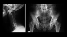
No significant sexual differences exist.

Age

Iritis may develop in patients of any age. The mean patient age is approximately 45 years.


History

Inquire about the patient's complete medical history, to include all medical conditions, surgeries, medications, and ocular history (eg, history of iritis). Perform a detailed review of systems. This is critical, as the history and the review of systems in many cases will suggest a diagnosis. 

Critical review questions include, but are not limited to, asking about recent ocular trauma, back stiffness, arthritis, rashes, shortness of breath, swollen lymph nodes, diarrhea, blood in stools, recent insect bites, sexually transmitted diseases (STDs), and tuberculosis (TB) exposure.

Inquire about the onset of the symptoms.  Most cases of acute anterior uveitis begin with the sudden onset of redness, pain, and photophobia.

Family history is important as well.  Inquire if there is a family history of uveitis or other symptoms in family members that might suggest associated diseases, such as a spondyloarthropathy or other human leukocyte antigen (HLA)-B27 processes.

Inquire in particular about the following symptoms:

  • Dull, aching eye pain occurs and may worsen when one touches the eye through the eyelid.
    • Pain may be referred to the temple or periorbital region.
    • Pain is usually abrupt in onset.
  • Photosensitivity to light, especially sunlight, worsens the patient's discomfort.
  • Redness with no mucopurulent discharge is seen. Patients rarely have a watery discharge or tearing.
  • Mild blurring of vision may be noted.
  • Most cases are unilateral.

Physical

  • Vision: Visual acuity is usually normal or only slightly decreased.
  • Intraocular pressure (IOP) is often lower in the eye with iritis when compared to the fellow eye. This is secondary to a decrease in aqueous production by the inflamed ciliary body. However, in some cases, the IOP may be elevated as a result of altered aqueous outflow.
  • Conjunctiva: Typically, the eye has a perilimbal injection termed ciliary flush. Less commonly, generalized redness of the bulbar conjunctiva may be present.
  • Cornea: Keratic precipitates (KPs) may be present. These clusters of WBCs collect on the endothelium. In nongranulomatous iritis, they tend to be small and are usually located over the inferior half of the cornea. Stellate-shaped KPs, uniformly spread over the endothelium, are typical of Fuchs heterochromic iridocyclitis. Corneal stromal edema may be present.
  • Anterior chamber: Flares, cells, and/or hypopyon may be present.
    • A flare, resulting from extra protein in the aqueous, is usually present and can be graded as follows:
      • 0 = Completely absent
      • 1+ = Barely present
      • 2+ = Moderate
      • 3+ = Marked (iris and lens detail hazy)
      • 4+ = Intense (formed fibrin in aqueous)
    • Cells, the hallmark of iritis, are present in the aqueous. They should be graded by severity under high-magnification slit lamp examination in a 1 X 3-mm field of light, as follows:
      • Trace = 1-5 cells
      • 1+ = 6-15 cells
      • 2+ = 16-25 cells
      • 3+ = 26-50 cells
      • 4+ = More than 50 cells
    • Hypopyon, if present, is highly suggestive of diseases associated with HLA-B27; with Behcet disease; or, less commonly, with an infection-associated iritis.
  • Iris: Posterior synechiae may be present. Inflammatory nodules are usually not present in nongranulomatous iritis. Sector atrophy of the iris, if present, suggests herpes zoster as the etiology of the inflammation. Heterochromia and loss of iris detail (eg, blurring of stroma) are suggestive of Fuchs heterochromic iridocyclitis.
  • Lens and anterior vitreous: Lenticular precipitates may be present on the anterior lens capsule. Posterior subcapsular cataracts may be present if the patient has had repeated episodes of iritis.
    • Cells are commonly seen in the anterior vitreous. They represent either an iridocyclitis or a spillover of cells from the anterior chamber into the retrolental space.
    • Occasionally, patients with HLA-B27 disease have an intense vitritis, papillitis, and cystoid macular edema.

Causes

  • Idiopathic causes
  • Diseases associated with HLA-B27
    • Ankylosing spondylitis
    • Reactive arthritis
    • Inflammatory bowel disease
    • Psoriasis
  • Sarcoidosis
  • Trauma
  • Infections
    • Herpes zoster and/or herpes simplex
    • Syphilis
    • Lyme disease
  • Juvenile idiopathic arthritis
  • Tubulointerstitial nephritis and uveitis syndrome (TINU)
  • Fuchs heterochromic iridocyclitis


Behcet Disease
Foreign Body, Intraocular
Herpes Simplex
Herpes Zoster
HLA-B27 Syndromes
Inflammatory Bowel Disease
Juvenile Xanthogranuloma
Leukemias
Lyme Disease
Ocular Manifestations of HIV
Ocular Rosacea
Psoriasis
Reactive Arthritis
Red Eye Evaluation
Retinoblastoma
Sarcoidosis
Uveitis, Fuchs Heterochromic

Other Problems to be Considered

Masquerade syndromes 
Pigment dispersion syndrome
Retinal detachment


Lab Studies

  • A comprehensive review of the patient's past medical history and a review of systems should guide the laboratory evaluation, and the workup should be tailored accordingly.
    • If a patient presents with a first episode of nongranulomatous iritis and if the medical history and the review of systems are unremarkable, laboratory studies are not indicated.
    • Iritis that is recurrent, unusual in severity, unresponsive to medical therapy, unusually persistent, or bilateral should be thoroughly evaluated.
  • The following list of possible laboratory studies may be requested. At minimum, chest radiography (see Imaging Studies) and a rapid plasma reagin (RPR) test with a fluorescent treponemal antibody absorption (FTA-ABS) test should be ordered.
    • The RPR test and the FTA-ABS test, or serologic tests for syphilis, should be ordered for each patient who undergoes a laboratory evaluation for uveitis.
    • The erythrocyte sedimentation rate (ESR), serum lysozyme level, and angiotensin-converting enzyme (ACE) test may help in evaluating the patient for sarcoidosis.
    • HLA-B27 typing may be useful.
    • Rheumatoid factor (RF) and antinuclear antibody (ANA) may be indicated if juvenile idiopathic arthritis is suspected.
    • Lyme serologic testing should be ordered if Lyme disease is suspected.
    • Serum creatinine and urinary beta-2 microglobulin levels should be obtained if TINU is suspected.

Imaging Studies

  • Chest radiography helps to rule out sarcoidosis and tuberculosis.
  • Chest CT is more sensitive for sarcoidosis than plain radiography and should be ordered if the radiographs are negative and if sarcoidosis is highly suspected as the etiology of the ocular inflammation.
  • Sacroiliac radiographs may be ordered if ankylosing spondylitis is suspected.

Procedures

  • If a patient presents with a secluded pupil from extensive posterior synechiae, iris bombe with angle-closure glaucoma may be present. In this case, an iridotomy may be necessary.


Medical Care

  • Cycloplegia: A long-acting cycloplegic agent, such as cyclopentolate or homatropine, should be used to help relieve both pain and photophobia and to prevent the formation of posterior synechiae.
  • Corticosteroids: Topical corticosteroids are the mainstays of therapy and should be used aggressively during the initial phases of therapy.
    • A subconjunctival injection of depot-steroids (eg, Celestone) may be used if the patient poorly complies with topical therapy or if the iritis is not responding to topical corticosteroids alone.
    • In severe cases of iritis, the addition of oral corticosteroids to the treatment regimen may be necessary.
  • Topical aqueous suppressant: If IOP is elevated, a topical aqueous suppressant should be used.

Consultations

Consultations with other subspecialists should be arranged, as warranted by the patient's history or based on the results of laboratory or radiographic investigations.


Topical corticosteroids and a cycloplegic agent should be started immediately, unless an infectious etiology is suspected. If the eye is not adequately responding to topical therapy within a week to 10 days, the addition of either oral corticosteroids or a periocular injection of corticosteroids to the treatment regimen may be necessary. The injection of steroids may be contraindicated in a known steroid responder or in a patient with an already elevated IOP.

Tapering of steroid therapy is guided by the clinical response on follow-up examination. Topical nonsteroidal anti-inflammatory drugs (NSAIDs) tend to be of little or no benefit in the treatment of iritis.

Drug Category: Corticosteroids

These are the mainstays of therapy for iritis and help to stabilize the blood-aqueous barrier.

Drug NamePrednisolone acetate 1% (Pred Forte, Econopred)
DescriptionDecreases inflammation by suppressing migration of polymorphonuclear leukocytes and by reversing increased capillary permeability.
Adult Dose1 gtt in affected eye; frequency based on severity of iritis but generally used as starting dose q1-2h
Pediatric DoseAdminister as in adults
ContraindicationsDocumented hypersensitivity; viral, fungal, or tubercular infections
InteractionsNone reported
PregnancyC - Fetal risk revealed in studies in animals but not established or not studied in humans; may use if benefits outweigh risk to fetus
PrecautionsProlonged use may contribute to glaucoma and cataract formation; close monitoring of IOP required

Drug NamePrednisone (Deltasone)
DescriptionCan be used if topical therapy inadequate to treat iritis. Decreases inflammation by suppressing migration of polymorphonuclear leukocytes and by reversing increased capillary permeability.
Adult DoseInitially 1 mg/kg/d or 40-80 mg qam depending on severity; taper over 2-4 wk after satisfactory response; tapering based on inflammation present on follow-up
Pediatric Dose0.5-1 mg/kg PO divided qd/qid
ContraindicationsDocumented hypersensitivity; viral infection; peptic ulcer disease; hepatic dysfunction; connective tissue infections; fungal or tubercular skin infections
InteractionsCoadministration with estrogens may decrease clearance; with digoxin, may increase digitalis toxicity due to hypokalemia; phenobarbital, phenytoin, and rifampin may increase metabolism of glucocorticoids (consider increasing maintenance dose); monitor for hypokalemia with coadministration of diuretics
PregnancyB - Fetal risk not confirmed in studies in humans but has been shown in some studies in animals
PrecautionsAbrupt discontinuation of glucocorticoids may cause adrenal crisis; hyperglycemia, edema, osteonecrosis, myopathy, peptic ulcer disease, hypokalemia, osteoporosis, euphoria, psychosis, myasthenia gravis, growth suppression, and infections may occur with glucocorticoid use

Drug Category: Cycloplegics

These agents help prevent or break posterior synechiae and reduce ciliary body–induced pain.

Drug NameCyclopentolate HCl 1% (Cyclogyl)
DescriptionPrevents spasm of ciliary muscle and iris sphincter. Induces mydriasis in 30-60 min and cycloplegia in 25-75 min.
Adult Dose1 gtt qd/tid
Pediatric DoseAdminister as in adults
ContraindicationsDocumented hypersensitivity; narrow-angle glaucoma
InteractionsDecreases effects of carbachol and cholinesterase inhibitors
PregnancyC - Fetal risk revealed in studies in animals but not established or not studied in humans; may use if benefits outweigh risk to fetus
PrecautionsCaution if IOP may be increased (eg, elderly persons); can cause toxic anticholinergic systemic adverse effects (common in children, especially infants) but rare when used sparingly; compressing lacrimal sac with digital pressure for 1-3 min after application may minimize systemic absorption


Further Outpatient Care

  • Patients require close follow-up care, with steroids tapered as the inflammation resolves.
  • Patients should be reexamined 2-3 weeks after all medications have been tapered to ensure that no residual inflammation is present.

Complications

  • Recurrent episodes of iritis and the subsequent therapy may lead to cataract formation and to glaucoma.

Prognosis

  • Most patients can expect to have a recurrence of iritis.
  • Overall, the visual prognosis for patients with recurrent iritis is good in the absence of either cataract formation or glaucoma.

Patient Education

  • If the patient has known or newly diagnosed HLA-B27 disease, the patient should be instructed to always keep a bottle of steroids handy.
    • The patient should instill the steroid at the first sign of an iritis flare.
    • The patient should come into the office as soon as possible to confirm the presence or absence of the iritis.
  • For excellent patient education resources, visit eMedicine's Eye and Vision Center. Also, see eMedicine's patient education articles Anatomy of the Eye and Iritis.


Medical/Legal Pitfalls

  • Because uveitis may be part of a systemic syndrome, exploring the patient's medical history and performing a full review of systems is critical. By failing to do so, a systemic process may be missed.
  • Failure to make the diagnosis and refer the patient to an appropriate subspecialist may result in undue morbidity.


Media file 1:  Fine keratic precipitates in a patient with ankylosing spondylitis–associated acute anterior uveitis.
Click to see larger pictureClick to see detailView Full Size Image
Media type:  Photo

Media file 2:  Small stellate keratic precipitates with fine filaments in a patient with Fuchs heterochromic iridocyclitis.
Click to see larger pictureClick to see detailView Full Size Image
Media type:  Photo

Media file 3:  Acute anterior uveitis with plasmoid aqueous and hypopyon in a patient with ulcerative colitis.
Click to see larger pictureClick to see detailView Full Size Image
Media type:  Photo

Media file 4:  Fuchs heterochromic iridocyclitis with cataract and iris heterochromia.
Click to see larger pictureClick to see detailView Full Size Image
Media type:  Photo

Media file 5:  Iris atrophy in a patient with herpes simplex virus–associated anterior uveitis.
Click to see larger pictureClick to see detailView Full Size Image
Media type:  Photo


  • Kump LI, Cervantes-Castaneda RA, Androudi SN, Foster CS. Analysis of pediatric uveitis cases at a tertiary referral center. Ophthalmology. Jul 2005;112(7):1287-92. [Medline].
  • Mackensen F, Smith JR, Rosenbaum JT. Enhanced recognition, treatment, and prognosis of tubulointerstitial nephritis and uveitis syndrome. Ophthalmology. May 2007;114(5):995-9. [Medline].
  • McCannel CA, Holland GN, Helm CJ, Cornell PJ, Winston JV, Rimmer TG. Causes of uveitis in the general practice of ophthalmology. UCLA Community-Based Uveitis Study Group. Am J Ophthalmol. Jan 1996;121(1):35-46. [Medline].
  • Menezo V, Lightman S. The development of complications in patients with chronic anterior uveitis. Am J Ophthalmol. Jun 2005;139(6):988-92. [Medline].
  • Nussenblatt RB, Whitcup SM. Uveitis: Fundamentals and Clinical Practice. 3rd ed. 2003.
  • Pepose JS, Holland GN, Wilhelmus KR. Ocular Infection and Immunity. 1996.
  • Rosenbaum JT, George RK. Uveitis. In: Current Ocular Therapy 5. 2000:519-21.

Uveitis, Anterior, Nongranulomatous excerpt

Article Last Updated: Sep 5, 2007