Background
Atopy is the hereditary predisposition to allergy or hypersensitivity. [1] Symptoms may present as a dermatitis, hay fever, or asthma. [1] According to Rapoza, Besnier first characterized atopic dermatitis, and many Europeans still use his name to describe the disease (prurigo Besnier). [1] This disease was labeled eczema for many years in the United States until Coca and Cooke coined the term atopy as a skin hypersensitivity seen in patients with hereditary allergies. Wise and Sulzberger have been credited with the term atopic dermatitis to describe a group of diseases associated with atopic conditions that may be seen in all age groups. [2]
Atopic dermatitis (AD) is a chronic and progressive skin disorder that includes an acute stage, characterized by erythema with edema and vesicles, and a chronic stage with skin lichenification. [3] The SCORAD (Severity Scoring of Atopic Dermatitis) Index characterizes the extent and severity of atopic dermatitis based on surface area involved, intensity of erythema, edema, oozing/crusting, excoriation, lichenification and dryness, as well as subjective symptoms of pruritis and sleep loss. [4] The purpose of this article is to describe the ophthalmologic complications of atopic dermatitis, specifically keratoconus, atopic keratoconjunctivitis (AKC), vernal keratoconjunctivitis (VKC), bacterial blepharoconjunctivitis, cicatricial entropion, cicatricial ectropion, retinal detachment, cataract and glaucoma. [3] These ophthalmologic complications can occur with or without other systemic complications including skin infections, gastrointestinal diseases, renal diseases, autoimmune diseases and psychological and psychiatric diseases. [3]
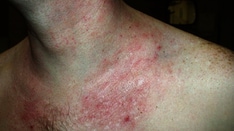
 Typical atopic dermatitis on the face of an infant.
Keratoconus (KC) is a non-inflammatory, progressive corneal thinning disorder that leads to corneal protrusion, irregular astigmatism, and corneal scarring in the final stages, that eventually leads to distorted and impaired vision. [3] The incidence of keratoconus is reported as approximately 0.005% in the general population, but from 0.5% to 39% in those with AD. [3] One of the symptoms of AD is pruritis, which stimulates eye rubbing, and many studies have demonstrated a link between eye rubbing and keratoconus. [5, 6, 7, 8] Lastly, there have been reports that individuals with KC are more likely to have atopic tendencies than the healthy control group (35% vs 12%), most commonly allergic rhinitis, but also asthma and AD. [9]
Atopic keratoconjunctivitis (AKC) is a chronic inflammatory allergic disease, with clinical characteristics including conjunctivitis, corneal ulceration, superficial punctate keratitis and corneal neovascularization. [3]
Vernal keratoconjunctivitis (VKC) is typically a self-limiting, atopic disease of the conjunctiva, limbus and/or cornea, with usual onset prior to age 10 years, in individuals with a personal or family history of atopy. [10]
The incidence of bacterial conjunctivitis and blepharitis in AD patients has reportedly been higher than those without AD (86% vs 25%), with S. Aureus identified as the most common pathogen amongst patients with AD (67%). [3] As a result, patients with AD may benefit from pre- and post-operative antibiotic regimens against S. Aureus to prevent infection and its sequelae.
Cicatricial entropion describes inward turning of the eyelid margin and its appendages secondary to chronic inflammatory changes and subsequent fibrosis, scarring and shortening of the posterior lamellae (e.g. conjunctiva, tarsus), and can lead to lash-cornea touch that may lead to corneal abrasion, scarring, thinning, neovascularization, or even corneal ulceration and perforation in severe cases. [11]
Cicatricial ectropion, in contrast, is caused by shortening of the anterior lamella (i.e. skin and orbicularis muscle), and is also secondary to chronic inflammatory changes including fibrosis and scarring. Similar to other types of ectropion, cicatricial ectropion can lead to exposure keratopathy, corneal abrasion, ulceration, and even thinning and perforation. [12]
Another serious ophthalmologic complication of AD includes retinal detachment, which may be due to eye rubbing as a result of atopic dermatitis-associated pruritis. A review of case studies and another retrospective study [13] , demonstrated that retinal breaks in patients with AD are most likely found in the peripheral fundus, near the ora serrata, which are similar to those caused by trauma (e.g. eye rubbing). [13, 14, 15] Serum IgE levels have not been reported to have an impact on RD. [14]
In patients with AD, cataracts are usually bilateral, symmetric, and occur in the anterior and/or posterior subcapsular regions. [3] Their progression has been linked to eye rubbing and severity of facial skin lesions, which are likely a sign of increased facial pruritis. [16] Some articles have not demonstrated a relationship with serum IgE levels, the duration of systemic or facial topical corticosteroid use and cataract development, [14] whereas other studies have proved an association between high serum IgE levels and cataract development in those with AD. [17]
The relationship with glaucoma and AD is rarely described. Some researchers have denied a correlation between the two conditions, [18] while Takakuwa et al. have focused on patients with severe AD and advanced glaucoma in order to propose a new clinical entity, "atopic glaucoma", that is thought to arise secondary to increased intraocular inflammatory responses, such as higher IL8 and CCL2 expression in the aqueous humor of these patients. [19] Takakuwa et al. excluded those cases showing an apparent relation between glucocorticoid use and IOP in order to propose this new clinical diagnosis. [19] These researchers believe that atopic glaucoma is associated with proinflammatory factors, is defined by vertical cup-disc ratio >0.7 and/or neuroretinal rim notching, compatible visual field loss and IOP >21 mmHg. [19]
Pathophysiology
Atopic dermatitis is primarily caused by cellular immune deficiency and elevated immunoglobulin E (IgE). The pathogenesis can be traced to a genetically inherited, bone marrow–derived cell associated with chromosome 11q. [20] Abnormal skin reactivity also plays a major role in the development of the disease. Irritants to the skin are believed to predispose an individual to develop dermatitis more often than simply exposure to an allergenic trigger. [20] Nonetheless, patients frequently have a history of food or inhalant allergies or eventually develop them. [20]
One of the symptoms of AD is pruritis, which is associated with keratoconus, though the exact mechanism remains unknown. Potential mechanisms of eye rubbing leading to KC include increased corneal temperature, released inflammatory mediators in the tear film and increased enzymatic activity, increased intraocular and hydrostatic tissue pressure, change in keratocytes and a decrease in corneal shear strength. [3]
Epidemiology
Frequency
United States
Atopic dermatitis affects approximately 17% of children and 5% of adults in the United States. [21]
Race
No clear racial predisposition appears to exist. [21]
Sex
Males appear to be affected more frequently by AKC and especially VKC than females, though this difference decreases with increasing age. [22]
Age
Children most commonly are affected, with 80% developing the disease before age 7 years. Less than 2% will have an onset after age 20 years. Most sources agree that persistence after age 20 years is uncommon. Only an estimated 10% of patients older than 20 years continue to be symptomatic. [23]
Specific ophthalmologic complications of AD occur at different ages. For example, the peak incidence of AKC is estimated between 30-50 years old, whereas the majority of VKC cases first occur between 10-12 years old. [10, 24]
However, in general, ophthalmologic complications of AD were far more frequent for adults than for children. Specifically, more severe complications leading to ocular morbidity or visual impairment were rare in younger children with mild atopic dermatitis. [25, 26]
Environmental
The highest incidence of atopic dermatitis is in urban areas. It seems there may be environmental factors in urban areas that predisposes the population to atopic dermatitis as compared to rural settings, but further evaluation of this relationship is needed for the exact cause. [27]
Specifically, VKC is primarily seen in hot and dry climates, such as West Africa, Middle East, Japan, India and South America, which is though to be secondary to a higher level of pollution by pollens and various other allergens in these areas. [10]
Prognosis
The prognosis is good if the inflammation can be kept under control with therapy. [3] Unfortunately, atopic disease can be controlled but not cured, and management focuses on improvement on symptoms, and prevention of complications and sequelae that can lead to vision loss, while limiting side effects of treatment. [3] Unfortunately, some cases are so severe that treatment will not prevent vision loss and other debilitating complications.
Patient Education
For excellent patient education resources, visit eMedicineHealth's Skin Conditions and Beauty Center. Also, see eMedicineHealth's patient education article Eczema.
-
Typical atopic dermatitis on the face of an infant.