Overview
Kawasaki disease is a systemic vasculitis of unknown etiology that affects the small- and medium-sized blood vessels of the body, particularly the coronary arteries.
Since its original description, in Japan in 1967, Kawasaki disease has been reported worldwide in children of all ethnic origins. The yearly incidence of endemic Kawasaki disease in the United States is 67 cases per 100,000 children who are younger than age 5 years. Approximately 3,000 hospitalizations occur annually in the United States as a result of Kawasaki disease.
The incidence of Kawasaki disease in Europe is comparable to that in the United States. In Japan, where Kawasaki disease has the highest incidence, an estimated 5000-6000 cases occur annually. Individuals of Asian descent are 20 times more likely to develop Kawasaki disease than Whites. [1] The exact etiology is unknown; however, recent evidence suggests a combination of genetic factors and a possible infectious source. [2]
Go to Kawasaki Disease and Dermatologic Manifestations of Kawasaki Disease for complete information on these topics.
See Kawasaki Disease: Do You Know the Signs?, a Critical Images slideshow, to help identify the specific criteria for diagnosis.
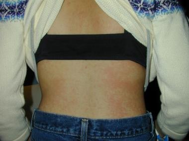 Patchy generalized macular erythema, which is also typical of some viral exanthems.
Ophthalmologic Presentation
The most common ophthalmologic finding in Kawasaki disease is bilateral conjunctival injection. Iridocyclitis, superficial punctate keratitis, vitreous opacities, papilledema, and subconjunctival hemorrhage also may occur.
In a study by Ohno et al, the incidence of superficial punctate keratitis, vitreous opacities, papilledema, and subconjunctival hemorrhage was, respectively, 22%, 11%, 11%, and 6%. [3]
Bilateral conjunctival erythema is one of the classic diagnostic criteria for Kawasaki disease, being present in more than 90% of children with the disorder. These findings occur in the acute phase of the disease and appear shortly after the fever. [3] It is mostly confined to the bulbar region and is nonexudative. Chemosis, follicles, and papillae are characteristically absent.
Conjunctival pathology obtained during the acute phase showed an increased number of neutrophils surrounding the conjunctival epithelial cells. This pattern, termed neutrophilic rosetting, is present in 36% of cases. [4] A 2016 study in Korea found that individuals with Kawasaki disease who did not initially present with anterior uveitis had a higher risk of coronary events and longer hospitalization. Therefore, one can hypothesize that the detection of anterior uveitis leads to an earlier diagnosis of Kawasaki disease, as opposed to a potentially delayed diagnosis in individuals who present without anterior uveitis. [5]
Burns et al found evidence of anterior uveitis in 83% of children who were diagnosed with Kawasaki disease in the first week of their illness. [6] The anterior uveitis, which usually is mild and bilateral, sometimes is associated with keratic precipitates. Anterior uveitis peaks about a week after fever onset. It generally resolves within 2-8 weeks after disease onset without any of the common sequelae that often are seen with many other infectious causes of uveitis.
Ryan and Walton described a case of a 10-month-old male infant in whom bilateral scarring of the superior and inferior fornices developed in association with Kawasaki disease. [7]
Less common posterior segment manifestations usually are limited to case reports. One such report, by Font et al, described bilateral inner retinal ischemia due to thrombotic occlusion as a result of systemic vasculitis. [8]
One case documented periorbital vasculitis in an 8-month-old infant with Kawasaki disease, a previously unreported manifestation of the disorder.
Although exceptionally rare, neuro-ophthalmic findings may be associated with Kawasaki disease. Forty-one reported cases of facial nerve palsies associated with Kawasaki disease have been reported in the current literature. Such patients are reported to have a higher risk of coronary aneurysms and therefore should be monitored with increased vigilance. [9] Other neuro-ophthalmic findings include cranial nerve palsies such as oculomotor palsy (reported by Thapa et al [10] ) and supranuclear vertical palsy (reported by Delafay et al [11] ). A few cases of papilledema have been reported. [12] These cranial nerve findings are presumed to result from inflammation of the nerves and increased intracranial pressure due to an aseptic meningitis component in some individuals. [9, 10, 12] Lin et al described eyelid ptosis and muscle weakness in a 3-year-old with typical Kawasaki disease. [13]
Diagnostic Considerations
Kawasaki disease is uncommon, whereas bilateral conjunctivitis is extremely common. The astute primary care physician, emergency room specialist, urgent care physician, or ophthalmologist should scrutinize cases of bilateral ocular redness associated with persistent fever, rash, and lymphadenopathy, particularly in the pediatric, Asian demographic groups most at risk. The lack of definitive autoimmune markers and laboratory tests stresses the importance of a prompt clinical diagnosis based on the constellation of symptoms. However, ongoing research is investigating the association with specific human leukocyte antigen (HLA) alleles. [2] Referral to the appropriate pediatric subspecialist will ascertain community standard care in selected cases. [14, 15, 16]
Treatment & Management
Consultation with a pediatrician, a pediatric intensivist, or a pediatric cardiologist is essential to the delivery of community standard care.
Treat Kawasaki disease uveitis with topical corticosteroids, such as prednisolone acetate, loteprednol etabonate, or dexamethasone, as well as with topical cycloplegics. Although it generally resolves within days of intravenous immunoglobulin therapy, conjunctival inflammation also may be treated with topical corticosteroids. [16, 17]
Keratitis is treated with supportive care using topical tear replacement, preservative-free tears, and ophthalmic ointments, and through the avoidance of irritants, tap water, rubbing behavior, and excessive use of topical preserved drops.
Cranial neuropathies are treated with supportive care and generally resolve after the initiation of intravenous immunoglobulin therapy. No long-term complications have been reported in these cases. [9, 11]
-
Patchy generalized macular erythema, which is also typical of some viral exanthems.
-
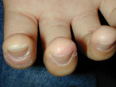
Peeling and erythema of the fingertips.
-
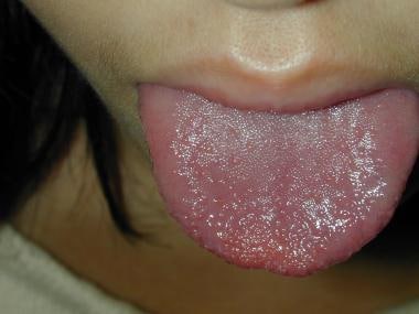
Strawberry tongue.


