You are in: eMedicine Specialties >
Ophthalmology > OPTIC NERVE
Optic Neuritis, Childhood
Article Last Updated: Jul 15, 2005
AUTHOR AND EDITOR INFORMATION
Section 1 of 11
Author: John E Carter, MD, Associate Professor, Department of Medicine, Assistant Professor, Department of Ophthalmology, University of Texas Health Science Center at San Antonio
Editors: Gerhard W Cibis, MD, Clinical Professor, Director of Pediatric Ophthalmology Service, Department of Ophthalmology, University of Kansas, Kansas City; Francisco Talavera, PharmD, PhD, Senior Pharmacy Editor, eMedicine; Brian R Younge, MD, Professor of Ophthalmology, Mayo Clinic School of Medicine; Ralph Garzia, OD, Assistant Dean for Clinical Programs, Associate Professor, School of Optometry, University of Missouri at St Louis; Hampton Roy Sr, MD, Associate Clinical Professor, Department of Ophthalmology, University of Arkansas for Medical Sciences
Author and Editor Disclosure
Synonyms and related keywords:
papillitis, retrobulbar optic neuritis, bilateral simultaneous optic neuritis, bilateral sequential optic neuritis, neuroretinitis, multiple sclerosis, MS, optic nerve, visual loss
Background
Optic neuritis implies an inflammatory process involving the optic nerve. In children, most cases are due to an immune-mediated process. These cases may be associated with a viral or other infection or with immunization. Less commonly, optic neuritis may be the first manifestation of multiple sclerosis (MS) or part of a more diffuse demyelinating disorder, including acute disseminated encephalomyelitis or Devic disease. Optic neuritis may be related to specific infections, diseases of the adjacent sinuses or orbital structures, and infectious and infiltrative diseases of the brain or meninges that involve the optic nerves.
The following definitions aid in further understanding optic neuritis:
Papillitis - Optic neuritis involving the optic disc with disc edema
Retrobulbar optic neuritis - Optic neuritis involving the optic nerve behind the globe. The optic disc appearance should be normal in first-time episodes of retrobulbar optic neuritis.
Bilateral simultaneous optic neuritis - Optic neuritis in both eyes occurring within 3 weeks of each other
Bilateral sequential optic neuritis - Optic neuritis occurring in both optic nerves but separated by a period of more than 3 weeks
Neuroretinitis - Inflammatory process involving the optic discs with exudative changes in the nerve fiber layer of the retina producing a partial or complete macular star. In the past, this condition was called Leber idiopathic stellate neuroretinitis, but now a number of underlying causes for this condition are known. Because this condition is not associated with demyelinating disease and does not imply a future risk of MS, the distinction is important.
Pathophysiology
Possible mechanisms of inflammation in immune-mediated optic neuritis are the cross-reaction of viral epitopes and host epitopes and the persistence of a virus in central nervous system (CNS) glial cells.
Frequency
United States
Optic neuritis is much less common in children than in adults but is not rare. In one combined series, children comprised 5% of cases.
Mortality/Morbidity
Patients with optic neuritis have a good prognosis, but a minority of patients experience persistent visual loss. When optic neuritis is associated with other CNS diseases, the morbidity and mortality of those disorders contribute substantially to the final outcome.
Race
Optic neuritis is more common in Caucasians than in other races.
Sex
In both children and adults, a female predominance exists. Females comprise 60-75% of patients.
Age
- Optic neuritis may occur at any age, including in infants younger than 1 year.
- A comparison of adult and childhood optic neuritis is presented in Table 1.These features are generally true but are not absolute, and they do overlap. Table 1. Comparison of features of optic neuritis in adults and children
| Adult Optic Neuritis | Pediatric Optic Neuritis |
|---|
| Unilateral | Bilateral | | Retrobulbar optic neuritis | Papillitis | | Commonly associated with pain on eye movements | Commonly associated with headache | | Most often idiopathic | Most often postinfectious or postimmunization | High probability of recurrent inflammatory demyelinating events in the CNS and a diagnosis of MS | Low probability of recurrent demyelinating events and a diagnosis of MS |
History
- In general, obtaining an accurate history from children may be difficult. Young children may not notice unilateral visual loss and may not report bilateral visual loss until their behavior indicates visual loss to parents or teachers.
- Optic neuritis produces a subacute loss of vision, reaching its maximum deficit in a few days to 2-3 weeks. In many cases, recovery is already underway at 2-3 weeks.
- Headache is common in children with optic neuritis. Periorbital pain, especially if it worsens with eye movements, supports a diagnosis of optic neuritis.
- Visual symptoms reflect the expected deficit observed in any optic neuropathy, including loss of visual acuity, change in color perception, change in brightness sense, and loss of portions of the visual field.
- In reviewing neurologic symptoms, prior resolved neurologic symptoms imply a recurrent process, such as MS, whereas ongoing neurologic symptoms may indicate MS, acute disseminated encephalomyelitis, or Devic disease.
- A review of systemic symptoms should be aimed at detecting recent vaccinations, infections, or vasculitis.
Physical
- Decreased visual acuity: Visual acuity is not the most sensitive indicator of optic nerve injury and may be normal; however, most children with optic neuritis have some loss of visual acuity. Occasionally, patients have severe loss of vision that includes no light perception.
- Decreased color acuity: A deficit in color vision is a more sensitive indicator of optic nerve injury, and, when checked, a deficit is expected that is out of proportion to any loss of visual acuity.
- Afferent pupil defect: In unilateral cases, an afferent pupil defect should be present. In bilateral cases, this sign is less reliable unless the 2 nerves are asymmetrically affected.
- Subjective light brightness difference between the 2 eyes is also common in asymmetric or unilateral cases.
- Fundus exam
- In children, most cases (60-70%) involve the optic disc with disc edema, as compared to 35% in adults (see Image 1).
- In retrobulbar optic neuritis, the optic disc should be normal. Atrophy of the disc implies a prior episode of optic neuritis or another more chronic process, such as an optic nerve glioma, a craniopharyngioma, or other compressive process.
- If macular edema or a macular star is associated, a diagnosis of neuroretinitis rather than optic neuritis should be made (see Image 2).
Causes
- Immune mediated
- In children, as many as 85% of cases are associated with a recent immunization or infection, usually a viral infection.
- Optic neuritis can be associated with a preceding nonviral infection, such as pertussis, infectious mononucleosis, toxoplasmosis, or brucella.
- MS
- Idiopathic
- Lyme disease in endemic areas
- Specific meningeal infections and infiltrations involving the optic nerves, including cryptococcus, tuberculosis, and sarcoidosis
- Vasculitis, such as systemic lupus erythematosus
- Syphilis
- Leukemia
- Associated with bee and wasp stings
- Causes of neuroretinitis
- Following a viral syndrome
- Cat scratch disease
- Toxoplasmosis
- Toxocariasis and helminths (The finding of a discrete white inflammatory mass overlying the optic disc is suggestive of toxocariasis and helminths.)
- Lyme disease, usually stage 2
- Syphilis, especially secondary syphilis as part of a meningitis
Other Problems to be Considered
Leber hereditary optic neuropathy
Papilledema from intracranial hypertension
Optic nerve glioma
Craniopharyngioma
Lab Studies
- Lumbar puncture with measurement of opening pressure excludes papilledema secondary to intracranial hypertension.
- Cerebrospinal fluid (CSF) studies may indicate the presence of a simultaneous meningitis or encephalitis.
- Systemic lab studies can be performed directed by features in the history and physical examination consistent with other non–immune-mediated causes of optic neuritis.
Imaging Studies
- An MRI of the brain and orbits with contrast should be performed.
- Enhancement of the optic nerve in the orbit (see Images 3-4) or the intracranial segment of the optic nerve or of the chiasm (see Images 5-6) is helpful in confirming the diagnosis. Some enlargement of the optic nerve is present in optic neuritis, and a diagnosis of optic nerve glioma should not be made unless the clinical course dictates reconsideration of the diagnosis of optic neuritis.
- An MRI should exclude extrinsic compressive lesions.
- Meningeal enhancement suggests some form of infectious or noninfectious meningitis and may merit additional workup and different therapy.
- Changes in the CNS white matter may confirm other neurologic involvement found on physical examination, may affect the prognosis of MS in the future, or may indicate the presence of acute disseminated encephalomyelitis. One third of children with optic neuritis will have asymptomatic white matter lesions of the brain as compared to one half of adults with optic neuritis (see Image 7).
- To either diagnose or exclude Devic disease, an MRI of the spinal cord with contrast is necessary if symptoms and signs consistent with a spinal cord process are present.
Medical Care
Consultations
The management of a child with optic neuritis is a combined effort by the ophthalmologist and the neurologist. Ophthalmologic input is required to distinguish between optic neuritis and neuroretinitis and to monitor visual response. Neurologic input is required to evaluate possible generalized CNS involvement and to make appropriate decisions and recommendations based on the future risk of MS.
The goals of pharmacotherapy are to reduce morbidity and to prevent complications.
Drug Category: Corticosteroids
A widely used group of drugs. They have anti-inflammatory properties and cause profound and varied metabolic effects. They modify the body's immune response to diverse stimuli. GI tract absorption is approximately 85% of intravenously administered doses. Corticosteroids are well tolerated. A brief course of steroids is not expected to produce significant adrenal complications. Many physicians prescribe an H2 blocker to prevent GI tract distress.
| Drug Name | Methylprednisolone (Medrol, Adlone, Medralone injection) |
| Description | Decreases inflammation by suppressing migration of polymorphonuclear leukocytes and reversing increased capillary permeability. |
| Pediatric Dose | 125-250 mg IV q6h for 3 d, followed by prednisone 1 mg/kg/d PO for 11 d |
| Contraindications | Documented hypersensitivity; viral, fungal, or tubercular skin infections |
| Interactions | Coadministration with digoxin may increase digitalis toxicity secondary to hypokalemia; estrogens may increase levels of methylprednisolone; phenobarbital, phenytoin, and rifampin may decrease levels of methylprednisolone (adjust dose); monitor patients for hypokalemia when taking medication concurrently with diuretics |
| Pregnancy | C - Safety for use during pregnancy has not been established.
|
| Precautions | Hyperglycemia, edema, osteonecrosis, peptic ulcer disease, hypokalemia, osteoporosis, euphoria, psychosis, growth suppression, myopathy, and infections are possible complications |
| Drug Name | Prednisone (Meticorten, Orasone, Deltasone, Sterapred) |
| Description | May decrease inflammation by reversing increased capillary permeability and suppressing PMN activity. |
| Pediatric Dose | 1 mg/kg/d PO for 11 d; take with food or milk |
| Contraindications | Documented hypersensitivity; viral infection; peptic ulcer disease; hepatic dysfunction; connective tissue infections; fungal or tubercular skin infections; GI tract disease |
| Interactions | Coadministration with estrogens may decrease prednisone clearance; concurrent use with digoxin may cause digitalis toxicity secondary to hypokalemia; phenobarbital, phenytoin, and rifampin may increase metabolism of glucocorticoids (consider increasing maintenance dose); monitor for hypokalemia with coadministration of diuretics |
| Pregnancy | B - Usually safe but benefits must outweigh the risks.
|
| Precautions | Abrupt discontinuation of glucocorticoids may cause adrenal crisis; hyperglycemia, edema, osteonecrosis, myopathy, peptic ulcer disease, hypokalemia, osteoporosis, euphoria, psychosis, myasthenia gravis, growth suppression, and infections may occur |
Further Outpatient Care
- Outpatient follow-up care should include monitoring both visual recovery and recovery from neurologic or systemic disorders that were associated with optic neuritis.
- Children who do develop MS may be more likely to develop disability at a younger age. Monitoring patients who had no associated neurologic signs or symptoms at the time of their optic neuritis will allow early diagnosis. Treatment of those patients for early onset MS with the disease modifying agents that are used in adults may be an option.
- Recurrence of optic neuritis during or shortly after the discontinuation of steroids indicates a steroid-dependent optic neuritis and requires reevaluation and a more prolonged taper of corticosteroids.
Prognosis
- The prognosis for visual recovery generally is considered excellent (see Medical Care).
- MS
Patient Education
- With Uhthoff symptom, patients who have had demyelinating lesions with recovery often have symptoms return with exercise, exposure to heat (eg, hot baths), or febrile illnesses; a rapid return to baseline occurs when body temperature returns to normal. Warning patients about Uhthoff symptom is important so that they do not think they are having a recurrence.
- For excellent patient education resources, visit eMedicine's Muscle Disorders Center. Also, see eMedicine's patient education article, Multiple Sclerosis.
Medical/Legal Pitfalls
- Children with optic neuritis need to be evaluated by both an ophthalmologist and a neurologist. Failure to diagnose and treat patients with optic neuritis who have additional CNS involvement could be a potential source of liability. Efforts to identify those patients who are at high risk of developing future episodes of demyelination and MS are motivated by the belief that early treatment with immunomodulator therapy is important. Although currently less of an issue with children, such decisions are in the purview of the pediatric neurologist who will be aware of the latest literature and treatment practices.
| Media file 1:
Optic disc swelling, right eye (Image 1a) and left eye (Image 1b), in a child with bilateral optic neuritis. |
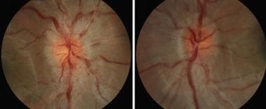
 | View Full Size Image | |
Media type: Image
|
| Media file 2:
Neuroretinitis in the right eye of an adolescent with cat scratch disease. The optic nerve is swollen, and a deposition of yellowish exudate in the nerve fiber layer of the macula produces a macular star. |
 | View Full Size Image | |
Media type: Image
|
| Media file 3:
T1 contrast enhanced axial section of an MRI of the orbital optic nerve of a child with optic neuritis on the left side. The arrows point to the left optic nerve that enhances along its entire orbital course. |
 | View Full Size Image | |
Media type: MRI
|
| Media file 4:
T1 contrast enhanced coronal section of the MRI of the orbital optic nerve, as seen in Image 3. The arrow points to the enhancing left optic nerve. |
 | View Full Size Image | |
Media type: MRI
|
| Media file 5:
T1 contrast enhanced axial section of an MRI of the intracranial optic nerves. Enhancement of both optic nerves is seen. The arrow indicates the left optic nerve. |
 | View Full Size Image | |
Media type: MRI
|
| Media file 6:
T1 contrast enhanced coronal section of the MRI, as seen in Image 5, showing the optic nerves that both enhance. The arrow points to the left optic nerve. |
 | View Full Size Image | |
Media type: MRI
|
| Media file 7:
T2 axial section of an MRI through the cerebral hemisphere of a boy with bilateral optic neuritis. Note high-signal abnormalities in the cerebral white matter that are most prominent in the posterior hemispheres. This is suspicious for mild acute disseminated leukoencephalitis. |
 | View Full Size Image | |
Media type: MRI
|
| Media file 8:
T2 axial sections of MRI of a man with optic neuritis showing scattered white matter lesions. The arrows point to 2 examples of the numerous lesions. |
 | View Full Size Image | |
Media type: MRI
|
- Banwell B, Tremlett H. Coming of age: the use of immunomodulatory therapy in children with multiple sclerosis. Neurology. Mar 8 2005;64(5):778-9. [Medline].
- Beck RW. Optic neuritis. In: Miller NR, Newman NJ, ed. Walsh and Hoyt's Clinical Neuro-ophthalmology. 5th ed. Baltimore:. Lippincott Williams & Wilkins;1998.
- Beck RW, Trobe JD. What we have learned from the Optic Neuritis Treatment Trial. Ophthalmology. Oct 1995;102(10):1504-8. [Medline].
- Bonhomme GR, Liu GT, Balcer LJ. Isolated pediatric optic neuritis. Brain MRI abnormalities and risk of multiple sclerosis. Paper Presented at: Annual Meeting of the North American Neuro-ophthalmology Society; 2005.
- Carroll DM, Franklin RM. Leber''s idiopathic stellate retinopathy. Am J Ophthalmol. Jan 1982;93(1):96-101. [Medline].
- Dreyer RF, Hopen G, Gass JD, et al. Leber''s idiopathic stellate neuroretinitis. Arch Ophthalmol. Aug 1984;102(8):1140-5. [Medline].
- Farris BK, Pickard DJ. Bilateral postinfectious optic neuritis and intravenous steroid therapy in children. Ophthalmology. Mar 1990;97(3):339-45. [Medline].
- Gass JD. Diseases of the optic nerve that may simulate macular disease. Trans Am Acad Ophthalmol Otolaryngol. Sep-Oct 1977;83(5):763-70. [Medline].
- Hierons R, Lyle TK. Bilateral retrobulbar optic neuritis. Brain. 1959;82:56-67.
- Keast-Butler J, Taylor D. Optic neuropathies in children. Trans Ophthalmol Soc U K. Apr 1980;100(Pt 1):111-8. [Medline].
- Kennedy C, Carroll FD. Optic neuritis in children. Arch Ophthalmol. 1960;73:747-755.
- King MH, Cartwright MJ, Carney MD. Leber''s idiopathic stellate neuroretinitis. Ann Ophthalmol. Feb 1991;23(2):58-60. [Medline].
- Koraszewska-Matuszewska B, Samochowiec-Donocik E, Rynkiewicz E. [Optic neuritis in children and adolescents]. Klin Oczna. Jun 1995;97(6):207-10. [Medline].
- Kriss A, Francis DA, Cuendet F, et al. Recovery after optic neuritis in childhood. J Neurol Neurosurg Psychiatry. Oct 1988;51(10):1253-8. [Medline].
- Lucchinetti CF, Kiers L, O''Duffy A, et al. Risk factors for developing multiple sclerosis after childhood optic neuritis. Neurology. Nov 1997;49(5):1413-8. [Medline].
- Maitland CG, Miller NR. Neuroretinitis. Arch Ophthalmol. Aug 1984;102(8):1146-50. [Medline].
- Meadows SP. Doyne memorial lecture (1969). Retrobulbar and optic neuritis in childhood and adolescence. Trans Ophthalmol Soc U K. 1970;89:603-38. [Medline].
- Morales DS, Siatkowski RM, Howard CW, et al. Optic neuritis in children. J Pediatr Ophthalmol Strabismus. Sep-Oct 2000;37(5):254-9. [Medline].
- Nakao Y, Omoto T, Shimomura Y. Optic neuritis in children. Folia Ophthalmol Jpn. 1983;34:496-498.
- Neetens A, Smets RM. Leber's neurogenic stellate maculopathy. Neuro Ophthalmol. 1987;7:315-328.
- Pohl D, Rostasy K, Gartner J. Treatment of early onset multiple sclerosis with subcutaneous interferon beta-1a. Neurology. Mar 8 2005;64(5):888-90. [Medline].
- Riikonen R, Donner M, Erkkila H. Optic neuritis in children and its relationship to multiple sclerosis: a clinical study of 21 children. Dev Med Child Neurol. Jun 1988;30(3):349-59. [Medline].
- Rothermel H, Hedges TR 3rd, Steere AC. Optic neuropathy in children with Lyme disease. Pediatrics. Aug 2001;108(2):477-81. [Medline].
- Rush JA. Idiopathic optic neuritis of childhood. J Pediatr Ophthalmol Strabismus. May-Jun 1981;18(3):39-41. [Medline].
Optic Neuritis, Childhood excerpt Article Last Updated: Jul 15, 2005
|