Continually Updated Clinical Reference
 
 
  All Sources     eMedicine     Medscape     Drug Reference     MEDLINE
 
eMedicine - Aniridia in the Newborn : Article by

Quick Find
Authors & Editors
Introduction
Clinical
Differentials
Workup
Treatment
Medication
Follow-up
Miscellaneous
Multimedia
References

Related Articles
Aniridia

Cataract, Congenital

Ectopia Lentis

Glaucoma, Complications and Management of Glaucoma Filtering

Glaucoma, Juvenile

Nystagmus, Congenital


Patient Education
Click here for patient education.


Author: Sophie Bakri, MD, Assistant Professor of Ophthalmology, Vitreoretinal Diseases and Surgery, Mayo Clinic

Sophie Bakri is a member of the following medical societies: American Academy of Ophthalmology

Coauthor(s): John W Simon, MD, Chair, Department of Ophthalmology, Professor, Departments of Ophthalmology and Pediatrics, Albany Medical College

Editors: Gerhard W Cibis, MD, Clinical Professor, Director of Pediatric Ophthalmology Service, Department of Ophthalmology, University of Kansas, Kansas City; Francisco Talavera, PharmD, PhD, Senior Pharmacy Editor, eMedicine; J James Rowsey, MD, Former Director of Corneal Services, St Luke's Cataract and Laser Institute, Florida; Lance L Brown, OD, MD, Ophthalmologist, Affiliated With Freeman Hospital and St John's Hospital, Regional Eye Center, Joplin, Missouri; Hampton Roy Sr, MD, Associate Clinical Professor, Department of Ophthalmology, University of Arkansas for Medical Sciences

Author and Editor Disclosure

Synonyms and related keywords: Wilms tumor, Wilm's tumor, nephroblastoma, WAGR complex, iris hypoplasia, absence of the iris, pediatric glaucoma, congenital aniridia

Background

In 1818, Barratta first described aniridia (Greek for absence of the iris). It is a rare, bilateral, panophthalmic disorder, of which iris hypoplasia is the most striking feature. Aniridia also has corneal, lens, optic nerve, and retinal manifestations. Foveal and optic nerve hypoplasia are often present, causing a reduction in visual acuity and congenital sensory nystagmus. Progressive worsening of vision may occur later in life as a result of cataracts, glaucoma, and corneal opacification.

Pathophysiology

Aniridia may be familial or sporadic. It is caused by a mutation in the PAX6 gene on chromosome 11.

Approximately 85% of cases are familial (designated AN1) with autosomal dominant inheritance with complete penetrance but variable expressivity. These familial cases usually have isolated ocular involvement. The 15% of cases that are sporadic have a deletion or mutation on the short arm of chromosome 11. The same deletion is responsible for the development of Wilms tumor (nephroblastoma).

The exact pathogenesis of aniridia is unknown. After early reports of ocular colobomas in patients with aniridia, some authors have proposed that it is a colobomatous disorder. Others have attributed it to a failure of mesodermal development with involvement of the rim of the optic cup, causing iris hypoplasia. Still others have advocated the neuroectodermal theory, which links the presence of retinal anomalies and iris muscular hypoplasia to a developmental failure of neuroectoderm.

Frequency

United States

Aniridia is a very rare disorder. According to a population-based study in Michigan, it has been estimated to affect approximately 1 per 61,000 newborns.

International

A more recent study from Denmark reported the incidence as 1 per 96,000 live births.

Mortality/Morbidity

Aniridia itself is not a lethal disorder. However, miscarriages and a stillborn child have been the only results of a consanguineous mating between individuals with aniridia. It is possible that a homozygous mutation of the aniridia gene may be lethal. The morbidity of aniridia is significant because of the decreased vision and nystagmus.

Race

No racial predilection has been described.

Sex

The incidence of aniridia is equal in males and females.

Age

Aniridia is a congenital disorder apparent at birth. It is never acquired.


History

An infant may come to medical attention with nystagmus or photophobia. Patients may present with strabismus and amblyopia. The family may notice an abnormal pupil, or they may describe a fixed, dilated pupil.

Physical

Visual acuity is reduced, usually to around the 20/100 level. Pendular nystagmus, strabismus, and amblyopia may be present. Nystagmus occurs in 85-92% of patients. Ocular examination reveals absence of the irides or dilated unresponsive pupils. Other findings may include small anterior polar cataracts, pupillary membrane remnants, ectopia lentis (in 18-35%), corneal pannus, progressive keratopathy, and optic nerve or foveal hypoplasia.

Poor vision in infancy often is due to foveal or optic nerve hypoplasia and secondary nystagmus. Acquired vision loss may be due to cataract (present in 85% of cases), glaucoma (present in 70%), keratopathy, or amblyopia.

Some have considered aniridia a misnomer because a rudimentary stump of tissue usually can be found behind the limbal sclera by gonioscopy. The diagnosis can be very difficult to make in patients with a substantial amount of iris tissue. However, characteristic absence of the pupillary rim, iris sphincter muscle, and central iris tissue is noted.

  • Syndromes associated with aniridia
    • Miller syndrome (WAGR complex, 11p- syndrome)
      • Approximately 30% of patients with sporadic aniridia develop Wilms tumor, usually before age 5 years. The association between aniridia and Wilms tumor is referred to as Miller syndrome, after his 1964 report of 6 cases of aniridia among 440 cases of Wilms tumor. It was reported that 1.4% of patients with Wilms tumor have aniridia compared to 1 in 64,000 to 1 in 100,000 of the general population.
      • Affected children have other genitourinary abnormalities, craniofacial dysmorphism, hemihypertrophy, and severe mental retardation. They also may have poorly lobulated low-set ears, prominent noses, and long narrow facies. The acronym WAGR (Wilms tumor, aniridia, genitourinary anomalies, and mental retardation) describes some of the features of Miller syndrome.
      • Miller syndrome has been attributed to a deletion of band 11p13. Patients with sporadic aniridia should undergo a thorough physical examination and workup by a family practitioner or pediatrician.
    • Gillespie syndrome
      • Gillespie syndrome is the association of aniridia, cerebellar ataxia, and mental retardation. It is autosomal recessive, occurring in approximately 2% of patients with aniridia. These patients have anatomical defects in the cerebellum and other parts of the brain, and they are not predisposed to developing Wilms tumor.
      • A study of the PAX6 gene in Gillespie syndrome failed to find a mutation, suggesting that abnormalities in the PAX6 gene are not responsible for this syndrome.
    • Aniridia and glaucoma
      • Glaucoma is one of the major causes of visual loss in patients with aniridia, with a reported incidence of 6-75%. This variation is explained by the referral patterns of the various institutions, the variable phenotypic expression of aniridia, and the definitions of glaucoma. Glaucoma in aniridia is rare in infants and newborns and typically is acquired in early adulthood. As a result of its delayed onset, the clinical findings of megalocornea, buphthalmos and Haab striae typically are not found.
      • Progressive apposition of the rudimentary stump of iris to the trabecular meshwork is believed to occur during the first 2 decades of life. In patients without glaucoma, the structures of the anterior chamber angle appear normal by gonioscopy. In older patients with aniridic glaucoma, the severity of the glaucoma is related to the proportion of the filtration angle covered by the iris stump. The periphery of the hypoplastic stump of iris may insert anteriorly, and even where it is more open, the scleral spur and ciliary body band may be defined poorly. In the rare cases of infants with aniridic glaucoma, filtration-angle defects are present at birth. In these cases, the angle usually is covered by vascularized sheets extending from the iris, rather than being blocked by the iris stump.
    • Cataracts
      • Cataracts occur in 50-85% of patients with aniridia, usually acquired during the first 2 decades of life. At birth, small lens opacities may be noted, but these typically are not visually significant. Types of cataracts described include anterior polar, pyramidal, nuclear, lamellar, and cortical.
      • Visually significant cataracts in neonates require prompt surgical extraction to prevent amblyopia. However, in patients whose cataracts develop later, the factor most strongly limiting visual acuity is the presence of foveal hypoplasia, not the cataract. Areas of clear space may be present in the cataractous lens, which allows for relatively good vision. In these patients, surgery is best delayed, since the risks of glaucoma developing or progressing in aniridic eyes outweighs the small potential for visual improvement.
    • Ectopia lentis: Some investigators have reported lens subluxations in as many as 56% of eyes with aniridia, while others have reported none. This finding may be due to failure to detect mild cases. The zonules are believed to be histologically normal, but their molecular structure is not understood clearly.
    • Corneal defects
      • Progressive corneal opacification and pannus occur in most patients, developing as early as age 2 years. Initially, fine radial vessels invade the superficial layers of the peripheral cornea at the 6- and 12-o'clock positions and, then, involve the whole circumference. This process may progress to involve the entire cornea, requiring corneal transplantation.
      • Pathologically, the pannus seen in aniridic keratopathy is quite unusual; the corneal epithelium may harbor ectopic conjunctival goblet cells, and inflammatory cells are usually present. The pathogenesis is uncertain, but many theories have been proposed. It has been attributed to a total absence of the limbal girdles of Vogt and corneal epithelial limbal stem cells. PAX6 has been found in corneal and conjunctival epithelia, and it is believed that this gene may play a role in the activity of corneal stem cells.
      • Microcornea also has been found in most cases of aniridia.
    • Optic nerve and foveal hypoplasia
      • Posterior segment abnormalities include both macular and optic nerve hypoplasia. Approximately 75% of patients with aniridia have some degree of optic nerve hypoplasia. The macula and optic nerve are usually normal in patients with Gillespie syndrome. Mild cases may be difficult to detect because of nystagmus and corneal or lens opacities. Poor macular or retinal development may be responsible for the optic nerve hypoplasia. Because the retina is derived from neuroectoderm, as are the iris epithelium and musculature, foveal hypoplasia often is associated with aniridia.
      • Decreased vision and nystagmus in patients with aniridia result from faulty development of the macula, not the iris. There appears to be no correlation between the degree of iris hypoplasia and foveal hypoplasia. In some patients, foveal hypoplasia may be so subtle that fluorescein angiography may be necessary to demonstrate lack of the foveal avascular zone.
    • Strabismus: This is a common finding in patients with aniridia, and esotropia most frequently is found. A full cycloplegic refraction is warranted because amblyopia may occur from anisometropia or strabismus.
    • Nystagmus: This is commonly pendular secondary to poor vision from foveal hypoplasia. It may improve with visual maturation.
  • Other findings
    • Peripheral retinal lipoidal deposits have been reported in at least 3 patients.
    • Electroretinogram (ERG) amplitudes are low in patients with worse than 20/60 vision, while patients with better vision may have high a:b wave ratios.

Causes

Aniridia may be familial or sporadic. It is caused by a mutation in the PAX6 gene on chromosome 11.


Aniridia
Cataract, Congenital
Ectopia Lentis
Glaucoma, Complications and Management of Glaucoma Filtering
Glaucoma, Juvenile
Nystagmus, Congenital


Histologic Findings

Histopathologic examination of advanced cases of aniridia reveals only stubs of iris tissue that lack dilator and sphincter muscles, both neuroectodermal derivatives.


Medical Care

  • Medical management of glaucoma associated with aniridia
    • Medical therapy of the glaucoma is the initial treatment of choice. It initially may be efficacious in reducing intraocular pressure, but most patients with aniridia who have glaucoma eventually require surgical therapy.
    • Miotics often are tried first; they improve aqueous outflow by contracting the ciliary muscle. However, the induced myopia may not be well tolerated by young patients. Adrenergic agonists, beta-blockers, and carbonic anhydrase inhibitors also may be tried, but they often are ineffective long term as the patient becomes refractory to them. Whenever a new medication is to be instituted, a trial should be performed, adding and removing only one medication at a time.
  • Optical correction
    • Patients with aniridia may be emmetropic, myopic, or hyperopic. Spectacle or contact lens correction of significant refractive errors should begin in the newborn. In cases of lens subluxation, the refractive error should be corrected through the aphakic portion of the pupil.
    • To lessen photophobia, patients may be fitted with tinted or iris contact lenses or with tinted spectacle lenses at any age.
  • Treatment of amblyopia and strabismus: In cases of strabismus, patching of the favored eye is indicated to treat amblyopia. Cycloplegic refraction should be performed, and appropriate correction should be given. Strabismus surgery may be indicated at an early age to enhance binocularity.

Surgical Care

No convincing opinion exists as to which surgical procedure is the treatment of choice for aniridic glaucoma; none has been considered reliable and predictable in efficacy. Surgical procedures for the treatment of aniridic glaucoma include the following:

  • Goniotomy
    • Although goniotomy has been suggested early in the management of aniridic glaucoma, reported series have shown disappointing results. The procedure is hazardous in these eyes, since the Barkan goniotomy knife is passed over the vulnerable lens and zonules in the anterior chamber. However, results of prophylactic goniotomy have been encouraging.
    • Two separate surgeries on each eye may be performed to strip the tissue extending over the trabecular meshwork, working on 180° of the angle each time. The benefit of this prophylactic procedure is not yet proven, and it should be delayed until after the first year of life. Because of the shallow anterior chamber angle, patients with aniridia should have gonioscopy performed yearly. If the iris processes are becoming more prominent, strong consideration should be given to performing a prophylactic goniotomy.
  • Trabeculotomy: This procedure is considered safer than goniotomy, since it relies on the posterior approach (ab externo), which avoids the lens and zonules. However, it is not without risk and has been associated with the need for repeat surgery.
  • Trabeculectomy: Most surgeons opt for trabeculectomy after a few attempts at goniotomy or trabeculotomy. However, the risk of vitreous loss is increased in aniridia, and cataract formation or progression may occur with inadvertent damage to the lens during surgery. Nelson et al (1984) reported that 5 of 14 patients needed reoperation or had failure with trabeculectomy; others have reported initial success rates from 0-9%.
  • Setons: The success rate of the Molteno implant has been reported as 83% of 6 eyes. This device cannot be recommended as the initial operation of choice because of the higher risk of complications.
  • Cyclophotocoagulation and cyclocryotherapy: Both these modalities are designed to destroy part of the ciliary body. Cyclophotocoagulation is performed with the yttrium-aluminum-garnet (YAG) laser. Both methods must be split into multiple treatment sessions to avoid complications such as uveitis, phthisis bulbi, uveal effusion, vitreous hemorrhage, and prolonged hypotony. Cryotherapy may accelerate peripheral corneal opacification or cataract progression. Therefore, these procedures must be approached with extreme caution, and they have been associated with hypotony and vitreous hemorrhage.
  • Cataract extraction: When the cataract is dense, removal may result in some improvement in visual acuity. Lensectomy performed with an aspiration-cutting instrument has been recommended. Capsule-supported intraocular lenses are not appropriate for patients with ectopia lentis.
  • Penetrating keratoplasty: This may be indicated for corneas that have opacified from pannus. However, the prognosis is guarded because of rejection and underlying amblyopia or other structural defects.

Consultations

  • Genetic counseling
    • All patients with aniridia should be referred for genetic counseling. A full family history should be sought, with specific attention to ocular abnormalities, low vision, genitourinary abnormalities, Wilms tumor, and mental retardation.
    • A full physical examination should be performed concentrating on the genitourinary system. Imaging of the abdomen and brain, preferably MRI, is indicated.
    • Chromosome analysis of the patient and family and genetic analysis of the PAX6 gene should be performed.
    • Parents and close relatives should have a careful ocular examination performed.
    • Fluorescein angiography of the iris and fundus may reveal subtle abnormalities not found clinically, for example, abnormalities of the iris collarette and foveal avascular zone.


Currently, no medications are approved by the US Food and Drug Administration (FDA) for the treatment of pediatric glaucoma. Medications, such as miotics, topical beta-blockers, and topical carbonic anhydrase inhibitors, have been tried; however, these medications must be prescribed only after a full discussion of their risks and benefits with the parents. The doses are not discussed here since the medications are not approved by the FDA.


Further Outpatient Care

  • Patients should be observed by the ophthalmologist as indicated, depending on the complications and their severity.

In/Out Patient Meds

Complications

  • The complications of aniridia are discussed throughout this article.

Prognosis

  • The prognosis and visual acuity level varies depending on the complications of aniridia. Generalizations on this topic cannot be made.

Patient Education

  • The patient's parents should be educated about the disease. Education should be geared toward maintaining careful follow-up care of the specific ocular complications of aniridia of the patient.


Medical/Legal Pitfalls

  • Medical/legal pitfalls relate to mismanagement of the specific complications of aniridia.


Media file 1:  
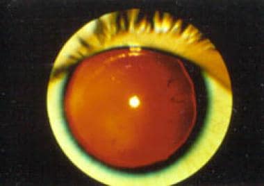
Aniridia. Note the almost complete absence of the iris.
Click to see larger pictureClick to see detailView Full Size Image
Media type:  Photo


  • Callahan A. Aniridia with ectopia lentis and secondary glaucoma. Am J Ophthalmol. 1949;32:28.
  • Chen TC, Walton DS. Goniosurgery for prevention of aniridic glaucoma. Arch Ophthalmol. Sep 1999;117(9):1144-8. [Medline].
  • Grant WM, Walton DS. Progressive changes in the angle in congenital aniridia, with development of glaucoma. Am J Ophthalmol. Nov 1974;78(5):842-7. [Medline].
  • Hamming N, Wilensky J. Persistent pupillary membrane associated with aniridia. Am J Ophthalmol. Jul 1978;86(1):118-20. [Medline].
  • Hittner HM, Riccardi VM, Ferrell RE, et al. Variable expressivity in autosomal dominant aniridia by clinical, electrophysiologic, and angiographic criteria. Am J Ophthalmol. Apr 1980;89(4):531-9. [Medline].
  • Jastaneiah S, Al-Rajhi AA. Association of aniridia and dry eyes. Ophthalmology. Sep 2005;112(9):1535-40. [Medline].
  • Jesberg DO. Aniridia with retinal lipid deposits. Arch Ophthalmol. 1962;68:331-336.
  • Koroma BM, Yang JM, Sundin OH. The Pax-6 homeobox gene is expressed throughout the corneal and conjunctival epithelia. Invest Ophthalmol Vis Sci. Jan 1997;38(1):108-20. [Medline].
  • Kremer I, Rajpal RK, Rapuano CJ, et al. Results of penetrating keratoplasty in aniridia. Am J Ophthalmol. Mar 15 1993;115(3):317-20. [Medline].
  • Layman PR, Anderson DR, Flynn JT. Frequent occurrence of hypoplastic optic disks in patients with aniridia. Am J Ophthalmol. Apr 1974;77(4):513-6. [Medline].
  • Mackman G, Brightbill FS, Optiz JM. Corneal changes in aniridia. Am J Ophthalmol. Apr 1979;87(4):497-502. [Medline].
  • Margo CE. Congenital aniridia: a histopathologic study of the anterior segment in children. J Pediatr Ophthalmol Strabismus. Sep-Oct 1983;20(5):192-8. [Medline].
  • Menezo JL, Martinez-Costa R, Cisneros A. Implantation of iris devices in congenital and traumatic aniridias: surgery solutions and complications. Eur J Ophthalmol. Jul-Aug 2005;15(4):451-7. [Medline].
  • Nelson LB, Spaeth GL, Nowinski TS, et al. Aniridia. A review. Surv Ophthalmol. May-Jun 1984;28(6):621-42. [Medline].
  • Nishida K, Kinoshita S, Ohashi Y, et al. Ocular surface abnormalities in aniridia. Am J Ophthalmol. Sep 1995;120(3):368-75. [Medline].
  • Ramaesh K, Ramaesh T, Dutton GN. Evolving concepts on the pathogenic mechanisms of aniridia related keratopathy. Int J Biochem Cell Biol. Mar 2005;37(3):547-57. [Medline].
  • Warburg M, Mikkelsen M, Andersen SR, et al. Aniridia and interstitial deletion of the short arm of chromosome 11. Metab Pediatr Ophthalmol. 1980;4(2):97-102. [Medline].
  • Wiggins RE, Tomey KF. The results of glaucoma surgery in aniridia. Arch Ophthalmol. Apr 1992;110(4):503-5. [Medline].

Aniridia in the Newborn excerpt

Article Last Updated: Dec 12, 2005