Continually Updated Clinical Reference
 
 
  All Sources     eMedicine     Medscape     Drug Reference     MEDLINE
 
eMedicine - Ectropion : Article by

Quick Find
Authors & Editors
Introduction
Clinical
Differentials
Treatment
Follow-up
Multimedia
References

Related Articles
Basal Cell Carcinoma, Eyelid

Bell Palsy

Floppy Eyelid Syndrome

Ichthyosis

Neuro-ophthalmic Examination

Neuro-ophthalmic History

Squamous Cell Carcinoma, Eyelid


Patient Education
Click here for patient education.


Author: Edsel Ing, MD, FRCSC, Assistant Professor, Department of Ophthalmology & Vision Sciences, University of Toronto: Consulting Staff, Toronto East General Hospital

Edsel Ing is a member of the following medical societies: American Academy of Ophthalmology, American College of Physician Executives, American Society of Contemporary Ophthalmology, Canadian Ophthalmological Society, Contact Lens Association of Ophthalmologists, North American Neuro-Ophthalmology Society, and Royal College of Physicians and Surgeons of Canada

Editors: Ron W Pelton, MD, PhD, Private Practice, Colorado Springs, Colorado; Simon K Law, MD, PharmD, Assistant Professor of Ophthalmology, Jules Stein Eye Institute; Chief of Section of Ophthalmology Surgical Services, Department of Veterans Affairs Healthcare Center, West Los Angeles; Mark T Duffy, MD, PhD, Consulting Staff, Division of Oculoplastic, Orbito-facial, Lacrimal, and Reconstructive Surgery, Green Bay Eye Clinic, BayCare Clinic; Lance L Brown, OD, MD, Ophthalmologist, Affiliated With Freeman Hospital and St John's Hospital, Regional Eye Center, Joplin, Missouri; Hampton Roy Sr, MD, Associate Clinical Professor, Department of Ophthalmology, University of Arkansas for Medical Sciences

Author and Editor Disclosure

Synonyms and related keywords: eversion, lid, lower lid, eyelid, corneal exposure, conjunctival exposure, tearing, lid deformity, irritated eyes, red eyes, lid laxity, punctal, medial, lateral, tarsal, cicatricial ectropion, punctal phimosis, paralytic ectropion, nocturnal lagophthalmos, Bell palsy

Background

Ectropion is an abnormal eversion (outward turning) of the lid margin away from the globe. Without normal lid globe apposition, corneal exposure, tearing, keratinization of the palpebral conjunctiva, and visual loss may result. Ectropion usually involves the lower lid and often has a component of horizontal lid laxity. Treatment is individualized based on the appropriate identification of the etiology.

Pathophysiology

The pathophysiology depends on the type of ectropion and is discussed within this article.

Mortality/Morbidity

The primary morbidity is associated with corneal/conjunctival exposure. Tearing may also cause significant patient complaints.

Race

No racial predilection has been described.

Sex

No sexual predilection has been described.

Age

Ectropion can affect patients of any age but is most commonly seen in older adults.


History

  • Patients may have a lid deformity for months or even years before they seek medical attention.
  • Patients often complain of irritated or red eyes with tearing. They may constantly wipe their eyes, thereby exacerbating lid laxity and the ectropion.
  • Advanced age or long-term use of eye drops may suggest the patient has involutional ectropion. A history of facial burns, lid surgery, or lid trauma is usually easily confirmed on cursory examination. It suggests cicatricial ectropion.
  • For patients with Bell palsy, the caregiver should be asked if nocturnal lagophthalmos occurs. These patients should also be examined for corneal problems.

Physical

  • Gestalt examination of the visage may reveal a connective tissue disorder, prior surgical scars or burns, cancerous skin conditions, or the physiognomy for floppy eyelid syndrome. All of these findings may be important in ectropion evaluation.
  • Documentation of visual acuity and examination of the cornea and the conjunctiva are part of any complete oculoplastic examination. Corneal exposure, corneal ulceration, and conjunctival keratinization may accompany ectropion.
  • Because of gravity, ectropion usually involves the lower lid and is described as punctal, medial, lateral, or tarsal (complete). Laxity-related ectropion typically begins medially; with time, the central lid margin and the lateral lid may evert.
  • Both the distraction test and the snap-back test are usually performed for abnormal horizontal lid laxity.
    • Anterior lid distraction of more than 6-8 mm from the globe suggests horizontal lid laxity.
    • If the lower lid is pulled inferiorly, the lid should quickly return to its previous position. If not, this may be interpreted as an abnormal snap-back test result. The patient should not be allowed to blink the eyelid back into position.
  • If cicatricial ectropion is suspected, superiorly displace the lower lid margin. If the lower lid margin does not extend 2 mm above the inferior limbus, then consider cicatricial ectropion.
  • The puncta should not be visible, unless the lid is everted. If this is not the case, punctal ectropion is present.
  • Chronic punctal ectropion may result in punctal phimosis.
  • Chronic ectropion may cause keratinization of the lid margin and the palpebral conjunctiva.
  • In patients with complete tarsal ectropion, a white line in the inferior fornix is often present, indicating a disinserted capsulopalpebral fascia.
  • In patients with suspected paralytic ectropion, the following should be documented:
    • Corneal integrity
    • Corneal sensation
    • Presence or absence of Bell phenomenon
    • Degree of lagophthalmos - To estimate nocturnal lagophthalmos, the patient should gently close the eyelids when in the supine position.
    • Disparity between spontaneous and voluntary lid closure
  • With a lower motor neuron seventh nerve palsy (eg, Bell palsy), the ipsilateral brow and the lower facial musculature are weak. With an upper motor neuron seventh nerve palsy, brow-elevation is relatively spared due to the bilateral innervation of the upper face.
  • In patients with suspected facial nerve palsy, orbicularis oris dysfunction can be tested for by asking them to show their teeth rather than smile. Compare the elevation of the angles of the lips; ptosis of the lateral lip on the affected side is often present.
  • If a slow-onset or nonresolving seventh nerve palsy is seen, perform the following:
    • Palpate the parotid gland for tumor.
    • Exclude prior malar skin cancer.
    • Check the patient's hearing to exclude a cerebellopontine angle tumor.
    • Perform a slit lamp examination for uveitis, which may suggest a disease process, such as sarcoidosis or Lyme disease.
  • Inferior scleral show should be distinguished from ectropion, especially in patients with prominent globes. Horizontal eyelid tightening will exacerbate the scleral show of a proptotic eye, because the shortest arc between the canthi lies inferior to the cornea.
  • Patients with involutional ectropion of the lower lid may also have involutional changes of the upper eyelid. Failure to recognize this prior to horizontal tightening of the lower lid may result in the upper lid prolapsing over the lower lid margin with the lower lid lashes rubbing the palpebral conjunctiva of the upper lid (ie, a form of floppy eyelid syndrome).

Causes

  • Ectropion may be congenital or acquired.
    • Congenital ectropion is rare and usually involves the lower lid. The cause often is a vertical deficiency of the anterior lamella.
    • Congenital ectropion is rarely an isolated anomaly. It may be associated with blepharophimosis syndrome, microphthalmos, buphthalmos, orbital cysts, Down syndrome, and ichthyosis (collodion baby).
    • Occasional congenital ectropion cases are on a paralytic basis.
  • Acquired ectropion may be involutional, paralytic, cicatricial, or mechanical.
  • Involutional ectropion is the most common form of ectropion.
  • A major factor is horizontal lid laxity, usually due to age-related weakness (most patients are elderly) of the canthal ligaments and the pretarsal orbicularis. Patients with involutional ectropion have been suggested to have an age-normal or larger than normal tarsal plate, which may mechanically overcome normal or decreased orbicularis tone, in conjunction with canthal tendon laxity.
  • Patients with an anophthalmic socket may have involutional ectropion due to chronic pressure of the ocular prosthesis.
  • Disinsertion of the capsulopalpebral fascia may lead to severe tarsal ectropion.
  • Paralytic ectropion may occur with seventh nerve palsy from diverse causes, such as Bell palsy, cerebellopontine angle tumors, herpes zoster oticus, and infiltrations or tumors of the parotid gland.
  • Cicatricial ectropion occurs from scarring of the anterior lamella by conditions, such as facial burns, trauma, chronic dermatitis, or excessive skin excision (or laser) with blepharoplasty.
  • Ectropion is not uncommon after orbital fracture repair with a transcutaneous approach.
  • Less common causes of cicatricial ectropion include cutaneous T-cell lymphoma.
  • Mechanical tumors may occur with lid tumors, such as neurofibromas that evert the lower lid.
  • Acute "idiopathic" bilateral lower lid ectropion has been described. An uncommon case of bilateral upper lid ectropion from blepharospasm has also been described.


Basal Cell Carcinoma, Eyelid
Bell Palsy
Floppy Eyelid Syndrome
Ichthyosis
Neuro-ophthalmic Examination
Neuro-ophthalmic History
Squamous Cell Carcinoma, Eyelid


Medical Care

  • Lubrication and moisture shields are helpful if significant corneal exposure exists from the ectropion. In patients with corneal exposure, plastic dressings are often superior to cloth patches. In some cases, taping the lateral canthal skin supertemporally provides temporary relief, especially in patients with new-onset seventh nerve palsy.
  • If the conjunctiva is markedly keratinized, a lubricating ointment should be used several days or weeks prior to ectropion repair. Corneal epithelial defects and prior herpes simplex infection are a relative contraindication to steroid-containing ointments.
  • Patients with tearing and incipient ectropion or early punctal ectropion should be instructed to wipe the eyelids in a direction up and in (toward the nose) to avoid worsening medial ectropion.
  • With cicatricial ectropion following trauma or lid surgery, digital massage may help stretch the scar. If not, steroid injection into the scar should be considered.
  • In patients with seventh nerve palsy, external paste-on upper lid weights are available and can be matched approximately for different skin colors.
    • A double-sided tape is used to apply the lid weight.
    • Removing the lid weight at night may avoid irritation of the lid skin.
    • The external lid weights are not a good option in patients with upper lid dermatochalasis or poor manual dexterity.
  • The use of steroids for Bell palsy remains controversial, because such a high likelihood of spontaneous recovery exists.
    • Steroid use early in the disease course may decrease the risk of subsequent aberrant regeneration (eg, crocodile tears).
    • Acyclovir in combination with prednisone has been shown to be of greater benefit than prednisone alone in Bell palsy.

Surgical Care

  • The correct surgical treatment of ectropion depends on the etiology.
    • Horizontal lid laxity is often seen with ectropion and usually can be corrected with a lateral tarsal strip procedure.
    • Mild-to-moderate cases of medial ectropion may respond to a medial conjunctival spindle procedure.
    • Tarsal ectropion requires reinsertion of the lower lid retractors.
    • Augmentation of the anterior lamellae (along with excision of any cicatrix) is required for cicatricial ectropion.
  • The use of a corneal protector during oculoplastic procedures is recommended. The surgeon must be wary of the remote possibility of flash burns whenever oxygen is on the surgical field. Failure to do so may transform an elective lid repair into a much more complicated problem.
  • Ensuring patient comfort during surgery is important. Because most cases of ectropion involve the lower lid, supplemental infraorbital nerve block is a useful adjunct to direct injection and subconjunctival injection.
  • Tarsorrhaphy can be performed as a temporary quick fix for corneal exposure, but most surgeons believe that better alternatives are available.
  • Electrocautery at the junction of conjunctiva and lower margin of the tarsus is not commonly advocated. It is usually only a temporary measure.
  • Suture repair is a temporary method of repair that is not advocated. Double-armed chromic sutures are passed through the inferior border of the tarsus, emerging at the skin surface near the orbital rim.
  • Congenital ectropion
    • Ensure corneal lubrication. If the condition does not resolve after a few days, consider placing lid margin sutures. A lateral tarsorrhaphy may be required if suture techniques do not work, but be careful of iatrogenic amblyopia. More severe cases of congenital ectropion may need a skin flap or graft.
    • Ichthyosis is a well-described cause of congenital ectropion. It is sometimes managed conservatively with lubrication, but skin grafts may be required.
  • Lateral tarsal strip: Horizontal lid laxity is a component of most ectropion cases, especially involutional ectropion. Numerous methods are available for correcting horizontal lid laxity. Older methods include wedge resections and the Kuhnt-Szymanowski procedure. Whenever feasible, a lateral canthal tightening procedure is preferred. Surgery at the lateral canthus avoids the possibility of lid notching with noncanthal procedures and decreases the risk of trichiasis. The most common variation of lateral canthal tightening is the lateral tarsal strip procedure.
    • The lateral canthus can be clamped prior to canthotomy, then inferior cantholysis is performed with Westcott scissors. The lower lid should now be freely mobile.
    • If excess lid skin is present, it can be draped over the lateral canthus, and an appropriate triangle of full-thickness lid is excised.
    • Then, about 3 mm of the lateral lid is split at the gray line with either sharp Westcott scissors or a 15 blade.
    • The meibomian orifices of the lateral strip are trimmed away.
    • The lateral conjunctiva is scraped to avoid epithelial inclusion cysts.
    • To secure the lateral strip of tarsus to the periosteum, 2 sutures (or a single horizontal mattress suture) can be placed about 4-5 mm posterior to the lateral orbital rim near the Whitnall tubercle (at or above the level of the inferior pupil). Suitable sutures with small semicircular needles include 5-0 Vicryl on a P2 needle or 4-0 Prolene on a PS-5 needle.
    • Retracting the upper lid supertemporally and placing a Q-tip at the lateral canthus to palpate the inner lateral orbital rim may help.
    • Before tying the suture, remove the corneal shield.
    • The orbicularis layer can be closed with 6-0 Vicryl. The skin can be closed with 6-0 plain gut. A stitch through the lateral-most gray line of the upper and lower lateral lid will help to keep the lateral canthus "sharp."
    • If the patient requires topical drops (eg, glaucoma therapy) postoperatively, do not retract the lower lid for the first month during drop instillation.
    • It is not uncommon for patients to complain of discomfort at the lateral canthus several weeks following this procedure.
  • Transconjunctival ectropion repair has been described.
  • Kuhnt-Szymanowski (Smith modification): When marked inferior dermatochalasis accompanies ectropion and the lateral canthal tendon is not dehisced, an inferior subciliary blepharoplasty skin incision can be combined with pentagonal wedge excision of the orbicularis and posterior lamellae. Precise closure is required to prevent a lid notch.
  • Tarsal ectropion
    • This complete eversion of the lower lid occurs when disinsertion of the capsulopalpebral fascia from the inferior tarsal border is present.
    • In addition to horizontal lid tightening, reinsert the retractors (ideally from a conjunctival approach).
    • A spindle of redundant conjunctiva, no more than 3 mm in vertical height, can be excised, if necessary.
    • A double-armed 5-0 chromic suture can be used to reattach the capsulopalpebral fascia to the inferior tarsus in a running fashion.
  • Medial ectropion: If tearing is the primary problem in patients with punctal ectropion, a 1-snip or 2-snip inferior punctoplasty may be beneficial. Easily performed with Vannas scissors and topical anesthetic, punctoplasty restores continuity between the lacus lacrimali and the medial canthal angle. For mild-to-moderate medial ectropion, a medial conjunctival spindle procedure (excision of the medial conjunctiva and retractors) can be performed.
    • Following anesthetic injection in the medial inferior fornix, the inferior canaliculus can be guarded with a lacrimal probe.
    • A horizontal ellipse or diamond of conjunctiva and underlying lid retractors is excised inferior to the punctum, approximately 3-4 mm high and 6-8 mm wide.
    • Then, the defect is closed with double-armed 5-0 chromic inverting suture. This can be accomplished by engaging the inferior lip of the wound, then the superior lip of the wound; the needle is then redirected from the inferior lid to the cutaneous surface. Alternatively, buried interrupted 6-0 polyglactin stitches can be used to close the medial conjunctival spindle.
  • The Byron Smith lazy-T procedure is a well-described procedure for repairing prominent medial ectropion. It combines a lower lid, full-thickness pentagonal wedge resection, 3-4 mm temporal to the punctum with resection of a medial triangle of conjunctiva and lower lid retractors (similar to medial conjunctival spindle).
    • Usually, 5-8 mm of lower lid is excised in the pentagonal wedge. When closed, the incisions resemble a "T" lying on its side, hence the name lazy T.
    • If marked medial canthal laxity is present, medial canthal tendon plication generally is performed with a lid shortening procedure.
    • A lacrimal probe is placed to guard the lower canaliculus. A skin incision, extending from just medial to the medial canthus to just temporal to the punctum, is made inferior to the canaliculus.
    • A double-armed 5-0 nylon suture is placed from the medial inferior tarsus to the medial canthal ligament near the anterior lacrimal crest.
    • The lacrimal probe is removed and the plication suture tightened enough to prevent lateral excursion of the puncta. Over-tightening the stitch may kink canalicular outflow. The skin incision can be closed with 6-0 fast-absorbing gut suture.
  • Paralytic ectropion
    • A tarsal strip procedure is often helpful. At least 5 mm of the lateral lower lid may have to be excised. With marked paralytic lower lid ectropion, a midface or suborbicularis oculi fat (SOOF) lift is a useful procedure.
    • In patients with extreme paralytic ectropion, a fascia lata (or Gortex) sling or temporalis transfer procedure may be required.
    • Upper lid gold weight implantation is a helpful adjunct for patients with lagophthalmos. Usually, a 1.0-1.2 g weight is implanted superior to the tarsus and inferior to the orbicularis. Extrusion of the gold weight occasionally occurs with time. Since the gold weight works by gravity, patients should sleep with their head slightly elevated. The gold weights are not a contraindication for MRI investigation.
  • Cicatricial ectropion
    • An enhanced tarsal strip (ie, a tarsal strip without the traditional lateral skin excision) may help correct some degree of cicatricial ectropion.
    • If an enhanced tarsal strip is insufficient, Z-plasties, V-Y plasty, skin grafts, or advancement flaps may be used to lengthen the anterior lamella.
    • Skin grafts may be obtained from the upper lid if dermatochalasis is present; preauricular or postauricular skin is another alternative. If facial skin is unavailable, the author prefers medial forearm skin. The skin graft should be thinned and buttonholed (for drainage). In patients with moderate lower eyelid cicatricial ectropion and upper eyelid dermatochalasis, the transfer of a bipedicle or monopedicle flap from the upper eyelid combined with canthopexy is an option.
    • A compressive bolster should be placed over the graft to enhance graft survival and to decrease hematoma formation. The bolster is left for 5 days. A superior traction suture decreases the risk of recurrent cicatrix postoperatively.


Further Outpatient Care

  • Postoperative care
    • For the lid sutures, an antibiotic steroid combination, used 3 times daily, such as Maxitrol (neomycin, polymyxin, bacitracin), is preferred.
    • Applying cold compresses to the eyelids every 15 minutes (as tolerated) while awake will decrease bruising and swelling. Frozen peas in a plastic bag are a useful alternative to traditional cloth compresses.
    • Generally, physicians do not prescribe narcotics postoperatively. The patient is asked to use acetaminophen (325-650 mg PO q4h prn) to manage pain. Patients are asked to avoid aspirin-containing products.
    • Patients are usually examined on the first postoperative day, then 5-7 days later for suture removal.

Complications

  • Complications are primarily related to corneal and conjunctival exposure. As listed above, these complications can involve conjunctival keratinization, corneal breakdown, epiphora, and pain.
  • Surgical complications may include bleeding, hematoma, infection, wound dehiscence, pain, and poor positioning of the tarsal strip.


Media file 1:  Medial cicatricial ectropion in a patient with extensive basal cell carcinoma.
Click to see larger pictureClick to see detailView Full Size Image
Media type:  Photo

Media file 2:  
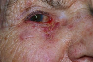
Complete (tarsal) paralytic ectropion in a patient with an ipsilateral parotid tumor.
Click to see larger pictureClick to see detailView Full Size Image
Media type:  Photo


  • Anderson RL, Gordy DD. The tarsal strip procedure. Arch Ophthalmol. Nov 1979;97(11):2192-6. [Medline].
  • Bashour M, Harvey J. Causes of involutional ectropion and entropion; age-related tarsal changes are the key. Ophthal Plast Reconstr Surg. Mar 2000;16(2):131-41. [Medline].
  • Ben Simon GJ, Molina M, Schwarcz RM, et al. External (subciliary) vs internal (transconjunctival) involutional entropion repair. Am J Ophthalmol. Mar 2005;139(3):482-7. [Medline].
  • Cook BE Jr, Bartley GB, Pittelkow MR. Ophthalmic abnormalities in patients with cutaneous T-cell lymphoma. Ophthalmology. Jul 1999;106(7):1339-44. [Medline].
  • Gupta B, Parmar B, Raina J, Chawla JS. Acute idiopathic bilateral lower lid ectropion. Indian J Ophthalmol. Sep 2006;54(3):212-4. [Medline].
  • Jordan DR, Bawazeer AM, Pelletier CR. Severe congenital ectropion secondary to lamellar ichthyosis. Can J Ophthalmol. Feb 1998;33(1):30-1. [Medline].
  • Jordan DR, Anderson RL. The lateral tarsal strip revisited. The enhanced tarsal strip. Arch Ophthalmol. Apr 1989;107(4):604-6. [Medline].
  • Koh S, Hosohata J, Tano Y. Bilateral upper eyelid ectropion associated with blepharospasm. Br J Ophthalmol. Nov 2006;90(11):1437-8. [Medline].
  • Manku K, Leong JK, Ghabrial R. Cicatricial ectropion: repair with myocutaneous flaps and canthopexy. Clin Experiment Ophthalmol. Sep-Oct 2006;34(7):677-81. [Medline].
  • Nowinski TS, Anderson RL. The medial spindle procedure for involutional medial ectropion. Arch Ophthalmol. Nov 1985;103(11):1750-3. [Medline].
  • Smith B. The "lazy-T" correction of ectropion of the lower punctum. Arch Ophthalmol. Jul 1976;94(7):1149-50. [Medline].
  • Tse DT, Kronish JW, Buus D. Surgical correction of lower-eyelid tarsal ectropion by reinsertion of the retractors. Arch Ophthalmol. Mar 1991;109(3):427-31. [Medline].
  • Uthoff D, Gorney M, Teichmann C. Cicatricial ectropion in ichthyosis: a novel approach to treatment. Ophthal Plast Reconstr Surg. Jun 1994;10(2):92-5. [Medline].

Ectropion excerpt

Article Last Updated: Dec 27, 2006