Wisse RP, Kuiper JJ, Gans R, Imhof S, Radstake TR, Van der Lelij A. Cytokine Expression in Keratoconus and its Corneal Microenvironment: A Systematic Review. Ocul Surf. 2015 Oct. 13 (4):272-83. [QxMD MEDLINE Link].
Jun AS, Cope L, Speck C, Feng X, Lee S, Meng H, et al. Subnormal cytokine profile in the tear fluid of keratoconus patients. PLoS One. 2011 Jan 27. 6 (1):e16437. [QxMD MEDLINE Link].
Lema I, Sobrino T, Durán JA, Brea D, Díez-Feijoo E. Subclinical keratoconus and inflammatory molecules from tears. Br J Ophthalmol. 2009 Jun. 93 (6):820-4. [QxMD MEDLINE Link].
Gănescu AM. Current approaches in the management of patients with keratoconus. Med Pharm Rep. 2022 Oct. 95 (4):385-392. [QxMD MEDLINE Link].
Vohra V, Tuteja S, Gurnani B, Chawla H. Collagen Cross Linking For Keratoconus. 2022 Jan. [QxMD MEDLINE Link]. [Full Text].
Asimellis G, Kaufman EJ. Keratoconus. 2022 Jan. [QxMD MEDLINE Link]. [Full Text].
Critchfield JW, Calandra AJ, Nesburn AB, Kenney MC. Keratoconus: I. Biochemical studies. Exp Eye Res. 1988 Jun. 46(6):953-63. [QxMD MEDLINE Link].
Sawaguchi S, Yue BY, Sugar J, Gilboy JE. Lysosomal enzyme abnormalities in keratoconus. Arch Ophthalmol. 1989 Oct. 107(10):1507-10. [QxMD MEDLINE Link].
Gondhowiardjo TD, van Haeringen NJ. Corneal aldehyde dehydrogenase, glutathione reductase, and glutathione S-transferase in pathologic corneas. Cornea. 1993 Jul. 12(4):310-4. [QxMD MEDLINE Link].
Bykhovskaya Y, Margines B, Rabinowitz YS. Genetics in Keratoconus: where are we?. Eye Vis (Lond). 2016. 3:16. [QxMD MEDLINE Link].
Rabinowitz YS. The genetics of keratoconus. Ophthalmol Clin North Am. 2003 Dec. 16 (4):607-20, vii. [QxMD MEDLINE Link].
Galvis V, Sherwin T, Tello A, Merayo J, Barrera R, Acera A. Keratoconus: an inflammatory disorder?. Eye (Lond). 2015 Jul. 29 (7):843-59. [QxMD MEDLINE Link].
Özalp O, Atalay E, Yıldırım N. Prevalence and risk factors for keratoconus in a university-based population in Turkey. J Cataract Refract Surg. 2021 Dec 1. 47 (12):1524-1529. [QxMD MEDLINE Link].
Papaliʼi-Curtin AT, Cox R, Ma T, Woods L, Covello A, Hall RC. Keratoconus Prevalence Among High School Students in New Zealand. Cornea. 2019 Nov. 38 (11):1382-1389. [QxMD MEDLINE Link].
Bak-Nielsen S, Ramlau-Hansen CH, Ivarsen A, Plana-Ripoll O, Hjortdal J. Incidence and prevalence of keratoconus in Denmark - an update. Acta Ophthalmol. 2019 Dec. 97 (8):752-755. [QxMD MEDLINE Link].
Torres Netto EA, Al-Otaibi WM, Hafezi NL, et al. Prevalence of keratoconus in paediatric patients in Riyadh, Saudi Arabia. British Journal of Ophthalmology. 2018. 102:1436-1441.
Gorskova EN, Sevost'ianov EN. [Epidemiology of keratoconus in the Urals]. Vestn Oftalmol. 1998 Jul-Aug. 114 (4):38-40. [QxMD MEDLINE Link].
Ihalainen A. Clinical and epidemiological features of keratoconus genetic and external factors in the pathogenesis of the disease. Acta Ophthalmol Suppl (1985). 1986. 178:1-64. [QxMD MEDLINE Link].
Godefrooij DA, de Wit GA, Uiterwaal CS, Imhof SM, Wisse RP. Age-specific Incidence and Prevalence of Keratoconus: A Nationwide Registration Study. Am J Ophthalmol. 2017 Mar. 175:169-172. [QxMD MEDLINE Link].
Krachmer JH, Feder RS, Belin MW. Keratoconus and related noninflammatory corneal thinning disorders. Surv Ophthalmol. 1984 Jan-Feb. 28 (4):293-322. [QxMD MEDLINE Link].
Zadnik K, Barr JT, Edrington TB, et al. Baseline findings in the Collaborative Longitudinal Evaluation of Keratoconus (CLEK) Study. Invest Ophthalmol Vis Sci. 1998 Dec. 39(13):2537-46. [QxMD MEDLINE Link].
Cristina Kenney M, Brown DJ. The cascade hypothesis of keratoconus. Cont Lens Anterior Eye. 2003 Sep. 26(3):139-46. [QxMD MEDLINE Link].
Sherwin T, Brookes NH. Morphological changes in keratoconus: pathology or pathogenesis. Clin Experiment Ophthalmol. 2004 Apr. 32(2):211-7. [QxMD MEDLINE Link].
Woodward MA, Blachley TS, Stein JD. The Association Between Sociodemographic Factors, Common Systemic Diseases, and Keratoconus: An Analysis of a Nationwide Heath Care Claims Database. Ophthalmology. 2015 Dec 16. [QxMD MEDLINE Link].
Flockerzi E, Xanthopoulou K, Goebels SC, Zemova E, Razafimino S, Hamon L, et al. Keratoconus staging by decades: a baseline ABCD classification of 1000 patients in the Homburg Keratoconus Center. Br J Ophthalmol. 2021 Aug. 105 (8):1069-1075. [QxMD MEDLINE Link].
Ertan A, Muftuoglu O. Keratoconus clinical findings according to different age and gender groups. Cornea. 2008 Dec. 27 (10):1109-13. [QxMD MEDLINE Link].
Millodot M, Ortenberg I, Lahav-Yacouel K, Behrman S. Effect of ageing on keratoconic corneas. J Optom. 2015 Jun 30. [QxMD MEDLINE Link].
Yeung KK et al. Where have all the keratoconics gone?. Int Cont Lens Clin. 1998. 25(4):109-13.
Popiela M, Young-Zvandasara T, Veepanat E, Saunders D. Demographics of older keratoconics in Wales and their mortality rates-Where are the older keratoconics?. Cont Lens Anterior Eye. 2016 Oct. 39 (5):365-8. [QxMD MEDLINE Link].
Moodaley LC, Woodward EG, Liu CS, Buckley RJ. Life expectancy in keratoconus. Br J Ophthalmol. 1992 Oct. 76(10):590-1. [QxMD MEDLINE Link]. [Full Text].
Sarezky D, Orlin SE, Pan W, VanderBeek BL. Trends in Corneal Transplantation in Keratoconus. Cornea. 2017 Feb. 36 (2):131-137. [QxMD MEDLINE Link].
Moschos MM, Nitoda E, Georgoudis P, Balidis M, Karageorgiadis E, Kozeis N. Contact Lenses for Keratoconus- Current Practice. Open Ophthalmol J. 2017. 11:241-251. [QxMD MEDLINE Link].
Dienes L, Kiss HJ, Perényi K, Nagy ZZ, Acosta MC, Gallar J, et al. Corneal Sensitivity and Dry Eye Symptoms in Patients with Keratoconus. PLoS One. 2015. 10 (10):e0141621. [QxMD MEDLINE Link].
Aldave AJ, Ann LB, Frausto RF, Nguyen CK, Yu F, Raber IM. Classification of posterior polymorphous corneal dystrophy as a corneal ectatic disorder following confirmation of associated significant corneal steepening. JAMA Ophthalmol. 2013 Dec. 131 (12):1583-90. [QxMD MEDLINE Link].
Shajari M, Eberhardt E, Müller M, Al Khateeb G, Friderich S, Remy M, et al. Effects of Atopic Syndrome on Keratoconus. Cornea. 2016 Nov. 35 (11):1416-1420. [QxMD MEDLINE Link].
Gupta PK, Stinnett SS, Carlson AN. Prevalence of sleep apnea in patients with keratoconus. Cornea. 2012 Jun. 31(6):595-9. [QxMD MEDLINE Link].
Rabinowitz YS. The genetics of keratoconus. Ophthalmol Clin North Am. 2003 Dec. 16(4):607-20, vii. [QxMD MEDLINE Link].
Rabinowitz YS, Li X, Canedo AL, Ambrósio R Jr, Bykhovskaya Y. Optical coherence tomography combined with videokeratography to differentiate mild keratoconus subtypes. J Refract Surg. 2014 Feb. 30 (2):80-7. [QxMD MEDLINE Link].
Mathew JH, Goosey JD, Söderberg PG, Bergmanson JP. Lamellar changes in the keratoconic cornea. Acta Ophthalmol. 2015 Dec. 93 (8):767-73. [QxMD MEDLINE Link].
Compañ V, Aguilella-Arzo M, Edrington TB, Weissman BA. Modeling Corneal Oxygen with Scleral Gas Permeable Lens Wear. Optom Vis Sci. 2016 Nov. 93 (11):1339-1348. [QxMD MEDLINE Link].
Rathi VM, Mandathara PS, Dumpati S. Contact lens in keratoconus. Indian J Ophthalmol. 2013 Aug. 61 (8):410-5. [QxMD MEDLINE Link].
Wollensak G, Spoerl E, Seiler T. Riboflavin/ultraviolet-a-induced collagen crosslinking for the treatment of keratoconus. Am J Ophthalmol. 2003 May. 135 (5):620-7. [QxMD MEDLINE Link].
Tomita M, Mita M, Huseynova T. Accelerated versus conventional corneal collagen crosslinking. J Cataract Refract Surg. 2014 Jun. 40 (6):1013-20. [QxMD MEDLINE Link].
Acar BT, Utine CA, Ozturk V, Acar S, Ciftci F. Can the effect of transepithelial corneal collagen cross-linking be improved by increasing the duration of topical riboflavin application? An in vivo confocal microscopy study. Eye Contact Lens. 2014 Jul. 40 (4):207-12. [QxMD MEDLINE Link].
Wittig-Silva C, Chan E, Islam FM, Wu T, Whiting M, Snibson GR. A randomized, controlled trial of corneal collagen cross-linking in progressive keratoconus: three-year results. Ophthalmology. 2014 Apr. 121 (4):812-21. [QxMD MEDLINE Link].
Sherif AM, Ammar MA, Mostafa YS, Gamal Eldin SA, Osman AA. One-Year Results of Simultaneous Topography-Guided Photorefractive Keratectomy and Corneal Collagen Cross-Linking in Keratoconus Utilizing a Modern Ablation Software. J Ophthalmol. 2015. 2015:321953. [QxMD MEDLINE Link].
Kymionis GD, Portaliou DM, Kounis GA, Limnopoulou AN, Kontadakis GA, Grentzelos MA. Simultaneous topography-guided photorefractive keratectomy followed by corneal collagen cross-linking for keratoconus. Am J Ophthalmol. 2011 Nov. 152(5):748-55. [QxMD MEDLINE Link].
Ferenczy PA, Dalcegio M, Koehler M, Pereira TS, Moreira H, Luciane Bugmann M. Femtosecond-assisted intrastromal corneal ring implantation for keratoconus treatment: a comparison with crosslinking combination. Arq Bras Oftalmol. 2015 Mar-Apr. 78 (2):76-81. [QxMD MEDLINE Link].
Kymionis GD, Grentzelos MA, Karavitaki AE, Zotta P, Yoo SH, Pallikaris IG. Combined corneal collagen cross-linking and posterior chamber toric implantable collamer lens implantation for keratoconus. Ophthalmic Surg Lasers Imaging. 2011 Feb 17. 42 Online:e22-5. [QxMD MEDLINE Link].
Alnawaiseh M, Rosentreter A, Eveslage M, Eter N, Zumhagen L. Changes in Corneal Transparency After Cross-linking for Progressive Keratoconus: Long-term Follow-up. J Refract Surg. 2015 Sep. 31 (9):614-8. [QxMD MEDLINE Link].
Raiskup F, Hoyer A, Spoerl E. Permanent corneal haze after riboflavin-UVA-induced cross-linking in keratoconus. J Refract Surg. 2009 Sep. 25 (9):S824-8. [QxMD MEDLINE Link].
Shalchi Z, Wang X, Nanavaty MA. Safety and efficacy of epithelium removal and transepithelial corneal collagen crosslinking for keratoconus. Eye (Lond). 2015 Jan. 29 (1):15-29. [QxMD MEDLINE Link].
Kymionis GD, Bouzoukis DI, Diakonis VF, Portaliou DM, Pallikaris AI, Yoo SH. Diffuse lamellar keratitis after corneal crosslinking in a patient with post-laser in situ keratomileusis corneal ectasia. J Cataract Refract Surg. 2007 Dec. 33 (12):2135-7. [QxMD MEDLINE Link].
Labiris G, Kaloghianni E, Koukoula S, Zissimopoulos A, Kozobolis VP. Corneal melting after collagen cross-linking for keratoconus: a case report. J Med Case Rep. 2011 Apr 16. 5:152. [QxMD MEDLINE Link].
Sharma A, Nottage JM, Mirchia K, Sharma R, Mohan K, Nirankari VS. Persistent corneal edema after collagen cross-linking for keratoconus. Am J Ophthalmol. 2012 Dec. 154 (6):922-926.e1. [QxMD MEDLINE Link].
Kymionis GD, Portaliou DM, Diakonis VF, Kounis GA, Panagopoulou SI, Grentzelos MA. Corneal collagen cross-linking with riboflavin and ultraviolet-A irradiation in patients with thin corneas. Am J Ophthalmol. 2012 Jan. 153 (1):24-8. [QxMD MEDLINE Link].
Zamora KV, Males JJ. Polymicrobial keratitis after a collagen cross-linking procedure with postoperative use of a contact lens: a case report. Cornea. 2009 May. 28 (4):474-6. [QxMD MEDLINE Link].
Ferrari G, Iuliano L, Viganò M, Rama P. Impending corneal perforation after collagen cross-linking for herpetic keratitis. J Cataract Refract Surg. 2013 Apr. 39 (4):638-41. [QxMD MEDLINE Link].
Raiskup F, Spoerl E. Corneal crosslinking with riboflavin and ultraviolet A. Part II. Clinical indications and results. Ocul Surf. 2013 Apr. 11 (2):93-108. [QxMD MEDLINE Link].
Sharif R, Fowler B, Karamichos D. Collagen cross-linking impact on keratoconus extracellular matrix. PLoS One. 2018. 13 (7):e0200704. [QxMD MEDLINE Link].
van Dijk K, Liarakos VS, Parker J, Ham L, Lie JT, Groeneveld-van Beek EA, et al. Bowman layer transplantation to reduce and stabilize progressive, advanced keratoconus. Ophthalmology. 2015 May. 122 (5):909-17. [QxMD MEDLINE Link].
Reddy JC, Hammersmith KM, Nagra PK, Rapuano CJ. The role of penetrating keratoplasty in the era of selective lamellar keratoplasty. Int Ophthalmol Clin. 2013 Spring. 53 (2):91-101. [QxMD MEDLINE Link].
Wisse RP, van den Hoven CM, Van der Lelij A. Does lamellar surgery for keratoconus experience the popularity it deserves?. Acta Ophthalmol. 2014 Aug. 92 (5):473-7. [QxMD MEDLINE Link].
Coster DJ, Lowe MT, Keane MC, Williams KA, Australian Corneal Graft Registry Contributors. A comparison of lamellar and penetrating keratoplasty outcomes: a registry study. Ophthalmology. 2014 May. 121 (5):979-87. [QxMD MEDLINE Link].
Arnalich-Montiel F, Alió Del Barrio JL, Alió JL. Corneal surgery in keratoconus: which type, which technique, which outcomes?. Eye Vis (Lond). 2016. 3:2. [QxMD MEDLINE Link].
Lowe MT, Keane MC, Coster DJ, Williams KA. The outcome of corneal transplantation in infants, children, and adolescents. Ophthalmology. 2011 Mar. 118 (3):492-7. [QxMD MEDLINE Link].
Pramanik S, Musch DC, Sutphin JE, Farjo AA. Extended long-term outcomes of penetrating keratoplasty for keratoconus. Ophthalmology. 2006 Sep. 113 (9):1633-8. [QxMD MEDLINE Link].
Choi JA, Lee MA, Kim MS. Long-term outcomes of penetrating keratoplasty in keratoconus: analysis of the factors associated with final visual acuities. Int J Ophthalmol. 2014. 7 (3):517-21. [QxMD MEDLINE Link].
Javadi MA, Motlagh BF, Jafarinasab MR, Rabbanikhah Z, Anissian A, Souri H, et al. Outcomes of penetrating keratoplasty in keratoconus. Cornea. 2005 Nov. 24 (8):941-6. [QxMD MEDLINE Link].
Niziol LM, Musch DC, Gillespie BW, Marcotte LM, Sugar A. Long-term outcomes in patients who received a corneal graft for keratoconus between 1980 and 1986. Am J Ophthalmol. 2013 Feb. 155 (2):213-219.e3. [QxMD MEDLINE Link].
Moschos MM, Gouliopoulos NS, Kalogeropoulos C, Androudi S, Kitsos G, Ladas D, et al. Psychological Aspects and Depression in Patients with Symptomatic Keratoconus. J Ophthalmol. 2018. 2018:7314308. [QxMD MEDLINE Link].
Kymes SM, Walline JJ, Zadnik K, Sterling J, Gordon MO, Collaborative Longitudinal Evaluation of Keratoconus Study Group. Changes in the quality-of-life of people with keratoconus. Am J Ophthalmol. 2008 Apr. 145 (4):611-617. [QxMD MEDLINE Link].
Mannis MJ, Ling JJ, Kyrillos R, Barnett M. Keratoconus and Personality-A Review. Cornea. 2018 Mar. 37 (3):400-404. [QxMD MEDLINE Link].
Weissman B, Chun MW, Barnhart LA. Corneal abrasion associated with contact lens correction of keratoconus--a retrospective study. Optom Vis Sci. 1994 Nov. 71(11):677-81. [QxMD MEDLINE Link].
Pearson AR, Soneji B, Sarvananthan N, Sandford-Smith JH. Does ethnic origin influence the incidence or severity of keratoconus?. Eye (Lond). 2000 Aug. 14 ( Pt 4):625-8. [QxMD MEDLINE Link].
Rabinowitz YS. Keratoconus. Surv Ophthalmol. 1998 Jan-Feb. 42(4):297-319. [QxMD MEDLINE Link].
Kennedy RH, Bourne WM, Dyer JA. A 48-year clinical and epidemiologic study of keratoconus. Am J Ophthalmol. 1986 Mar 15. 101(3):267-73. [QxMD MEDLINE Link].
Sawaguchi S, Twining SS, Yue BY, et al. Alpha 2-macroglobulin levels in normal human and keratoconus corneas. Invest Ophthalmol Vis Sci. 1994 Nov. 35(12):4008-14. [QxMD MEDLINE Link].
Fukuchi T, Yue BY, Sugar J, Lam S. Lysosomal enzyme activities in conjunctival tissues of patients with keratoconus. Arch Ophthalmol. 1994 Oct. 112(10):1368-74. [QxMD MEDLINE Link].
Kenney MC, Nesburn AB, Burgeson RE, Butkowski RJ, Ljubimov AV. Abnormalities of the extracellular matrix in keratoconus corneas. Cornea. 1997 May. 16(3):345-51. [QxMD MEDLINE Link].
Behndig A, Karlsson K, Johansson BO, Brännström T, Marklund SL. Superoxide dismutase isoenzymes in the normal and diseased human cornea. Invest Ophthalmol Vis Sci. 2001 Sep. 42(10):2293-6. [QxMD MEDLINE Link].
Buddi R, Lin B, Atilano SR, Zorapapel NC, Kenney MC, Brown DJ. Evidence of oxidative stress in human corneal diseases. J Histochem Cytochem. 2002 Mar. 50(3):341-51. [QxMD MEDLINE Link].
Atilano SR, Coskun P, Chwa M, Jordan N, Reddy V, Le K. Accumulation of mitochondrial DNA damage in keratoconus corneas. Invest Ophthalmol Vis Sci. 2005 Apr. 46(4):1256-63. [QxMD MEDLINE Link].
Kenney MC, Chwa M, Atilano SR, Tran A, Carballo M, Saghizadeh M. Increased levels of catalase and cathepsin V/L2 but decreased TIMP-1 in keratoconus corneas: evidence that oxidative stress plays a role in this disorder. Invest Ophthalmol Vis Sci. 2005 Mar. 46(3):823-32. [QxMD MEDLINE Link].
Meek KM, Tuft SJ, Huang Y, Gill PS, Hayes S, Newton RH, et al. Changes in collagen orientation and distribution in keratoconus corneas. Invest Ophthalmol Vis Sci. 2005 Jun. 46(6):1948-56. [QxMD MEDLINE Link].
Määttä M, Heljasvaara R, Sormunen R, Pihlajaniemi T, Autio-Harmainen H, Tervo T. Differential expression of collagen types XVIII/endostatin and XV in normal, keratoconus, and scarred human corneas. Cornea. 2006 Apr. 25(3):341-9. [QxMD MEDLINE Link].
Määttä M, Väisänen T, Väisänen MR, Pihlajaniemi T, Tervo T. Altered expression of type XIII collagen in keratoconus and scarred human cornea: Increased expression in scarred cornea is associated with myofibroblast transformation. Cornea. 2006 May. 25(4):448-53. [QxMD MEDLINE Link].
Ku JY, Niederer RL, Patel DV, Sherwin T, McGhee CN. Laser scanning in vivo confocal analysis of keratocyte density in keratoconus. Ophthalmology. 2008 May. 115(5):845-50. [QxMD MEDLINE Link].
Lema I, Durán JA. Inflammatory molecules in the tears of patients with keratoconus. Ophthalmology. 2005 Apr. 112(4):654-9. [QxMD MEDLINE Link].
Lema I, Durán JA, Ruiz C, Díez-Feijoo E, Acera A, Merayo J. Inflammatory response to contact lenses in patients with keratoconus compared with myopic subjects. Cornea. 2008 Aug. 27(7):758-63. [QxMD MEDLINE Link].
Macsai MS, Varley GA, Krachmer JH. Development of keratoconus after contact lens wear. Patient characteristics. Arch Ophthalmol. 1990 Apr. 108(4):534-8. [QxMD MEDLINE Link].
Li X, Yang H, Rabinowitz YS. Keratoconus: classification scheme based on videokeratography and clinical signs. J Cataract Refract Surg. 2009 Sep. 35 (9):1597-603. [QxMD MEDLINE Link].
Steinberg J, Aubke-Schultz S, Frings A, Hülle J, Druchkiv V, Richard G, et al. Correlation of the KISA% index and Scheimpflug tomography in 'normal', 'subclinical', 'keratoconus-suspect' and 'clinically manifest' keratoconus eyes. Acta Ophthalmol. 2015 May. 93 (3):e199-207. [QxMD MEDLINE Link].
Zadnik K, Barr JT, Gordon MO, Edrington TB. Biomicroscopic signs and disease severity in keratoconus. Collaborative Longitudinal Evaluation of Keratoconus (CLEK) Study Group. Cornea. 1996 Mar. 15(2):139-46. [QxMD MEDLINE Link].
Wagner H, Barr JT, Zadnik K. Collaborative Longitudinal Evaluation of Keratoconus (CLEK) Study: methods and findings to date. Cont Lens Anterior Eye. 2007 Sep. 30(4):223-32. [QxMD MEDLINE Link].
Zadnik K, Barr JT, Gordon MO, Edrington TB. Biomicroscopic signs and disease severity in keratoconus. Collaborative Longitudinal Evaluation of Keratoconus (CLEK) Study Group. Cornea. 1996 Mar. 15(2):139-46. [QxMD MEDLINE Link].
Ridley F. EYE-RUBBING AND CONTACT LENSES. Br J Ophthalmol. 1961 Sep. 45(9):631. [QxMD MEDLINE Link].
Karseras AG, Ruben M. Aetiology of keratoconus. Br J Ophthalmol. 1976 Jul. 60(7):522-5. [QxMD MEDLINE Link].
Jaynes J, Weissman BA, Edrington T. Predicting scleral GP lens entrapped tear layer oxygen tensions. Cont Lens Anterior Eye. 2015 Oct. 38 (5):392. [QxMD MEDLINE Link].
Bykhovskaya Y, Li X, Epifantseva I, Haritunians T, Siscovick D, Aldave A. Variation in the lysyl oxidase (LOX) gene is associated with keratoconus in family-based and case-control studies. Invest Ophthalmol Vis Sci. 2012. 53(7):4152-7. [QxMD MEDLINE Link].
Nielsen K, Hjortdal J, Pihlmann M, Corydon TJ. Update on the keratoconus genetics. Acta Ophthalmol. 2012 Apr 4. [QxMD MEDLINE Link].
Chang CY, Hersh PS. Corneal collagen cross-linking: a review of 1-year outcomes. Eye Contact Lens. 2014 Nov. 40 (6):345-52. [QxMD MEDLINE Link].
Tuft SJ, Hassan H, George S, Frazer DG, Willoughby CE, Liskova P. Keratoconus in 18 pairs of twins. Acta Ophthalmol. 2012 Sep. 90(6):e482-6. [QxMD MEDLINE Link].
Farjadnia M, Naderan M. Corneal cross-linking treatment of keratoconus. Oman J Ophthalmol. 2015 May-Aug. 8 (2):86-91. [QxMD MEDLINE Link].
Georgiou T, Funnell CL, Cassels-Brown A, O'Conor R. Influence of ethnic origin on the incidence of keratoconus and associated atopic disease in Asians and white patients. Eye (Lond). 2004 Apr. 18(4):379-83. [QxMD MEDLINE Link].
Alió JL, Shabayek MH. Corneal higher order aberrations: a method to grade keratoconus. J Refract Surg. 2006 Jun. 22(6):539-45. [QxMD MEDLINE Link].
American Academy of Ophthalmology 2013 Annual Meeting. Presented November 16, 2013.
Colin J, Malet FJ. Intacs for the correction of keratoconus: two-year follow-up. J Cataract Refract Surg. 2007 Jan. 33(1):69-74. [QxMD MEDLINE Link].
Fontana L, Parente G, Tassinari G. Clinical outcomes after deep anterior lamellar keratoplasty using the big-bubble technique in patients with keratoconus. Am J Ophthalmol. 2007 Jan. 143(1):117-124. [QxMD MEDLINE Link].
Goodman A. Crosslinking safe in children, adolescents with keratoconus. Medscape Medical News. November 28, 2013. Available at http://www.medscape.com/viewarticle/815184. Accessed: December 7, 2013.
Jafri B, Li X, Yang H, Rabinowitz YS. Higher order wavefront aberrations and topography in early and suspected keratoconus. J Refract Surg. 2007 Oct. 23(8):774-81. [QxMD MEDLINE Link].
Kelly TL, Williams KA, Coster DJ. Corneal transplantation for keratoconus: a registry study. Arch Ophthalmol. 2011 Jun. 129(6):691-7. [QxMD MEDLINE Link].
Kosaki R, Maeda N, Bessho K, Hori Y, Nishida K, Suzaki A. Magnitude and orientation of Zernike terms in patients with keratoconus. Invest Ophthalmol Vis Sci. 2007 Jul. 48(7):3062-8. [QxMD MEDLINE Link].
Rabinowitz YS. Intacs for keratoconus. Curr Opin Ophthalmol. 2007 Jul. 18(4):279-83. [QxMD MEDLINE Link].
Raiskup-Wolf F, Hoyer A, Spoerl E, Pillunat LE. Collagen crosslinking with riboflavin and ultraviolet-A light in keratoconus: long-term results. J Cataract Refract Surg. 2008 May. 34(5):796-801. [QxMD MEDLINE Link].
Schlegel Z, Hoang-Xuan T, Gatinel D. Comparison of and correlation between anterior and posterior corneal elevation maps in normal eyes and keratoconus-suspect eyes. J Cataract Refract Surg. 2008 May. 34(5):789-95. [QxMD MEDLINE Link].
Shimmura S, Tsubota K. Deep anterior lamellar keratoplasty. Curr Opin Ophthalmol. 2006 Aug. 17(4):349-55. [QxMD MEDLINE Link].
Sorbara L, Dalton K. The use of video-keratoscopy in predicting contact lens parameters for keratoconic fitting. Cont Lens Anterior Eye. 2010 Jun. 33(3):112-8. [QxMD MEDLINE Link].
Rathi VM, Mandathara PS, Dumpati S. Contact lens in keratoconus. Indian J Ophthalmol. 2013 Aug. 61 (8):410-5. [QxMD MEDLINE Link].
Raiskup F, Spoerl E. Corneal crosslinking with riboflavin and ultraviolet A. Part II. Clinical indications and results. Ocul Surf. 2013 Apr. 11 (2):93-108. [QxMD MEDLINE Link].
Nattis A, Donnenfeld ED, Rosenberg E, Perry HD. Visual and keratometric outcomes of keratoconus patients after sequential corneal crosslinking and topography-guided surface ablation: Early United States experience. J Cataract Refract Surg. 2018 Aug. 44 (8):1003-1011. [QxMD MEDLINE Link].
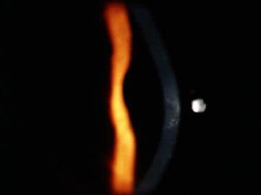 An optic section of a keratoconic cornea shows corneal thinning. Vogt striae and some scarring can also be seen centrally; superiorly, a small (brown) section of the Fleischer ring is noted.
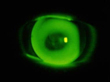 The fluorescein pattern of a rather flat-fitted rigid contact lens on an advanced keratoconic cornea.

