Background
Exophthalmos is defined in Dorland's Medical Dictionary as an "abnormal protrusion of the eyeball; also labeled as proptosis." Proptosis in the same reference is defined as exophthalmos. Another resource suggests that the terms exophthalmos and proptosis can be used to describe eyes appearing to bulge out of the face due to an increase in the volume of the tissue behind the eyes. Proptosis can describe any organ that is displaced forward, while exophthalmos refers to only the eyes. [1] Proptosis can include any directional forward displacement.
Henderson reserves the use of the word exophthalmos for those cases of proptosis secondary to endocrinological dysfunction. [2] Therefore, this dictum will be followed, and non–endocrine-mediated globe protrusion will be referred to as proptosis and exophthalmos will be reserved for protrusion secondary to endocrinopathies. See the image below.
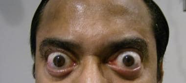 Exophthalmos due to thyroid dysfunction. The patient has significant forward protrusion of the eyes with bilateral upper- and lower-lid retraction. Image courtesy of Adam J. Cohen, MD.
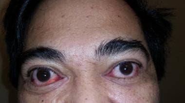 Exophthalmos due to thyroid dysfunction. The patient has significant forward protrusion of the eyes and bilateral upper- and lower-lid retraction. Also note conjunctival injection, especially of the right inferomedial conjunctiva. Image courtesy of Adam J. Cohen, MD.
Pathophysiology
The etiological basis of proptosis can include inflammatory, vascular, infectious, cystic, neoplastic (both benign and malignant, metastatic disease), and traumatic factors. Some representative examples (not a complete list of proptosis from different causes) include infectious causations such as orbital cellulitis and subperiosteal abscesses. Traumatic causations could be orbital emphysema, retro-orbital hemorrhage, and carotid-cavernous fistula. Vascular causations not traumatically related would be orbital arteriovenous malformation (AVM) varices and aneurysms. Neoplastic causations include adenocarcinoma of the lacrimal gland, pleomorphic adenoma of the lacrimal gland, meningioma, lymphoma, and metastatic disease.
For instance, lymphangiomas, by their histologic nature, can increase in size during viral illnesses and result in an increase in orbital volume. A ruptured lymphangioma can enlarge after its rupture and sequestering of heme, which pathologically is described as a chocolate cyst. Orbital varices can result in proptosis with increased venous pressure in the orbit, as seen with a Valsalva maneuver or change in postural position.
Unusual cases are also encountered, such as bilateral proptosis due to orbital hemorrhage caused by factor IX deficiency (hemophilia B). [3]
In children, unilateral proptosis is often due to an orbital cellulitis–type picture, and, in bilateral cases, neuroblastoma and leukemia are more likely. Other causes in children include rhabdomyosarcoma, retinoblastoma, capillary hemangioma, dermoid cyst, glioma of the optic nerve, and metastatic disease.
Thyroid orbitopathy, also referred to as thyroid ophthalmopathy, is categorized as an inflammatory process that is autoimmune-mediated. [4, 5, 6] As it affects the orbit, the author prefers to use thyroid orbitopathy and, more particularly, thyroid-associated orbitopathy, commonly referred to as Graves disease. In adults, it is the most common cause of unilateral and bilateral exophthalmos. Noninflammatory thyroid orbitopathy has also been reported. [7]
The etiology of the thyroid-related orbitopathy is an autoimmune-mediated inflammatory process of the orbital tissues, predominantly affecting the fat and the extraocular muscles. Lymphocytes, plasma, and mast cells are the cellular constituents in this process. The deposition of glycosaminoglycans and the influx of water increase the orbital contents. Over time, fibrosis can occur. [8, 9] Genetic factors have been associated with Graves disease. [10] Obstruction of the superior ophthalmic vein with resultant diminished venous outflow also contributes to the orbital engorgement.
Nunery has segregated patients with thyroid-related orbitopathy into type I and type II. [11] Those with type I do not have restrictive myopathy, whereas those with type II do. Type I was believed to be caused by a profundity of hyaluronic acid manufactured by the orbital fibroblasts, stimulating lipoid hyperplasia and edema. Patients with type II experience restrictive myopathy and have diplopia within 20° of fixation.
Orbital emphysema can be a significant cause of proptosis and requires emergency treatment.
No matter what the etiology may be, globular protrusion is secondary to the increase in volume within the fixed bony orbital confines. Since the orbit is widest toward its anterior aspect, the orbital contents are displaced anteriorly, resulting in proptosis and exophthalmos.
Epidemiology
Frequency
Five percent of the general population is affected by thyroid autoimmunity problems. [5]
United States
Bartley et al had reported a frequency of 2.9 cases per 100,000 population per year in men and 16 cases per 100,000 population per year in women. They also observed a bimodal distribution in both sexes, with women showing one peak at age 40-44 years and the other peak at age 60-64 years. In men, the bimodal occurrence was at age 45-49 years and age 65-69 years. Both peaks incidences in men were 5 years later than in women. [12]
International
Tellez et al, in another small study consisting of 155 patients who were newly diagnosed with Graves ophthalmopathy, 26% were male and 36% were female; however, the prevalence was higher in the Europeans, at a rate of 42% versus 7.7% in Asians. Analysis of the data indicated that Europeans were 6.4 times more likely to have Graves ophthalmopathy than Asians. [13] In northern India, the prevalence of Graves orbitopathy was reported as being similar to that in European patients but with less severity. [14]
Predictive
In a report from the European Group on Graves’ Orbitopathy (EUGOGO), 15% of patients who did not have orbitopathy at the time of diagnosis of Graves hyperthyroidism went on to develop orbitopathy. [15]
Mortality/Morbidity
Proptosis due to any cause can compromise visual function and the integrity of the eye.
A proptotic eye not adequately protected by the lids, as with lagophthalmos, can develop exposure punctuate keratopathy. Such disruption of the finely orchestrated homeostatic mechanism to protect the eye will result in corneal compromise, epithelial death, ulceration, and possible corneal perforation in severe cases. At a minimum, the disruption of the tear film layer and incomplete moisturizing of the eye will adversely affect vision and ocular comfort.
Proptosis secondary to a space-occupying process can result in a compressive optic neuropathy. Impeded optic nerve blood flow results in irreversible neuronal death and diminished optic nerve function. Such manifestations as depression of visual and color acuities, pupillary dysfunction, and constriction of visual field can occur.
Proptotic compressive effects are remedied initially by forward protrusion of the eye, thereby reducing the compressive effect within the orbit. However, the eye can extend only so far, and severe stretching can adversely affect the eye and compromise the optic nerve.
A difference of more than 2 mm between the 2 eyes of any given patient is considered abnormal.
Race
Epstein et al state that proptosis is a globe that protrudes 18 mm or less and exophthalmos is protrusion of greater than 18 mm. The upper limit of normal was 21 mm. [16]
In adult white males, the average distance of globe protrusion is 16.5 mm, with the upper limit of normal at 21.7 mm. [17]
In adult African American males, it averages 18.5 mm, with the upper limit of normal reported as 24.7 mm. [17] A separate study reported the average as 18.2 mm, with an upper normal limit of 24.14 mm in males and 22.74 mm in females. [18]
In Mexican adults, males averaged 15.18 mm and females averaged 14.83 mm. [19]
In Tehran, Iran, for the age group of 20-70 years, the average was 14.7 mm. [20]
In Taiwanese adults, comparing normal subjects to those with Graves disease, the normal group had an average reading of 13.91 mm versus 18.32 mm for the Graves disease group. [21]
Even within a group of people, there can be variability. Four ethnic groups in Southern Thailand had exophthalmometry measurement averages ranging from 15.4 mm to 16.6 mm. [22]
In 2477 Turkish patients, the median Hertel measurement was 13 mm, with an upper limit of 17 mm. [23]
In a Dutch study, the upper limit by Hertel measurement was 20 mm in males and 16 mm in females. [24]
Sex
Females also show racial variation. The average in white women was 15.4 mm and the average was 17.8 mm in African American women. The upper limits of normal in each group were 20.1 mm and 23 mm, respectively.
In general, adult females across all races have lower exophthalmometry readings than adult males.
Thyroid orbitopathy has a female preponderance, with a female-to-male ratio of 5:1. However, these differences diminish for the more serve cases of ophthalmopathy. The incidence of severe ophthalmopathy is 3-5%. [25] The female to male ratio in this subgroup is 1.4:1. [26, 8, 27]
Age
Proptosis occurs in both adults and children at any age. Thyroid orbitopathy and the resultant exophthalmos show a predilection for females aged 30-50 years.
Ahmadi et al showed that with increasing age occurs a "linear reduction in ocular protrusion." With advancing age, there was no asymmetries between the eyes noted. [28]
A US pediatric population showed exophthalmometry measurements that increased with increasing age, as one would expect. The results were stratified into age groups with the following corresponding averages:
-
Younger than 4 years: 13.2 mm
-
Aged 5-8 years: 14.4 mm
-
Aged 9-12 years: 15.2 mm
-
Aged 13-17 years: 16.2 mm
Of the 673 subjects in this study, only 2 had a 2-mm difference between the eyes. [29]
In Tehran, Iran, for the age group 6-12 years, the average was 14.2 mm and for the age group 13-19 years, the average was 15.2 mm. [20]
In Chinese children and adolescents from Xiamen, in the age range from 5-17 years, the average exophthalmometry reading was 14.48 mm. [30]
Note that CT scanning and exophthalmometry yield measurements that are not identical, especially when proptosis is present. [31] In addition, parallax errors exist with most commonly used measuring devices. [32]
-
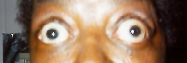
Bilateral exophthalmos and upper lid retraction secondary to Graves disease.
-
Exophthalmos due to thyroid dysfunction. The patient has significant forward protrusion of the eyes with bilateral upper- and lower-lid retraction. Image courtesy of Adam J. Cohen, MD.
-
Exophthalmos due to thyroid dysfunction. The patient has significant forward protrusion of the eyes and bilateral upper- and lower-lid retraction. Also note conjunctival injection, especially of the right inferomedial conjunctiva. Image courtesy of Adam J. Cohen, MD.


