Overview
The inherited Charcot-Marie-Tooth peripheral neuropathies (CMT) were first described independently by Charcot and Marie in France [1] and by Tooth in England. [2]
The heterogeneous nature and different forms of inheritance of the condition were soon recognized. Dejerine and Sottas described more severe infancy-onset cases, [3] and Roussy and Levy described cases associated with tremor. [4]
In the late 1960s, neurophysiologic testing allowed the classification of CMT into 2 groups, one with slow nerve conduction velocities and histologic features of a hypertrophic demyelinating neuropathy (hereditary motor and sensory neuropathy type 1 or CMT1) and another with relatively normal velocities and axonal and neuronal degeneration (hereditary motor and sensory neuropathy type 2 or CMT2). [5]
Since the early 1990s, patients with both CMT1 and CMT2, while often clinically similar, were found to be genetically heterogeneous. Now a large and ever increasing number of genetic subtypes has been described, and major advances in molecular and cellular biology have clarified the understanding of the role of different proteins in the physiology of peripheral nerve conduction in health and in disease. Dejerine-Sottas disease is also known as CMT3. Autosomal recessive forms can be also divided into demyelinating (CMT4 or AR-CMT1) and axonal forms (AR-CMT2). Subtypes with velocities within the intermediate range are called DI-CMT. Better understanding of new mutations and the wide range of possible phenotypes led to the development of a new nomenclature proposal, based on the gene and inheritance pattern. However, there is no unanimous agreement, less especially about the nomenclature of the recessive and intermediate-conduction velocity subtypes.
This article is a brief introduction to the study of CMT and correlates the most common different genotypes and phenotypes. Inherited neuropathies in which autonomic or sensory features predominate, conditions in which the neuropathy is part of a multiple-organ disturbance, and neuropathies with specific metabolic dysfunction are not discussed.
Pathophysiology
Traditionally, CMT pathophysiology has been categorized into 2 processes: a predominant demyelinating process resulting in low conduction velocities (CMT1) and a predominant axonal process resulting in low potential amplitudes (CMT2). However, even for CMT1 a heated debate has focused on the relative contribution of axonal versus demyelinative damage to the disease manifestations and progression.
In some hereditary neuropathies discussed below, focal asymmetric features (eg, hereditary neuropathy with liability to pressure palsy [HNPP]) predominate; in others (eg, certain cases of Charcot-Marie-Tooth disease type 1A (CMT1A) and inherited brachial plexus neuropathy [IBPN]/hereditary neuralgic amyotrophy [HNA]), proximal weakness predominates. Typically, a predilection exists for distal limbs as the site of disease onset and more severe symptoms and signs. Furthermore, while significant variation in nerve conduction velocities exists between and within families, this parameter does not predict severity, with the exception of the very low (ie, < 5 m/s) velocities observed in Dejerine-Sottas syndrome (DSS) and congenital hypomyelination neuropathy (CHN).
Axonal degeneration is a prediction of disability. This suggests that, in most cases, axonal damage is the root cause of the neuropathy, not demyelination. [6] However, the gene mutations responsible for the different forms of CMT1 are clearly myelin genes. Through mechanisms that at present can only be speculated about, myelin disturbances result in axonal damage. This is not surprising given the strong evidence for interaction between myelin and axon gene expression in development and after experimental nerve lesions. On the other hand, axonal damage can result in secondary demyelination.
Myelinating Schwann cells form a myelin sheath around a single axon and express high levels of myelin-related proteins and messenger RNA (mRNA). Axonal degeneration leads to Wallerian degeneration, in which myelin sheaths are phagocytosed, previously myelinating Schwann cells dedifferentiate, and mRNA expression is down-regulated. When Schwann cells re-ensheathe axons, levels of proteins or mRNA, or both, increase. Myelin genes and products affected in this manner include myelin protein zero (MPZ, P0), peripheral myelin protein 22 (PMP22), connexin 32 (CX32, Cx32), myelin-associated glycoprotein (MAG, Mag), myelin basic protein (MBP, Mbp), early growth response gene 2 (EGR2), periaxin (PRX), and others.
Whether Schwann cells differentiate into myelinating or nonmyelinating (a misnomer because such Schwann cells myelinate > 1 axon) phenotypes depends on axonal characteristics, which are determined at least in part by transcription factors such as Oct-6 (POU domain family) and EGR2.
Mutations in structural myelin genes or the transcription factors responsible for their expression lead to demyelination (ie, PMP22, P0, CX32, EGR2). P0, linked to Charcot-Marie-Tooth disease type 1B (CMT1B), is the most abundant protein in compact myelin and is a cell adhesion molecule (CAM). PMP22, linked to CMT1A, is a protein of unclear function that has features of both a channel protein and a CAM. Schwann cells also express Cx32, linked to X-linked Charcot-Marie-Tooth Disease type 1 (CMTX1), which belongs to the connexin family. EGR2 is a transcription factor linked to Charcot-Marie-Tooth disease type 1D (CMT1D).
More recently, mutations in the LITAF (lipopolysaccharide-induced tumor necrosis factor-alpha) complex were found to account for CMT1C; these mutations are involved in the proliferation and apoptosis in Schwann cells carrying PMP22 alterations. [7]
Frequency
In the United States CMT is the most common inherited neurologic disorder, affecting approximately 150,000 Americans (1 CMT/2500 inhabitants). Estimates of the frequency of each CMT subtype in the United States are variable. [8]
Internationally, CMT is among the most common heritable neurologic disorders. An exhaustive Norwegian study indicated a prevalence of 36 cases per 100,000 population, [9] whereas a worldwide meta-analysis estimated a prevalence of 10 cases per 100,000 population. [8] A Japanese epidemiologic study demonstrated an incidence of 10.8 cases per 100,000 population. [10]
A Finnish study of 435,000 individuals found a prevalence of 16 cases per 100,000 population for HNPP and 20 cases per 100,000 population for CMT in general, indicating that HNPP is not rare. [11] Because both diseases result from unequal crossover during meiosis, similar epidemiologic data are not surprising. Conditions associated with reduced PMP22 expression are underdiagnosed because of the variable phenotype, the insidious nature of the disease, and the many mildly affected patients.
Overall, CMT1A accounts for about 60% of all autosomal dominant neuropathies, CMT2 accounts for about 22%, X-linked Charcot-Marie Tooth disease (CMTX) for about 16%, and CMT1B for approximately 1.6%. The other forms are rarer, except in populations with high consaguinity, where AR phenotypes are significantly more common.
Race-, sex-, and age-related demographics
CMT is found world wide in people of all races and ethnic groups. [12]
Some rare CMT subtypes, in particular autosomal recessive, are largely restricted to particular racial groups.
In the United States, CMT may be less common among black people. Whether this represents a lower frequency of the specific mutations or protection from disease manifestation through unknown disease-modifying genes remains unclear.
CMT subtypes may be inherited in an autosomal dominant or recessive or an X-linked pattern.
Importantly, CMTX (similar to other X-linked diseases), while certainly more severe in men, is not uncommonly associated with clear disease in women, which may even be severe, most likely because of unequal inactivation of the X chromosome, which may result in predominant expression of the abnormal CX32 allele in nerves.
Reportedly, CMT may have a more severe phenotype in men, possibly because of environmental (nerve trauma) or X-linked neuroprotective factors, but, in practice, this impression is of little value because of the great phenotypic variability between and within families.
Age of onset is variable according to subtype, penetrance, familial phenotype, and ascertainment bias. Most symptoms start during childhood but may go unnoticed. In the authors' experience, onset of disease is noticed much earlier in large families with a lot of knowledge of their disease.
Diagnosis is usually not made until late adolescence or early or late adulthood. Exceptions are more severe phenotypes such as DSS and CHN.
Mortality/morbidity
Life expectancy is normal in most patients with CMT. The degree of disability varies according to the CMT subtype, and it is unpredictable between and within families; even identical twins may be differently affected.
Typically, HNPP patients have a good quality of life between episodes of nerve damage. About 10% of patients have an incomplete recovery from episodes of nerve palsy. Cases of respiratory failure have been reported.
Rarely, patients with CMT may have laryngeal dysfunction with aspiration and voice problems.
Patients with DSS are often disabled in early childhood. CHN can lead to early death.
Whenever a patient is unexpectedly severely affected, a search for additional neurologic diseases is warranted.
Per definition, CMT is a disease of the peripheral nervous system (PNS), but CNS features can be found.
Clinical Presentation
Signs and Symptoms
Patients with CMT can present with many symptoms and signs. Disease expression varies between and within kindreds and even among identical twins. CNS features may be part of the disease or indicate an independent coexisting condition; CNS features warrant further investigation in all cases. Nonneurologic manifestations, including endocrine disturbances, have been described. In typical CMT, symptoms are chronic and slowly progressive, but they may be episodic and asymmetric in patients with HNPP and IBPN/HNA.
Most patients with CMT have autosomal dominant forms involving weakness, muscle wasting, and sensory loss predominantly in the distal legs, with onset in the first 2 decades of life. Autosomal recessive inheritance with late onset in earlier generations must be considered when a family history is apparently absent. Importantly, CMT1A has one of the highest de novo mutation rates, similar to neurofibromatosis type 1, as discussed in Pathophysiology. Consequently, cases occur in which a family history is truly absent.
Enlarged and excessively firm nerves are found in more than 25% of patients with CMT1 or DSS and are often visible in the superficial cervical nerves and are palpable in the arms. Gait may be compromised by distal weakness, foot deformities, and poor proprioception. Ankle sprains and fractures are frequent. Reports of cold feet, hair loss, or leg edema are common. Some patients report faster deterioration during pregnancy that is usually, but not always, associated with recovery. As in surgical procedures, prolonged arrangement of the body and limbs in particular positions can result in nerve compression, which could make any underlying neuropathy worse.
Onset
Because of the insidious onset, some patients are unaware of their disease and seek medical attention only late in life. Parents, caregivers, or teachers may notice clumsiness, frequent sprains, poor athletic performance, or toe walking in a child. For example, one of the authors' patients noticed that her father's feet slapped the ground when he walked around the house. Later, having observed her personal series of 20 relatives, she was able to accurately diagnose CMT in her grandchildren and in her siblings' grandchildren by watching their feet point down when sitting on the ground. Not infrequently, asymptomatic individuals are detected during screening of families after one relative has been diagnosed. Depending on the age of onset, children may have normal examination findings. Occasionally, the only finding is impaired heel gait.
Motor symptoms and signs
Motor symptoms usually predominate over sensory symptoms. Motor and sensory symptoms may be exclusive in the subgroup of hereditary motor neuropathies (HMN) and hereditary sensory neuropathies (HSN), respectively, which are not discussed in this article.
Proximal weakness is rarely present except in the most severely affected patients, in some unusual pedigrees, and with brachial plexus neuropathies, which means that even patients with marked weakness are able to walk. Manipulating small objects such as zippers, forks, or pencils may be difficult because of motor and sensory impairment. On examination, most patients with CMT exhibit distally dominant weakness, hyporeflexia, and muscle atrophy affecting the legs earlier and more significantly than the arms.
Patients report loss of balance, tripping over objects because of foot-drop and impaired sensation, weakness, and foot deformities resulting in unequal wearing of shoes and trouble finding shoes that fit. Foot deformities include pes cavus, high arches or flat feet, hammertoes, and shortened Achilles tendons. Such deformities become more prevalent with age but are variable even among relatives of the same age. Wasting of foot and distal lower extremity muscles develops over time and may produce the classic "inverted champagne bottle" appearance. Some patients with CMT, and in particular patients with HNPP, find that wider shoes are more comfortable because of high arches and hammertoes or because local nerve compression may be less (in the case of HNPP).
Families with CMT and pyramidal features have also been reported. In 2003, Vucic et al found 2 families with dominantly inherited axonal neuropathies, distal wasting, weakness, pes cavus, sensory loss, and mild pyramidal signs (including extensor plantar responses, mild increase in tone, and preserved or increased reflexes but no spastic gait). [13] Linkage studies excluded Charcot-Marie-Tooth disease type 2A (CMT2A), Charcot-Marie-Tooth disease type 2B (CMT2B), Charcot-Marie-Tooth disease type 2D (CMT2D), Charcot-Marie-Tooth disease type 2E (CMT2E), CMT2, amyotrophic lateral sclerosis 4 (ALS4), hereditary motor neuropathy type 2 (HMN II) and mutations in the PMP22, MPZ/Po, or EGR2 genes.
Sensory symptoms and signs
Sensation may be normal until adulthood. Distal mild pansensory loss is common and, with semiquantitative methods, can often be documented in children. At times, sensation is severely impaired. Sensory changes dominate in HSN, which are not a subject of this article.
Paresthesias are typically less severe and rarely a presenting symptom, in marked contrast to acquired neuropathies. However, upon further questioning, paresthesias are mentioned. Patients may deny sensory symptoms despite marked loss of sensation on examination. Radicular pain resulting from CMT1 is rare but well described and is caused by nerve roots that are enlarged (and sometimes visible by MRI) because of ongoing demyelination/remyelination with connective tissue proliferation. Back and radicular pain unrelated to CMT1 is likely to be more common in patients with CMT.
Nonneuropathic pain
Patients often have multiple nonneuropathic pain symptoms. Pain can result from pressure or strain of various structures associated with bones, joints, and tendons and abnormal posture at the knees, hips, and back, resulting from foot weakness and fixed foot deformities such as Achilles tendon shortening. Because of hammertoes and high arches, patients experience painful calluses. Scoliosis is common and leads to back pain. Patients experience leg and hand cramps that are often worse with fatigue and relieved by wearing ankle-foot orthoses. [14] The following paragraphs focus on distinguishing features of CMT subtypes.
Charcot-Marie-Tooth disease type 1A
Approximately 85% of patients develop clinical evidence of disease before age 20 years. In one large series, 34 patients were classified as having a classic CMT phenotype, whereas 27 patients had additional features such as CNS features, diabetes mellitus, and prominent muscle cramps. [15] Forty-five of 61 patients had deficits in their hands. Loss of large- and small-fiber sensory function, ranging from mostly mild to moderately severe, was reported in 43 patients. Tremor occurred in up to 25% of patients. Whether it was incidental or part of the syndrome is controversial. Tremor with CMT is referred to as Roussy-Levy syndrome.
One Austrian family with CMT1A had slow progression and predominantly proximal arm weakness and wasting. [16] Sleep apnea was described in a French family with CMT1A (a feature previously reported in Charcot-Marie-Tooth disease type 2C [CMT2C]); most family members were asymptomatic, but sleep apnea correlated with the severity of the neuropathy, possibly because of pharyngeal neuropathy. Two sisters had an atypical CMT1A phenotype consisting of prominent sensory symptoms, tremor, and episodes of acute paralysis. [17]
Recent studies of DSS or hereditary motor and sensory neuropathy type 3 demonstrated that point mutations in PMP22 are a common cause, as are homozygous PMP22 deletions.
Clinical progression is slow in the second to fourth decades of life; therefore, any change in pace must lead to a consideration of superimposed acquired or possibly independently inherited forms of neuromuscular diseases. The authors have reported the association of McArdle disease and CMT1A in 2 patients who had subacute dramatic worsening of their CMT.
The severity of the CMT1A phenotype can also possibly increase over time, being more severe in new generations for unknown reasons.
Charcot-Marie-Tooth disease type 1B
As expected, instances exist of heterozygous CMT1B parents with children with DSS or CHN due to a homozygous or compound heterozygous myelin protein zero mutation. Particular severe forms with onset at age 2 years have been reported with 2 mutations in different domains of MPZ. Some point mutations result in a surprisingly mild phenotype with late onset and relatively faster nerve conduction velocities. A mild phenotype, which was recurrent in response to intense manual labor, not unlike HNPP, has been reported.
Some MPZ mutations are associated with a steroid-responsive neuropathy. Others lead to CMT with relatively faster conduction velocities, dysphagia, deafness, and pupillary abnormalities.
A clinical vignette is as follows:
Two patients, father and son, presented with slowly progressive weakness since childhood, affecting the arms more than the legs, and numbness in the hands and feet. They denied recurrent focal weakness, liability to pressure palsies, or pain. Multiple living male and female relatives from 4 generations were affected.
Findings were similar in father and son but were more pronounced in the former. Both had pes cavus. The father had enlarged firm peripheral nerves. Muscle strength was reduced to four fifths and was worse distally. Deep tendon reflexes were absent. Plantar responses were flexor. All sensory modalities were impaired. Laboratory studies in the father revealed vitamin B-12, folate, and lead levels that were within the reference range and normal findings on myelin-associated glycoprotein and GM1 antibody titers and serum protein electrophoresis. No sensory or motor responses were obtained with surface recordings. Needle examination of the left median nerve revealed motor nerve conduction velocities of 11 m/s (lower reference range value is 49 m/s) and a compound motor action potential (CMAP) amplitude of 0.3 mV (lower reference range value is 5 mV). Sensory responses and F waves were absent. Electromyography (EMG) revealed minimal spontaneous activity with high-amplitude motor unit potentials.
Sural nerve biopsy findings were similar in father and son. Semithin cross-sections of nerve showed a reduction of myelinated fiber density. Many remaining fibers had thin myelin sheaths. Frequent small onion bulbs and scattered tomaculae (as shown below) were found. The myelinated fiber density was 250/mm2 in the father and 3147/mm2 in the son. Histometric measurements showed a unimodal distribution of myelinated fibers with a shift of the peak to diameters between 1 µm and 4 µm in the father and a bimodal distribution with one peak between 1 µm and 4 µm and a second peak at 6 µm in the son.
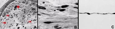 Light microscopy of tomaculae, which are typical of hereditary neuralgic amyotrophy (HNA) and hereditary neuropathy with predisposition to pressure palsies (HNPP) but are also associated with certain MPZ mutations in Charcot-Marie-Tooth disease type 1B (CMT1B). (A) Semithin cross-section shows myelinated nerve fiber loss with scattered onion bulbs (arrowheads). Several myelinated fibers have a thick myelin sheath and an irregular contour (large arrow). (B) Semithin longitudinal section demonstrating tomaculae in continuity with myelinated fibers. The myelin sheath is inappropriately thin, probably as a result of segmental remyelination or hypomyelination. (C) Teased myelinated nerve fiber containing tomaculae that consist of globular expansions of myelin measuring 30-50 µm in length.
Most of the fibers larger than 5 µm in diameter had tomaculae. Teased fibers and longitudinal semithin sections revealed sausage-shaped expansions of myelin, located in both the paranodal and internodal regions in virtually all fibers. Segmental remyelination was found in all teased myelinated nerve fibers. Ultrathin sections demonstrated that the tomaculae consisted of closely apposed redundant loops of myelin sheath wound around or layered on one side of a thinly myelinated fiber. MPZ mutation was found in both father and son.
X-linked Charcot-Marie-Tooth disease type 1
In men, symptoms typically begin in childhood or adolescence. CMTX1 may have asymmetric features. Women are often asymptomatic, but they may have late onset of mild symptoms. Rarely, they may have more severe disease, probably because of predominant inactivation of the X chromosome that bears the normal CX32 allele.
According to a large series, atrophy, particularly of intrinsic hand muscles; paresthesias; and sensory loss may be more common in CMTX1 than other subtypes.
The nomenclature reflects the fact that conduction is often not as slow as in CMT1A and CMT1B and that a second CMT locus must exist on the X chromosome because some pedigrees do not have a mutation in the CX32 gene.
Dejerine-Sottas syndrome
DSS was originally described as a hypertrophic polyneuropathy with onset in infancy or early childhood, distal sensory loss with ataxia, pes cavus with progression towards the proximal limbs, and Argyll-Robertson pupils. Such patients must be tested for mutations in MPZ, PMP22, EGR2, and PRX. Milder and unrecognized hereditary neuropathies in the parents must be considered and investigated.
Congenital hypomyelination neuropathy
Patients with CHN present with neonatal hypotonia, areflexia, distal weakness, slow nerve conduction velocities, and at times with contractures or arthrogryposis. Just like DSS, it may result from MPZ or EGR2 mutations, as well as separate mutations in the same or different genes inherited from both parents or from one mutation inherited from one parent combined with a de novo mutation in the same or another gene.
Charcot-Marie-Tooth disease type 2
Patients tend to present with symptoms later in life than patients with CMT1. They may have greater atrophy and distal leg weakness with relatively less hand weakness. Nerve hypertrophy is absent but is variable in CMT1 as well. CMT2 often presents a greater diagnostic dilemma because characteristic features such as enlarged nerves and near-pathognomonic neurophysiologic findings are absent. With later onset, this condition may be very difficult to differentiate from a late-life acquired neuropathy when the family history is unclear. While several disease genes have been identified, not all can be tested for commercially yet, and together they explain as many cases of CMT2 as is the case for disease genes in CMT1. In practice, CMT2 and CMT1 can rarely be differentiated by history and examination findings alone. As with other forms of CMT, phenotypic variation is common between and within families.
In CMT2A, the clinical presentation is typical. Affected members of a large southern Italian pedigree had distal weakness, wasting, hyporeflexia, and mild panmodal sensory loss. Nerve biopsies revealed a loss of large myelinated fibers but no myelin abnormalities.
CMT2B is a predominantly sensory neuropathy to the point that its classification with hereditary motor and sensory neuropathy (HMSN) is unclear. While neurophysiologic findings are established early in life, clinical onset may be much later. CMT2B was mapped to chromosome arm 3q13-q22, and 2 missense mutations (Leu129Phe and Val162Met) in the small guanosine triphosphatase (GTPase) late endosomal protein RAB7 were recently found. [18] RAB7 is ubiquitously expressed, and the authors found expression in sensory and motor neurons.
CMT2C usually starts in the first decade of life. Mild sensory loss is combined with weakness in the limbs, diaphragm, intercostal muscle, and vocal cords, which can lead to early death. Vocal cord dysfunction also occurs occasionally in several other CMT subtypes.
Patients with CMT2D may have worse hand than leg weakness and slow progression. Onset is usually in people aged 16-30 years. Tendon reflexes are usually absent in the arms and decreased in the legs. Progression is slow. It has been linked to chromosome arm 7p14.
CMT2E is characterized by worse leg weakness, slow progression, and onset in the second and third decades of life. Pes cavus was reported in 100% of patients older than 20 years, and some develop hyperkeratosis, although this association remains unclear. Missense mutations (Gln333Pro, protein rod domain and Pro8Arg, head domain) affecting the self-assembly of neurofilament light chain protein were found (chromosome arm 8p21).
Patients with Charcot-Marie-Tooth disease type 2F (CMT2F) have slow progression and worse distal weakness. In 2001, Ismailov et al reported a 6-generation family with autosomal dominant CMT of the axonal type from the Voronezh province of Russia. [19] Disease onset occurred in people aged 15-25 years. Patients had symmetric progressive weakness and atrophy of the lower limb muscles, resulting in foot drop and steppage gait. Wasting of upper limb muscles caused clawing of the hands several years later. Depressed or absent deep tendon reflexes were observed at an early stage. Mild-to-moderate sensory impairments occurred in the feet and hands in all the patients. The course of the disease was slowly progressive, with disability after 15-20 years, but reproductive fitness was conserved and life span not restricted. Linkage to chromosome arm 7q11-q21 was established.
HNPP and other phenotypes of PMP22 deletions and certain point mutations
Onset of neuropathies associated with PMP22 deletions or mutations is typically in the third or fourth decades of life, but it ranges from the first to the eighth decades of life; palsy may be present at birth. Because of its insidious onset, some patients are unaware of their disease or seek medical attention only late in life, whereas others remain asymptomatic.
In one study of patients with PMP22 deletions, 40% were unaware of their condition; 25% were essentially without symptoms. Monozygotic twins have been reported of which only 1 was affected clinically, although both and their father had slowed nerve conduction.
The most common presentation is recurrent acute mononeuropathy often related to minor nerve compression. In typical HNPP, motor symptoms predominate over sensory symptoms. Patients often report that after resting on a limb in an awkward position, the resulting weakness and dysesthesias last weeks to months, rather than seconds to minutes.
Slight compression of peripheral nerves and repeated local exercise leads to episodes of weakness with decreased perception to touch and pain. Most attacks are of sudden onset, are painless, and are initially followed by recovery. Attacks most often occur with a single nerve involvement, with onset on awakening. They are usually triggered by mild compression that resolves in days to months. Patients with frequent episodes may have persistent neurologic abnormalities. Precipitating trauma, such as carrying heavy loads, writing, or playing musical instruments, may be minimal and unavoidable. Other precipitating events include prolonged immobilization during surgery or childbirth. Typically, no neuropathic pain is present, but tingling is a frequent symptom.
In patients with the recurrent pattern, the examination findings between episodes may be normal or mildly abnormal. Distal and mild pansensory loss may be present. Reflexes are normal in approximately two thirds of patients. In one study, ankle jerks were absent in 37.5%, and areflexia occurred in 12.5%.
Sites of compression are often those of anatomic vulnerability, including the fibular neck for the peroneal nerve, the cubital tunnel for the ulnar nerve, the spiral groove of the humerus for the radial nerve, and the carpal tunnel for the median nerve. However, many other sites may exist, as evidenced by the recent report of a lesion of the anterior branch of the axillary nerve, likely caused by compression against the surgical neck of the humerus.
In a study of 70 patients, the average number of nerve palsies over a lifetime was approximately 2; the most commonly affected structure was the peroneal nerve, followed by the ulnar, the brachial plexus, and the radial and median nerves; weakness persisted for over 3 months in 15%. The second most frequent presentation in 2 series was a largely symmetric slowly progressive polyneuropathy, causing a misdiagnosis of CMT in the narrow sense. With this subtype, high arches and hammertoes are common. Scoliosis is rarely observed.
Some patients have recurrent sensory symptoms lasting minutes to hours triggered by limb position, nerve compression, or holding a phone, whereas others present with a chronic sensory polyneuropathy. Rarely, patients can present with subacute recurrent or confluent demyelinating multiple mononeuropathies, and they are misdiagnosed with acute or chronic inflammatory demyelinating polyneuropathy. Cranial neuropathies, including deafness, are infrequent and may sometimes represent chance events. One of the authors' patients provided a history of 3 episodes of Bell palsy over 3 years.
The brachial plexopathy form of HNPP may be recurrent, isolated, or part of other multiple mononeuropathies, and it is painless, which serves to differentiate it from HNA. Furthermore, no genetic overlap exists between the 2 conditions.
Rare associations of PMP22 deletions include CNS demyelination, moving toes and myoclonus, and fulminant 4-limb weakness possibly related to nerve compression. Some patients are oligosymptomatic or asymptomatic, but their examination findings during family evaluations may reveal subtle abnormalities such as distal hyporeflexia or Tinel signs. Not surprisingly, conditions may evolve, with patients expressing one phenotype and progressing to another, and different phenotypes may coexist in a given family.
The condition may occasionally manifest in later life when individuals develop an acquired unrelated neuropathy due to metabolic derangements, autoimmunity, or neurotoxic drugs. Undiagnosed PMP22 deletions in an oligosymptomatic patient may complicate the treatment and diagnosis of an acquired neuromuscular disease. For instance, a person with the disorder may, in the seventh decade of life, become diabetic and develop diabetic amyotrophy. Alternately, such a patient may require neurotoxic drugs such as platinum or vincristine in the treatment of a malignancy. Some HNPP patients might develop chronic inflammatory demyelinating polyneuropathy. Such patients' clinical and electrical presentations may be unusual and difficult to understand, unless a diagnosis of HNPP is also considered. Even more importantly, such a patient's spontaneous recovery or positive treatment response may be disappointing and far less complete than expected if the acquired neuropathy were the only condition.
A clinical vignette is as follows:
A 42-year-old man was referred for evaluation of diffuse body aches. He had a history of episodic bilateral wrist drop; intermittent limb numbness, often after repetitive movements; and muscle cramps since age 20 years. On examination, he had subtle abnormalities. Panmodal sensory loss was found in a right ulnar and peroneal and left median and ulnar distribution. Power, bulk, gait, and coordination were intact, but distal hyporeflexia was present. Tinel signs were present at the wrists and elbows. No nerves could be palpated. Hammertoes and high arches were present.
Findings on routine laboratory studies, such as erythrocyte sedimentation rate, vitamin B-12, folate, rapid plasma reagin, antinuclear antibodies, rheumatoid factor, thyroid function, HgA1c, creatine kinase, and HIV antibodies, were noncontributory, but hepatitis C serology was positive. His 65-year-old mother provided a history of finger numbness and postpartum arm weakness, and she had distal hyporeflexia and decreased sensation. His 81-year-old great-aunt was similarly affected.
His daughter, aged 18 years, had numbness and tingling in the back of her legs during gym class and middle finger numbness when holding a pen or playing the flute, but she denied episodic weakness; symptom onset was at age 13 years. She had high arches and hammertoes, slightly weak finger extension, mild distal pansensory impairment, and a Romberg sign. Her 15-year-old sister was reluctant to attend gym class because of foot numbness; symptoms improved when she shook her legs. Either hand occasionally went numb, especially the right thumb when writing. Once after sitting on her cousin's shoulders, both legs were weak for 20 minutes. Her examination findings were remarkable only for weak interossei in both hands. Two younger children were asymptomatic but had distal pinprick and vibratory sense loss with normal proprioception, strength, and reflexes.
Electrodiagnostic studies in 1987, 1988, and 1996 in the father revealed multiple nerve entrapments at both median and ulnar nerves at the wrists, the ulnar nerves at the elbows, the right radial nerve at the spiral groove, and the right peroneal nerve at the fibular head. Conduction velocities were normal or low-normal except for slowing in the demyelinative range at areas of nerve entrapment, where amplitudes were also low. No significant change occurred over time in nerves studied repeatedly.
Light and electron microscopy of the father's sural nerve biopsy specimen showed prominent sausage-shaped expansion of the myelin sheath in the perinodal and internodal regions, the classic finding that gives this condition its other name—tomaculous neuropathy; thinly myelinated fibers; and reduction in large myelinated fiber density with a shift of fiber diameters to small sizes. Areas of incomplete myelin compaction and redundant loops of the myelin sheath were present. Genetic testing revealed a deletion of the 1.5-Mb gene region that harbors the PMP22 gene in all affected family members as well as in the 2 asymptomatic younger daughters.
This family illustrates the typical presentation of PMP22 deletions, but the diagnosis is not always as straightforward.
Neuritis with brachial predilection/hereditary neuralgic amyotrophy/hereditary brachial plexus neuropathy
Dreschfeld in 1886 may have been the first to recognize this condition when he described a woman with 3 episodes of painful arm weakness, whose sister had experienced 7 such attacks. Jacob et al in 1961 described in 7 patients of 2 unrelated families 14 similar episodes of recurrent brachial neuritis with incapacitating pain, weakness, wasting, depression of reflexes, and sensory loss. The legs were involved, and arm involvement was severe. [20] In 1973, Guillozet and Mercer described a similar condition in 3 generations of a family. [21] At times, the lower cranial nerves and sympathetic nervous system are involved as well.
HNA is an autosomal dominant form of recurrent focal neuropathy. Individuals experience episodic brachial plexus neuropathy with weakness, atrophy, and sensory disturbances, preceded almost always by severe pain in the affected arm. Age of onset is in the second and third decades of life and rarely in the first. Recovery is usually complete and begins weeks to months after the onset of symptoms. Recurrent episodes may affect either arm. The right arm is involved more often.
Phenotypic variation may occur, with some patients following the classic relapsing-remitting course and others following a chronic undulating course. Common dysmorphic features include hypotelorism, short stature, cleft palate, unusual skin folds, and creases in the neck or scalp referred to as cutis verticis gyrata.
Causes
The Table below provides a summary of the current classification of CMT disorders based on updated knowledge of molecular biology and genetics. For completeness, several conditions are included that are discussed in this article.
In 1991, 2 groups showed that CMT1A, the most common form of CMT1, was associated with a 1.5-Mb duplication within chromosome arm 17p11.2. Some 70% of CMT1 cases and 90% of CMT1A cases result from this duplication, while the deletion of the same DNA region causes HNPP and related disorders. Importantly, other cases of CMT1A and HNPP result from point mutations in the PMP22 gene. [22] HNA is linked to chromosome arm 17q25.
CMT1B, which was linked to chromosome 1, was found to be associated with mutations in the myelin protein zero glycoprotein.
Usually, CMTX1 results from mutations in coding sequences of the gap junction protein beta 1/connexin 32 on chromosome arm Xq13.1, but some mutations occur in regulatory sequences such as the promotor or in introns. X-linked Charcot-Marie-Tooth disease type 2 (CMTX2) has been linked to an unknown gene on chromosome arm Xq24-26.
Mutations in the zinc-finger domain containing transcription factor EGR2 were linked to CMT1D and CHN.
DSS, a severe infantile neuropathy, was associated with PMP22 and MPZ mutations. Autosomal recessive DSS can be caused by mutations in the PRX gene on chromosome arm 19q13.13-2, which is regulated by EGR2.
CMT2A was linked to a loss-of-function mutation in the KIF1B gene on chromosome arm 1p36-35, which appears to be a motor protein involved in anterograde mitochondrial transport.
The gene responsible for CMT2B remains unknown, but linkage has been established to a 10-cM interval locus on chromosome arm 3q13-q22.
A linkage has yet to be identified for CMT2C. [23]
CMT2D is linked to an unknown gene on chromosome arm 7p14. [24]
Mutations in the neurofilament light subtype gene on chromosome arm 8p21 are the cause of CMT2E.
Several new disease genes have recently been identified: 2 signal transduction genes, the N-myc downstream-regulated gene-1 on chromosome arm 8q24.3 for the Lom form of autosomal recessive HSMN; the gene for the phosphatase myotubularin–related protein-2 on chromosome arm 11q22 for autosomal recessive HSMN; and the gene for a serine palmitoyltransferase subunit on chromosome arm 9q22 for HSN1.
Two different loci have been associated with the so-called intermediate nerve conduction forms of CMT: 10.7-Mb interval on chromosome arm 10q24.1-q25.1 and chromosome arm 19p12-p13.2.
Table 1. Genetics of CMT and Other HMSN Types (Open Table in a new window)
Disorder |
Gene |
Locus |
CMT1: Dominant; Demyelinating |
||
CMT 1A |
PMP-22 |
17p11 |
CMT 1B |
P0 |
1q22 |
CMT 1C |
LITAF |
16p13 |
CMT 1D |
EGR2 |
10q21 |
CMT 1F |
NF-L (NF-68) |
8p21 |
CMTX1 |
Connexin-32 |
Xq13 |
CMTX2 |
Unknown |
Xp22.2 |
CMTX3 |
Unknown |
Xq26 |
HNPP |
PMP-22 |
17p11 |
Dejerine-Sottas (HMSN 3) |
PMP-22 8q23 EGR2 |
17p11 8q23 10q21 |
DI-CMT (Intermediate nerve conduction velocities) |
DNM2 10q24 1p34 P0 CMT-X |
19p12 10q24 1p34 1q22 Xq13 |
CMT2: Dominant; Axonal |
DNM2 |
19p13.2-p12 |
CMT 2A |
KIF1Bß |
1p36 |
CMT 2A2 |
Mitofusin 2 |
1p36 |
CMT 2B |
RAB7 |
3q13-q22 |
CMT 2B2 |
Unknown |
19q13.3 |
CMT 2C |
TRPV4 |
12q24.13 |
CMT 2D |
GARS |
7p14 |
CMT 2E |
NF-68 |
8p21 |
CMT 2F |
HSPB1 (HSP 27) |
7q11 |
CMT 2G |
Unknown |
12q12 |
SMT 2H |
GDAP1 |
8q21.3 |
CMT 2L |
HSPB8 |
12q24 |
CMT 2 P0 |
P0 |
1q22 |
AR-CMT2: Recessive; Axonal |
|
|
AR-CMT2A |
Lamin A/C |
1q21 |
AR-CMT2B |
|
19q13 |
CMT 4A |
GDAP1 |
8q13-21 |
CMT 4B1 |
MTMR2 |
11q23 |
CMT 4B2 |
SBF2/MTMR13 |
11q15 |
CMT 4C |
SH3/TPR |
5q23 |
CMT 4D (Lom) |
NDRG1 |
8q24 |
CMT 4E |
EGR2 |
10q21 |
CMT 4F |
Periaxin |
19q13 |
CMT 4G |
HK1 |
10q23.2 |
CMT 4H |
FIG4 |
6q21 |
Dejerine-Sottas (HMSN 3) |
P0 CMT 4F |
Autosomal |
Congenital hypomyelinating neuropathy |
P0 EGR2 PMP-22 |
Autosomal |
CCFDN |
CTDP1 |
18q23 |
Giant axonal neuropathy |
Cytoskeletal protein gigaxonin |
Unknown |
Diagnostic Considerations
The first challenge for the clinician is to demonstrate that a patient's weakness and sensory loss result from peripheral nerve disease and not from abnormalities elsewhere in the nervous system.
This can usually be accomplished by a clinical examination revealing distal weakness and muscle wasting, stocking-glove type sensory loss, and hyporeflexia.
Pes cavus and hammertoes are nonneurologic stigmata of the disease, and although not specific (can occasionally be observed in other forms of chronic acquired neuropathies), should raise the suspicion of CMT if clinical context is appropriate.
If the patient has a neuropathy and a positive family history, CMT becomes likely.
Pedigree analysis can clarify inheritance patterns.
Slow nerve conduction velocities distinguish CMT1 from CMT2, although significant variation may exist within families.
Although exceptions exist, uniform conduction slowing distinguishes most type 1 cases from acquired disorders such as chronic inflammatory demyelinating polyneuropathies, in which conduction slowing typically varies along the same nerve and between nerves. The Guillain-Barré syndrome also has asymmetric slowing and a more rapid onset. Dispersion and conduction block are rarely described in CMT and are more compatible with acquired neuropathies.
Finally, acquired disorders may cause neuropathy, including diabetes mellitus; alcohol abuse; monoclonal gammopathy; infections such as HIV, hepatitis C, leprosy, and Lyme disease; and renal disease. Medications should be evaluated when considering the possibility of inherited neuropathies in patients.
Differentials
Acute Inflammatory Demyelinating Polyradiculoneuropathy
Chronic Inflammatory Demyelinating Polyradiculoneuropathy
Diseases of Tetrapyrrole Metabolism: Refsum Disease and the Hepatic Porphyrias
Emery-Dreifuss Muscular Dystrophy
HIV-1 Associated Multiple Mononeuropathies
Workup
Lab Studies
When considering an inherited neuropathy, the goal is to prove or refute this diagnosis and possibly discover coexisting treatable condition such as nerve entrapment and acquired neuropathy. Thus, the workup must address causes of neuropathies such as endocrine, infectious, immunologic, vitamin and nutritional abnormalities, deficiencies, and nerve compression. Required screening tests include rapid plasma reagin, vitamin B-12, folate, antinuclear antibodies, erythrocyte sedimentation rate, thyroid-stimulating hormone (TSH), and serum protein electrophoresis (SPEP) and urine protein electrophoresis (UPEP).
Recognize that standard SPEP is too insensitive to identify small quantities of monoclonal proteins that may well be pathogenic, and it must routinely be replaced by serum immunofixation electrophoreses (IFE). Similarly, clinically relevant vitamin B-12 deficiency is not excluded by a reference range vitamin B-12 level equal to or above 170 ng/L (111 pM/L) for the radioimmunoassay and 250 ng/L (184 pM/L) for the chemiluminescent assay. When suspected, this test must be supplemented by methylmalonic acid and homocysteine levels.
Findings on cerebrospinal fluid (CSF) analysis are usually normal in CMT, but protein is often high in DSS; protein was high in a patient with PMP22 deletion and recurrent polyradiculoneuropathy.
Perform pedigree analysis. Establishing inheritance patterns, if available, can narrow the differential diagnosis and eliminate the need for some genetic tests. For instance, CMT1X becomes unlikely with well-documented male-to-male transmission.
Genetic testing
When the clinical and neurophysiologic phenotype and the family history suggest CMT, the patient should undergo genotyping. This is important because clinical examination and electrodiagnostic study findings often cannot definitively establish a precise diagnosis because of the overlap between clinical syndromes and the significant variability between family members with an identical genotype. Genotyping permits sound genetic and prognostic counseling and advances the scientific understanding of phenotypes. To date, there are at least 50 different genes linked to CMT subtypes
Although fresh blood samples are routinely required for DNA analysis, a recent report documented that chromosomal changes of the PMP22 gene can be diagnosed in highly degraded DNA from sural nerve biopsy specimens that are up to 12 years old.
The limitations of genotyping must be recognized because they do not exclude mutations with 100% certainty. Laboratory errors such as mislabeling occur. When a result is counterintuitive, the test should be repeated with a new blood sample, and, typically, the testing laboratory does not charge for a second test. The PMP22 gene, until recently, was only tested for deletion and duplication, not for point mutation, as is now the case, but this test must be requested separately. Point mutation analysis is limited to the open reading frame, ie, the protein coding sequences. It does not include search for changes in promoter, enhancer, and silencer and other nontranslated sequences, which could result in too much or too little RNA.
As stated before, de novo mutations are particularly common with PMP22, but they can occur with any gene. In other words, a general tenet holds for the family history: absence of evidence is not evidence of absence. Testing is possible only for mutations in known genes that are sufficiently common to make commercialization feasible, usually after 3 independent pedigrees have been identified. As indicated by the ever-increasing number of mutations, which makes a publication instantaneously obsolete, many additional genes exist that will be discovered in the future. Updated information is readily available at Victor McKusick's web site National Center for Biotechnology Information.
Electrodiagnostic studies
Compared with acquired neuropathies, CMT1 is typically associated with diffuse and uniform conduction slowing. Nerve conduction is stable and secure, so conduction block or dispersion is rare, contrasting with acute or chronic inflammatory demyelinating polyradiculoneuropathies. Little difference exists between proximal and distal findings on nerve conduction studies (NCS). F-wave responses are usually prolonged and EMG shows evidence of denervation. Brainstem auditory evoked potentials can demonstrate delay in wave 1.
Median motor nerve conduction velocities are below 38 m/s in CMT1 and above in CMT2, although some studies have proposed a cut-off of 42 m/s. Nerve conduction studies in CMT2 typically reveal mild slowing, with median nerve velocities above 38 m/s, reduced CMAP amplitudes to less than 4 mV, and reduced sural nerve sensory action potential (SNAP) amplitudes to less than 10 mV. Sural nerve sensory responses can be absent. Phrenic CMAP also shows reduced amplitudes. EMG reveals signs of chronic denervation.
Although the separation of neuronal and nonneuronal forms is an important etiologic and pathogenic distinction, even in CMT1 the clinical deficits appear to correlate better with progressive axonal degeneration than with slowed nerve conduction velocities (see below). This is not surprising because demyelination disturbs axonal structure and transport.
The distinction between demyelinating and nondemyelinating CMT is not always clear. Relatively normal nerve conduction velocities have been reported in younger members of a family with an MPZ mutation, whereas older relatives had severely slowed nerve conduction velocities. Conduction values are symmetric in CMT1, and few differences exist between proximal and distal nerve segments. Nerves often are refractory to stimulation or require higher amplitude and prolonged stimulation. Sensory nerve conduction velocities in all forms of CMT1 are reduced and often unrecordable. Sensory loss correlates with median sensory nerve conduction velocities and CMAP amplitudes.
In one large study of people with CMT1A, mean motor nerve conduction velocities in the median and peroneal nerves were 20 m/s (5-34) and 17 m/s (10-20). Responses in the legs are often absent because of complete denervation of small foot muscles. In other studies, nerve conduction velocities have ranged from 10-42 m/s, again illustrating that the distinction of CMT1 and CMT2 cannot rest on NCS findings alone. In general, nerve conduction velocities are slower in CMT1A because of PMP22 point mutations versus duplication. In a study of 42 patients with CMT1A, weakness correlated with axonal loss as measured by CMAP amplitudes but not with nerve conduction velocities. This suggested that disability results from loss or damage to large-caliber motor and sensory axons.
In CMT1B, a similar nerve conduction velocity range of 4-59 m/s has been documented, although typical nerve conduction velocities are 5-15 m/s. In a study of a single pedigree, nerve conduction velocities were significantly slower than in CMT1A patients, whereas in a comparison of 119 CMT1A and 10 CMT1B patients, no differences were found.
In CMT1D due to EGR2 mutations, the nerve conduction velocity range is 16-41 m/s.
In CMTX1 due to CX32 mutations, NCS findings are more variable and axonal features are more common. Asymmetry may be prominent, and conduction block and dispersion have been observed. The nerve conduction velocity range is 25-43 m/s in men and 31-50 m/s in women. Not surprisingly, because of the CNS expression of CX32, subclinical CNS involvement has been documented in some patients who have abnormal visual, motor, and brainstem auditory evoked potentials. MRI abnormalities have been reported as well.
In children, NCS findings are normal at birth, except for children with CHN and DSS. As the PNS matures, abnormal nerve conduction velocities develop and then are stable for life. Changes are fully manifest at age 2-4 years, even in asymptomatic patients.
In HNPP, background polyneuropathy independent of superimposed entrapment neuropathy, which becomes more prevalent with age, is typically present. The variability within families may be considerable. In a recent series of 99 patients with a PMP22 deletion, a multifocal polyneuropathy with diffusely increased distal motor latencies (DML) was typical, with more normal motor conduction velocities, diffuse reduction of sensory nerve action potentials, and multiple instances of focal slowing at anatomic entrapment sites. These features, including focal slowing, were also observed in several patients with a clinical CMT rather than an HNPP phenotype; this indicates that NCS findings suggest PMP22 deletion, even when the clinical features do not suggest this. Neurophysiologic findings were similar in oligosymptomatic and asymptomatic patients and became characteristic as early as the second decade of life.
EMG findings are normal in proximal muscles but may show distal changes with increased duration and amplitude motor unit potentials. Signs of active denervation such as increased insertional activity and fibrillation potentials are not prominent in muscles unaffected by weakness. Diffusely slow sensory nerve conduction velocities independent of nerve entrapment were found in another study, consistent with a background dysmyelinative polyneuropathy. Slowed motor conduction was less common in HNPP, although DML were frequently prolonged, indicating that a distal motor polyneuropathy is present, similar to that observed in immunoglobulin M (IgM) monoclonal gammopathy against myelin-associated glycoprotein or sulfated glucuronyl paragloboside.
HNPP may be characterized electrically by a profile of slow nerve conduction velocities in most sensory nerves, relatively less frequent and more minor motor slowing, prolonged DML, and F-wave latencies. Other authors proposed diagnostic criteria for the disorder based on bilaterally delayed median DML, slowed median sensory nerve conduction velocities at the wrist, and prolonged DML or motor conduction slowing in the peroneal nerves. Bilaterally normal median DML and sensory nerve conduction velocities at the wrist appear to exclude HNPP.
In HNA, evidence for a generalized neuropathy is absent.
Imaging Studies
MRI can demonstrate enlarged nerves, not only at the level of the spinal roots but also in the limbs. More recent studies have documented involvement of the foot muscles early in the disease course, with subsequent variable involvement of the proximal leg musculature. Subclinical central nervous system disease has been described in several subtypes of CMT. These forms are associated with abnormalities on brain MRI, somatosensory evoked potentials and transcranial magnetic stimulation. MRI of the muscles may also help in the differentiation between CMT1A and CMT2A. CMT1A patients have more significant involvement of peroneal nerve innervated muscles while fatty infiltration involving superficial posterior compartment muscles is more common in CMT2A. [25]
Neuropathologic Studies
Interpreting histologic studies from the era prior to the modern genetic classification is difficult. Some autopsy studies reported demyelination and sclerosis of the dorsal column, which was more severe in the cervical fasciculus gracilis, and atrophy of anterior horn cells. With the advent of genetic testing, muscle and nerve biopsy are now rarely performed for diagnostic purpose. Most nerve biopsies from patients with CMT1 show evidence of a hypertrophic demyelinating neuropathy with onion bulbs as evidence of chronic remyelination and loss of myelinated fibers, preferentially those of large diameter.
Charcot-Marie-Tooth disease type 1A
Light microscopy of sural nerve biopsies from patients with a PMP22 gene region duplication reveal normal epineurium and perineurium; increased fascicular area, endoneurial collagen, and numbers of Schwann cell nuclei; and loss of large myelinated fibers that correlates with age and clinical severity. Small fibers resulting from axonal degeneration and regeneration are increased. A variable degree of granular degeneration of the myelin sheath is present. Onion bulbs are found around myelinated internodes, demyelinated internodes, or former sites of demyelinated fibers. Vacuolated fibroblasts may be observed in advanced cases. PMP22 expression in nerve biopsies is increased. Most teased fibers are abnormal with widespread segmental demyelination and frequent paranodal loss of myelin. Internode length is decreased and variable with numerous short internodes.
Axonal changes are usually mild, except for attenuation of axons of intermediate- and large-diameter myelinated fibers. The mean g ratio (axon diameter–to–fiber diameter) is low, despite the presence of demyelinated fibers, indicating an above-normal thickness of myelin sheaths (ie, hypermyelination). In contradistinction to PMP22 duplication, patients with PMP22 missense mutations who present with CMT1A or DSS have a high g ratio, which is indicative of hypomyelination and resembles Trembler-J and Trembler mice. Nerve biopsies from infants already show myelinated fiber loss with increase of the total transverse fascicular area. Onion bulbs are infrequent in the first years. During late childhood, active demyelination diminishes, demyelinated fibers become infrequent, and many onion bulbs appear.
Charcot-Marie-Tooth disease type 1B
Several groups (including one of the authors) have reported hypermyelination typical of tomaculae previously considered pathognomonic of HNPP (see below) with MPZ mutations in certain gene domains, while mutations in other domains result in hypomyelination indistinguishable from findings in CMT1A.
Hereditary neuropathy with predisposition to pressure palsies
Both motor and sensory fibers in most nerves show segmental demyelination and remyelination; variable, secondary, and axonal loss; and focal thickening of the myelin sheath or tomaculae. [26, 27, 28, 29] Outside of tomaculae, the ratio of axon and fiber diameter (g ratio) is normal. Onion bulb formation and increase in endoneurial connective tissue are limited. Tomaculae are more often perinodal than internodal. Nodes of Ranvier are often obscured, probably by transnodal myelin. Branching and duplication of the mesaxons may be present, and more than one Schwann cell may participate in myelination. [27] Ultrastructurally, tomaculae appear as redundant myelin loops, both external and internal, with intramyelinic folds. Uncompacted myelin is typically rare.
A teased fiber analysis of 37 biopsies found features of demyelination and remyelination as well as tomaculae in all cases. About 23% of fibers were normal, and 52% showed evidence of demyelination or remyelination; 54% had tomaculae with a mean diameter and length of 16.3 µm and 83.7 µm, respectively.
Focal myelin thickening, although a hallmark of HNPP, is not restricted to this condition but occurs to varying degrees with CMT1B, immune-mediated neuropathies, hereditary neuropathies with myelin outfolding, and others, and it can occur experimentally from pressure injury. An issue with important implications for the molecular pathogenesis is the reason for tomaculae formation. In 1971, Ochoa and colleagues explained pressure-induced myelin abnormalities with slippage of myelin lamellae. In HNPP, the genetic defect may disturb adhesion of myelin lamellae and make them susceptible to displacement. The susceptibility of nerve biopsies from patients and PMP22 deletion transgenic mice to artifacts supports the exquisite sensitivity to mechanical forces. Findings suggest that tomaculae are accumulated because of everyday injuries that patients may not notice, whereas more intense or longer injuries displace sufficient myelin lamellae to cause demyelination and conduction block with palsies.
Interestingly, several conditions in which tomaculae occur result from either antibodies binding to proteins in compact myelin carrying the L2/HNK-1 epitope or from genetic defects in the proteins themselves. This is the case for myelin protein zero, PMP22, and myelin-associated glycoprotein; myelin protein zero may be a binding partner for L2/HNK-1. Mutations in these genes and myelin-associated glycoprotein antibodies are also associated with widening of the myelin lamellae. Although altered PMP22 function may play a role in some mutations, PMP22 deletion results in underexpression of PMP22 mRNA that correlates with disease severity, axon diameter, and g ratio but not with myelin thickness, number of tomaculae, or nerve conduction parameters. Mutations of the CX32 gene that are not expressed in compact myelin and do not carry the L2/HNK-1 epitope are not associated with tomacula formation in CMTX.
Myelin proteins expressing or reacting with HNK-1 are crucial in myelin adhesion and tomacula formation. However, why tomaculae, indistinguishable histologically, are associated with liability to pressure palsies in some conditions but not others remains unexplained. Additional genes expressed in peripheral nerve are likely to also be involved because the genes in 2 other conditions with focal myelin thickening, hereditary neurologic amyotrophy, and hereditary motor and sensory neuropathy with myelin outfolding have not been identified, although gene linkages have been established to chromosome arms 17q25 and 11q23, respectively.
Congenital hypomyelination neuropathy type A
Tomaculae are typically observed.
Dejerine-Sottas syndrome and congenital hypomyelination neuropathy
Findings include more severe hypomyelination and demyelination and axonal loss.
Charcot-Marie-Tooth disease type 2
Sural nerve pathology usually reveals reduced numbers of myelinated axons, especially the larger ones. Rare myelin changes can be observed, none on teased fibers. CMT2B biopsies reveal evidence of degeneration and regeneration, with the presence of occasional onion bulbs.
Treatment & Management
Medical care
Currently no medical therapy is capable of preventing the progression of CMT. Therefore, therapy should be focused on the management and prevention of the development of the physical disability related to CMT.
Experimental approaches that may benefit humans in the future include the introduction of recombinant DNA encoding nl wild-type versions of mutated CMT genes into the nerves of knockout mice. Another approach explores neurotrophin gene transfer into the spinal cord to prevent secondary axonal changes in models of CMT.
Two recent studies of transgenic rodent models of PMP22 mutations revealed promising results. Ascorbic acid reduced PMP22 overexpression and ameliorated the phenotype in a transgenic CMT1A mouse model. A trial is currently underway to address the possible role of high doses of ascorbic acid in CMT1A patients. In a rat model of CMT1A, a selective progesterone antagonist improved the CMT phenotype, whereas administration of progesterone increased PMP22 and MPZ mRNA expression and Schwann cell pathology and led to clinical progression.
In a randomized, double-blind, placebo-controlled trial, Burns et al tested high-dose ascorbic acid (30 mg/kg/d) in 81 children with CMT1A. Although previous studies in nonhuman models have shown decreased PMP22 expression, in this study, no measurable neurophysiologic effect was observed in strength, function, or quality of life outcomes in children treated with high-dose ascorbic acid. [30]
For discussion of the treatment of neuropathic, musculoskeletal, cramping, and other pain, the reader is referred to the many articles that deal with the use of antidepressants and anticonvulsants for dysesthetic pain, nonsteroidal anti-inflammatory agents, and muscle relaxants.
Surgical care
Depending on the degree of foot deformities, patients may benefit from Achilles tendon lengthening, tendon transfers, hammertoe correction, and release of the plantar fascia. However, such surgeries can often be prevented by conservative measures and lifelong follow-up with physical therapists. Patients should only be referred to orthopedic surgeons or podiatrists with specific training with foot surgery and experience with CMT. Similarly, because of concerns that the median and ulnar nerves may be more sensitive to manipulation in CMT patients, special caution must be exercised during entrapment surgery. The authors routinely refer patients for separate opinions from more than one surgeon. Orthopedic surgeons also play a role in the treatment of secondary joint problems at more proximal sites and in the evaluation and treatment of scoliosis.
In a series of 161 surgical procedures performed on 86 patients with CMT, the patients had no difficulties tolerating anesthetics, even with succinylcholine. However, in patients who are rapidly becoming weak from CMT, using succinylcholine may be inadvisable. Nitrous oxide, by inactivating the cobalamin-dependent enzyme methionine synthase, may be neurotoxic. Other risks, including sensitivity to neuromuscular blocking agents and malignant hyperthermia, are reported to be minimal.
Prolonged body and limb positions can result in nerve compression. Regional anesthesia is relatively contraindicated in CMT.
Consultations
Referrals to physical therapists and prosthetics/orthotics specialists are often required to prevent and treat joint deformities. Orthotics and ankle-foot orthoses (AFO) frequently enable patients to continue performing activities they enjoy while preventing falls that might result in broken ankles and other injuries that can severely limit future independence for the patients. In addition, orthotics and AFO can prevent Achilles tendon shortening and extend near-normal ambulation. At times, boots can delay the need for such ankle braces. Multiple types of AFOs of different weight and sturdiness exist. With moderate foot drop, a lighter AFO may be more appropriate than with severe foot drop. Thick-handle tools and cutlery can render certain activities of daily living easier.
Dealing with a life of initially mild, but typically worsening, disability can increase the risk of depression and lead to maladjustment. This should be borne in mind and addressed in patients of all ages, including in teenagers.
Follow-up
Prevention
Inherited neuropathies cannot be prevented at present unless affected parents choose not to have children. Because CMT does not usually affect life span, intellect, or independent living, most patients have children. In the future, prenatal detection of a band 17p11.2 duplication could become available commercially. Because CMT1A occurs relatively frequently as a new mutation, it will remain a prevalent condition, even if affected patients have no children.
Secondary prevention focuses on awareness and avoidance of intercurrent medical problems or interventions that can lead to systemic or focal neuropathies, such as diabetes mellitus, hypothyroidism, vitamin deficiencies, neurotoxic drugs, carpal tunnel syndrome, and prolonged immobilization of limbs during surgery.
Intercurrent illnesses
Disease awareness on the part of the patient and health care providers is important. For example, development of cancer may lead to the consideration of neurotoxic drugs such as platinum compounds or vincristine. At times, other treatment protocols may be equally effective, at other times not. Also, colchicine for the treatment of gout may be avoided in favor of the much less (if at all) neurotoxic allopurinol.
Lifestyle and diet
Patients should maintain a well-balanced diet and avoid obesity, which can contribute to back pain, spinal root disease, and certain entrapment neuropathies (meralgia paresthetica); naturally, carrying an overweight body is more of a strain on weakened muscles. Obesity and other causes of glucose intolerance are also particularly undesirable because of the risk of diabetic neuropathies.
Avoiding excessive alcohol use is important. Whether alcohol abuse alone without associated nutritional deficits leads to neuropathy is unclear; however, intoxication with alcohol or other drugs can result in nerve or other trauma. While neurotoxic alcohol intake in patients with preexisting neuropathy has not been studied, common sense suggests that patients with CMT should consume less alcohol than unaffected individuals.
Activity
Patients with CMT should lead as much of a full lifestyle as they can manage. As long as patients feel capable of performing activities, then no clear reason exists for them not to do so. However, avoidance of exhaustion is important because of recent evidence that the intrinsic hand muscles of the dominant, and thus more active hand, may be weaker in CMT than those of the nondominant hand. Another caveat concerns HNPP patients, who, as much as possible should avoid work and recreational activities that can compress or otherwise injure nerves.
Prognosis
In most inherited neuropathies, a person's life span is not altered. Disability is highly variable and difficult to predict in young individuals, even among siblings. In general, CMT is slowly progressive. If progression accelerates, other causes, such as acquired neuropathies or other inherited neuromuscular conditions, should be sought. Often, males are affected more than females, possibly because of a greater likelihood of nerve trauma. Most patients need some kind of ankle support at some time in their life. Weakness rarely spreads to proximal leg or arm muscles. Patients with DSS are more likely to lose their ability to ambulate independently. One study reported significant disability in 44% of patients with CMT1A and depression in 18% of patients with CMT1A. In addition, 36% of patients opted against childbearing.
In general, patients with HNPP have excellent quality of life. About 10% of patients make an incomplete recovery from episodes of nerve palsy. The above-mentioned fulminant forms are extremely rare.
Patient education
This is an important aspect of the long-term management of patients with CMT. Education helps them to cope with the progression of disability, and education leads to prevention of further nerve damage (eg, avoiding exposure to drugs/toxins with known deleterious effect on the peripheral nerves). Internet resources include sites such as the Charcot-Marie-Tooth Association in North America, which has patient support groups in most major cities, and the National Organization for Rare Diseases.
Patients are also well advised to acquaint themselves with the Americans with Disabilities Act (ADA). In most regions, organizations exist that advise patients on their rights in the work place and offer mediation between employees and employers.
Parents should also be advised to address issues of disability in their affected children with their teachers and school counselors. A fine line exists between expecting too much from a child and being overprotective, both for well-intentioned parents and for teachers alike.
Questions & Answers
Overview
What is Charcot-Marie-Tooth (CMT) disease?
What is the pathophysiology of Charcot-Marie-Tooth (CMT) disease?
What is the prevalence of Charcot-Marie-Tooth (CMT) disease?
Which patient groups have the highest prevalence of Charcot-Marie-Tooth (CMT) disease?
What is the mortality and morbidity associated with Charcot-Marie-Tooth (CMT) disease?
What are the signs and symptoms of Charcot-Marie-Tooth (CMT) disease?
Which clinical history findings are characteristic of Charcot-Marie-Tooth (CMT) disease?
What are the motor symptoms and signs of Charcot-Marie-Tooth (CMT) disease?
What are the sensory symptoms and signs of Charcot-Marie-Tooth (CMT) disease?
What are the nonneuropathic pain symptoms of Charcot-Marie-Tooth (CMT) disease?
What is Charcot-Marie-Tooth (CMT) disease type 1A?
What is Charcot-Marie-Tooth (CMT) disease type 1B?
What is a case study of Charcot-Marie-Tooth (CMT) disease type 1B?
What is X-linked Charcot-Marie-Tooth (CMT) disease type 1?
What is Dejerine-Sottas syndrome in Charcot-Marie-Tooth (CMT) disease?
What is congenital hypomyelination neuropathy in Charcot-Marie-Tooth (CMT) disease?
What is Charcot-Marie-Tooth (CMT) disease type 2?
What is neuritis with brachial predilection in Charcot-Marie-Tooth (CMT) disease?
What is hereditary neuralgic amyotrophy (HNA) in Charcot-Marie-Tooth (CMT) disease?
What causes Charcot-Marie-Tooth (CMT) disease?
How is Charcot-Marie-Tooth (CMT) disease diagnosed?
Which conditions are included in the differential diagnoses of Charcot-Marie-Tooth (CMT) disease?
What is the role of lab tests in the workup of Charcot-Marie-Tooth (CMT) disease?
What is the role of genetic testing in the workup of Charcot-Marie-Tooth (CMT) disease?
What is the role of EMG and NCS in the workup of Charcot-Marie-Tooth (CMT) disease?
What is the role of imaging studies in the workup of Charcot-Marie-Tooth (CMT) disease?
What is the role of nerve biopsy in the workup of Charcot-Marie-Tooth (CMT) disease?
Which nerve biopsy findings are characteristic of Charcot-Marie-Tooth (CMT) disease type 1A?
Which nerve biopsy findings are characteristic of Charcot-Marie-Tooth (CMT) disease type 1B?
Which nerve biopsy findings are characteristic of Charcot-Marie-Tooth (CMT) disease type 2?
How is Charcot-Marie-Tooth (CMT) disease treated?
What is the role of surgery in the treatment of Charcot-Marie-Tooth (CMT) disease?
Which specialist consultations are beneficial to patients with Charcot-Marie-Tooth (CMT) disease?
How is Charcot-Marie-Tooth (CMT) disease prevented?
What is the focus of secondary prevention of Charcot-Marie-Tooth (CMT) disease?
How does Charcot-Marie-Tooth (CMT) disease affect the treatment of other disorders?
Which lifestyle modifications are used the treatment of Charcot-Marie-Tooth (CMT) disease?
Which activity modifications are used in the treatment of Charcot-Marie-Tooth (CMT) disease?
What is the prognosis of Charcot-Marie-Tooth (CMT) disease?
What is included in patient education about Charcot-Marie-Tooth (CMT) disease?
Which drugs should be used with caution in patients with Charcot-Marie-Tooth (CMT) disease?
-
Light microscopy of tomaculae, which are typical of hereditary neuralgic amyotrophy (HNA) and hereditary neuropathy with predisposition to pressure palsies (HNPP) but are also associated with certain MPZ mutations in Charcot-Marie-Tooth disease type 1B (CMT1B). (A) Semithin cross-section shows myelinated nerve fiber loss with scattered onion bulbs (arrowheads). Several myelinated fibers have a thick myelin sheath and an irregular contour (large arrow). (B) Semithin longitudinal section demonstrating tomaculae in continuity with myelinated fibers. The myelin sheath is inappropriately thin, probably as a result of segmental remyelination or hypomyelination. (C) Teased myelinated nerve fiber containing tomaculae that consist of globular expansions of myelin measuring 30-50 µm in length.
Tables
Disorder |
Gene |
Locus |
CMT1: Dominant; Demyelinating |
||
CMT 1A |
PMP-22 |
17p11 |
CMT 1B |
P0 |
1q22 |
CMT 1C |
LITAF |
16p13 |
CMT 1D |
EGR2 |
10q21 |
CMT 1F |
NF-L (NF-68) |
8p21 |
CMTX1 |
Connexin-32 |
Xq13 |
CMTX2 |
Unknown |
Xp22.2 |
CMTX3 |
Unknown |
Xq26 |
HNPP |
PMP-22 |
17p11 |
Dejerine-Sottas (HMSN 3) |
PMP-22 8q23 EGR2 |
17p11 8q23 10q21 |
DI-CMT (Intermediate nerve conduction velocities) |
DNM2 10q24 1p34 P0 CMT-X |
19p12 10q24 1p34 1q22 Xq13 |
CMT2: Dominant; Axonal |
DNM2 |
19p13.2-p12 |
CMT 2A |
KIF1Bß |
1p36 |
CMT 2A2 |
Mitofusin 2 |
1p36 |
CMT 2B |
RAB7 |
3q13-q22 |
CMT 2B2 |
Unknown |
19q13.3 |
CMT 2C |
TRPV4 |
12q24.13 |
CMT 2D |
GARS |
7p14 |
CMT 2E |
NF-68 |
8p21 |
CMT 2F |
HSPB1 (HSP 27) |
7q11 |
CMT 2G |
Unknown |
12q12 |
SMT 2H |
GDAP1 |
8q21.3 |
CMT 2L |
HSPB8 |
12q24 |
CMT 2 P0 |
P0 |
1q22 |
AR-CMT2: Recessive; Axonal |
|
|
AR-CMT2A |
Lamin A/C |
1q21 |
AR-CMT2B |
|
19q13 |
CMT 4A |
GDAP1 |
8q13-21 |
CMT 4B1 |
MTMR2 |
11q23 |
CMT 4B2 |
SBF2/MTMR13 |
11q15 |
CMT 4C |
SH3/TPR |
5q23 |
CMT 4D (Lom) |
NDRG1 |
8q24 |
CMT 4E |
EGR2 |
10q21 |
CMT 4F |
Periaxin |
19q13 |
CMT 4G |
HK1 |
10q23.2 |
CMT 4H |
FIG4 |
6q21 |
Dejerine-Sottas (HMSN 3) |
P0 CMT 4F |
Autosomal |
Congenital hypomyelinating neuropathy |
P0 EGR2 PMP-22 |
Autosomal |
CCFDN |
CTDP1 |
18q23 |
Giant axonal neuropathy |
Cytoskeletal protein gigaxonin |
Unknown |