Overview
This article describes the most common features of the normal awake EEG. The images at the end of the article show representative examples of the features discussed here. [1, 10]
The alpha rhythm is the most prominent feature of the normal mature EEG. It typically is identified first during the review.
Beta activity refers to a frequency band rather than a distinct (specific) rhythm such as alpha or mu. Beta activity is commonly present in the EEG of healthy people. However, it is often difficult to see because of its low amplitude.
Gastaut initially described the mu rhythm in 1952. This morphologically distinct activity is observed in approximately 17-19% of young adults. [11]
Waveform Description
Alpha rhythm
The normal alpha rhythm has the following characteristics:
-
Frequency of 8-12 Hz - Lower limit of normal generally accepted in adults and children older than 8 years is 8 Hz
-
Location - Posterior dominant; occasionally, the maximum may be a little more anterior, and it may be more widespread
-
Morphology - Rhythmic, regular, and waxing and waning
-
Amplitude - Generally 20-100 mV
-
Reactivity - Best seen with eyes closed; attenuates with eye opening
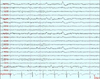
 A 10-second segment showing a well-formed and well-regulated alpha rhythm at 9 Hz. Note that it is very regular, rhythmic, waxing and waning, and posterior dominant. The contrast between the first and second halves of the page illustrates the reactivity of a normal alpha rhythm, with attenuation upon eye opening.
 Fleeting alpha. At times, as shown here, the alpha rhythm can be identified only in very brief bursts and often immediately after eye closure. If normal in frequency, this is normal.
 This is an example of an alpha rhythm with a wider distribution than is typical. If frequency and reactivity are normal, this is another variation of normal. A similar EEG pattern can be seen in patients in a coma (ie, alpha coma), but in these situations it is usually unreactive.
 This is an example of "slow alpha variant." The patient's alpha rhythm at 12 Hz is seen in the second half of the sample. The first half shows a subharmonic at half that frequency, and this is the "slow alpha variant."
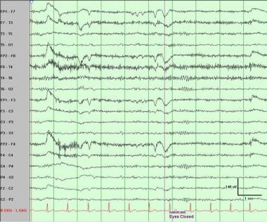
 An example of a typical normal alpha rhythm, showing clear attenuation upon eye opening (second half of page).
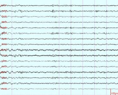
 Alpha rhythm with somewhat "spiky" or sharply contoured morphology. When fragmented (eg, in drowsiness), this can be misinterpreted as sharp waves.


Beta activity
Normal beta activity has the following characteristics:
-
Frequency (by definition) greater than 13 Hz - Common 18-25 Hz, less common 14-16 Hz, and rare 35-40 Hz [2]
-
Location - Mostly frontocentral but somewhat variable; some describe various types according to location and reactivity: generalized, precentral, and posterior
-
Morphology - Usually rhythmic, waxing and waning, and symmetric
-
Amplitude - Usually range of 5-20 mV
-
Reactivity - Most common 18- to 25-Hz beta activity enhanced during stages I and II sleep and tends to decrease during deeper sleep stages; central beta activity may be reactive (attenuates) to voluntary movements and proprioceptive stimuli; in infants older than 6 months, onset of sleep marked by increased beta activity in central and postcentral regions [3]
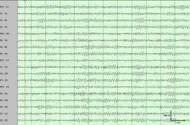
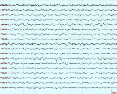
 A sample of awake EEG showing the normal or usual amount of beta activity. As shown here, beta activity is often easier to identify during relaxed wakefulness or early drowsiness.
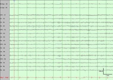
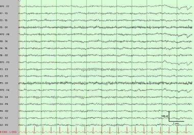
 This is the normal amount of beta activity, frontally predominant, with waxing and waning amplitude.


Mu rhythm
Characteristics of the mu rhythms are as follows:
-
Frequency of 7-11 Hz - Generally in alpha frequency band (8-12 Hz)
-
Location - Centroparietal area
-
Morphology - Archlike shape or like an "m"; most often asymmetric and asynchronous between the 2 sides and may be unilateral
-
Amplitude - Generally low to medium and comparable to that of the alpha rhythm
-
Reactivity - Most characteristic feature defining the mu rhythm; mu rhythm attenuates with contralateral extremity movement, the thought of a movement, or tactile stimulation; contrary to the alpha rhythm, does not react to eye opening and closing
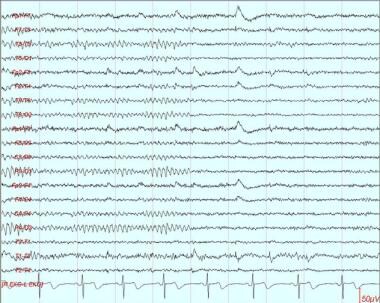
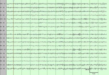
 Mu rhythm over the left (greater than right) central region. To be absolutely certain that this is a mu rhythm, reactivity should be tested. However, morphology (not absolutely typical but fairly so), frequency, and distribution strongly suggest that this is a mu rhythm.

The mu rhythm has been documented on subdural recording of both sensory and motor cortex and shows the same characteristics as that seen on surface EEG, including distribution, morphology, and reactivity. [4] Furthermore, some correspondence exists between functional mapping of sensorimotor function and somatotopic distribution of mu reactivity. [3, 5, 6]
Clinical Correlation
Alpha rhythm
Occasionally the alpha rhythm is of very low amplitude or even not identifiable. This is not abnormal. In addition to amplitude, other characteristics can vary somewhat without being abnormal, including morphology (eg, spiky), distribution (eg, widespread), and harmonic frequency (eg, slow or fast alpha variant).
Beta activity
In healthy individuals, beta activity commonly can be mildly different (< 35%) in amplitude between the 2 hemispheres, which may be caused by differences in skull thickness. Definite focal, regional, or hemispheric difference (at least 50%) in amplitude may be significant and may suggest either skull defect (side with higher amplitude) or a structural lesion (side with lower amplitude). [7] The amount and voltage of beta activity is enhanced by commonly used sedative medications (benzodiazepines, barbiturates).
Mu rhythm
Asymmetry, unilaterality, or asynchrony of the mu rhythm is generally not abnormal unless associated with other abnormalities. Very high-voltage mu activity may be recorded in the central regions over skull defects and may become sharp in configuration, and thus can be mistaken for epileptiform discharges. [8] When mu rhythm is detected in an EEG, it should be verified by testing its reactivity.
Patient Education
For excellent patient education resources, see eMedicineHealth's patient education article Electroencephalography (EEG).
Questions & Answers
Overview
What are the features of a normal awake EEG?
What are the characteristics of normal alpha rhythm on EEG?
What are the characteristics of normal beta activity on EEG?
What are the characteristics of the mu rhythms on EEG?
What are the variances in normal alpha rhythm on EEG?
What causes differences in normal beta activity on EEG?
What are the variances in normal mu rhythm on EEG?
-
A 10-second segment showing a well-formed and well-regulated alpha rhythm at 9 Hz. Note that it is very regular, rhythmic, waxing and waning, and posterior dominant. The contrast between the first and second halves of the page illustrates the reactivity of a normal alpha rhythm, with attenuation upon eye opening.
-
Fleeting alpha. At times, as shown here, the alpha rhythm can be identified only in very brief bursts and often immediately after eye closure. If normal in frequency, this is normal.
-
This is an example of an alpha rhythm with a wider distribution than is typical. If frequency and reactivity are normal, this is another variation of normal. A similar EEG pattern can be seen in patients in a coma (ie, alpha coma), but in these situations it is usually unreactive.
-
This is an example of "slow alpha variant." The patient's alpha rhythm at 12 Hz is seen in the second half of the sample. The first half shows a subharmonic at half that frequency, and this is the "slow alpha variant."
-
A sample of awake EEG showing the normal or usual amount of beta activity. As shown here, beta activity is often easier to identify during relaxed wakefulness or early drowsiness.
-
Mu rhythm over the left (greater than right) central region. To be absolutely certain that this is a mu rhythm, reactivity should be tested. However, morphology (not absolutely typical but fairly so), frequency, and distribution strongly suggest that this is a mu rhythm.
-
An example of a typical normal alpha rhythm, showing clear attenuation upon eye opening (second half of page).
-
This is the normal amount of beta activity, frontally predominant, with waxing and waning amplitude.
-
Alpha rhythm with somewhat "spiky" or sharply contoured morphology. When fragmented (eg, in drowsiness), this can be misinterpreted as sharp waves.