Overview Cardioembolic Stroke
The heart was established as an important source for the development of emboli when Gowers, in 1875, described a case of left middle cerebral artery and retinal artery emboli. Cardiogenic embolism accounts for approximately 20% of ischemic strokes each year. New diagnostic techniques (transesophageal echocardiography, cardiac magnetic resonance imaging) have allowed clinicians to better characterize well-established sources of embolism and to discover other potential etiologies of cardioembolic stroke (see the following image). [1]

Cardioembolic stroke is largely preventable, warranting efforts at primary prevention for major-risk cardioembolic sources. Once stroke due to cardiac embolism has occurred, the likelihood of recurrence is relatively high for most cardioembolic sources; consequently, secondary prevention is also important. [2]
For patient education information, see eMedicineHealth's Brain and Nervous System Center, as well as Stroke.
Go to Medscape Reference articles Acute Management of Stroke, Ischemic Stroke in Emergency Medicine, Hemorrhagic Stroke in Emergency Medicine, and Stroke Anticoagulation and Prophylaxis [Ischemic Stroke] for more information on these topics.
Overview of Cardiac Sources of Emboli
Definitions of major- and minor-risk cardioembolic sources
More than 20 specific cardiac disorders have been implicated in leading to brain embolism. Dividing cardiac sources of emboli into major- and minor-risk categories is clinically useful (see below).
Major-risk sources carry a relatively high risk of initial and recurrent stroke convincingly linked to a cardioembolic mechanism. When a major-risk cardioembolic source is present, efforts at primary prevention of stroke usually are indicated; stroke in patients with any of these causes is most often cardioembolic.
Minor-risk sources are frequent in the general population, and the associated risk of initial and recurrent stroke with any of these conditions is either low or uncertain. When a minor-risk cardioembolic source is present in a patient with cerebral ischemia, the etiologic role must be viewed with skepticism and considered in the context of other diagnostic information.
Sources of cardioembolic embolism include valvular diseases, left ventricular and atrial thrombi, cardiac tumors, and paradoxical emboli, as well as other sources. In the following subsections, the asterisks [*] indicate a major risk source; the daggers [†] indicate emboli originating in the venous circulation or right heart that cause ischemic stroke via abnormal cardiac or pulmonary shunting around the pulmonary capillary bed.
Valvular diseases
Valvular diseases include rheumatic mitral stenosis,*prosthetic valves,* infective endocarditis,* nonbacterial thrombotic (marantic) endocarditis* associated with malignancies and prothrombic states, calcific aortic stenosis, bicuspid aortic valves, mitral annulus calcification, myxomatous mitral valvulopathy with prolapse, inflammatory valvulitis (ie, Libman-Sacks endocarditis, Behçet disease, syphilis), and Lambl excrescences and/or strands.
Left ventricular thrombi
Left ventricular thrombi associated with prothrombotic states,* as well as ischemic heart disease,* acute myocardial infarction,* left ventricular akinesis or aneurysm,* nonischemic cardiomyopathies* (eg, idiopathic dilating, viral myocarditis –associated, echinococcal, peripartum, amyloid-associated, hypereosinophilia syndrome–associated, rheumatic myocarditis–associated, sarcoidosis-related, neuromuscular disorder–associated, alcoholism-related, catecholamine-induced, Chagas disease–associated, doxorubicin-induced, mitoxantrone-related, crack cocaine–related, cardiac oxalosis–associated), idiopathic hypertrophic subaortic stenosis,trauma(myocardial contusion), and ventricular noncompaction, contribute to cardioembolic emboli.
Left atrial thrombi
Atrial fibrillation,*atrial flutter,* sinus node dysfunction/atrial asystole, arrhythmias, atrial septal aneurysms, and the Chiari network are associated with left atrial thrombi.
Cardiac tumors
Atrial myxoma,*cardiac sarcoma, endocardial fibroelastoma, and metastatic disease are associated with cardiac tumors and cardioembolic emboli.
Paradoxical emboli
Parodoxica emboli have been associated with atrial septal defects, patent foramen ovale (PFO), ventricular septal defects, and pulmonary arteriovenous fistulas.
Other sources
Antiphospholipid antibodies, disseminated intravascular coagulation (DIC), essential thrombocytosis, and myeloproliferative diseases, as well as postcardiac catheterization, postvalvuloplasty, and esophageal-atrial fistula can also contribute as sources of cardioembolic emboli.
Overview of the Disease Process
Mechanism of cardioembolic stroke
The underlying mechanism of cardioembolic stroke is occlusion of cerebral vessels with debris from a cardiac source. An embolus may consist of platelet aggregates, thrombus, platelet-thrombi, cholesterol, calcium, bacteria, etc. Most embolic debris contains platelet aggregates.
However, no single mechanism is responsible for the development of cardiac emboli. The specific underlying cardiac disease determines the pathophysiology and natural history, and hence each cardioembolic source must be considered individually. Emboli secondary to chamber abnormalities (eg, atrial fibrillation, acute myocardial infarction) are induced mainly by stasis, whereas those secondary to valve involvement are the result of endothelial abnormalities with attachment of material (eg, platelets, bacteria) to their free borders.
The nature of the embolus differs depending on the source (eg, calcified particles from calcific valves, neoplastic cells from myxomas). This must be considered when choosing specific therapies; no single treatment covers the wide variety of heart disease that can cause embolism to the brain.
Distribution of cardiac emboli
Emboli from the heart are distributed evenly throughout the body according to cardiac output, but more than 80% of symptomatic or clinically recognized emboli involve the brain. Of emboli to the brain, approximately 80% involve the anterior circulation (ie, carotid artery territory), whereas 20% involve the vertebrobasilar distribution, proportional to the distribution of cerebral blood flow.
Once emboli have reached the cerebral circulation, they obstruct brain-supplying arteries, causing ischemia to the neurons and to the blood vessels within the area of ischemia. In contrast to thrombi, emboli are attached loosely to the vascular walls and thus commonly migrate distally. When this occurs, reperfusion of the damaged capillaries and arterioles allows blood to leak into the surrounding infarcted tissue. This explains the more frequent association of hemorrhagic infarction with cardiogenic embolism than with other causes of ischemic stroke. In the great majority of patients with hemorrhagic infarcts, the hemorrhagic transformation does not cause clinical worsening because the bleeding involves necrotic tissue.
In short, cardioembolic stroke is not one disease with a single natural history. Many different types of cardiac disorders lead to cardioembolic stroke, each with unique clinical features, risks of initial and recurrent stroke, and optimal therapy.
Prevalence of Cardioembolic Strokes
Approximately 20% of ischemic strokes are considered cardioembolic. The annual incidence is estimated at approximately 146,000 cases. In recent years, there has been an increased prevalence of cardioembolic stroke compared to stroke from large-artery atherosclerosis in high-income countries. This is likely the result of improved management of hypertension, hyperlipidemia, and hyperglycemia in these countries. [3, 4]
In the United States, atrial fibrillation represents the most common cause of cardioembolic stroke and is a major cause of stroke in the elderly. Worldwide, the estimated frequency of cardioembolic strokes varies from 12%–31% of ischemic strokes, depending on the criteria applied for definition, extent of the evaluation, and study design (see Table 1, below). Consistent geographic variation is not evident, and the frequency is likely similar throughout the world if adjusted for mean population age.
The risk of a cardioembolic event increases with age. The older the cohort, the higher the estimated frequency of cardioembolic stroke because of the rapidly increasing prevalence of atrial fibrillation in elderly persons. Elderly women are particularly affected, whereas black and Hispanic individuals reportedly have a lower frequency of cardioembolic strokes than white persons, reflecting the respective prevalence of atrial fibrillation among these groups.
Table 1. Frequency of Cardioembolic Stroke/All Ischemic Stroke (Open Table in a new window)
Frequency of Cardioembolic Stroke* |
|||
Study |
N |
Patient Age (Mean) |
Presumed Cardioembolic, % |
Oxfordshire, UK (1989) |
224 |
73 |
20† |
Melbourne, Australia (1989) |
353 |
-- |
19 |
Lausanne, Switzerland (1991) |
1311 |
65 |
18 |
Klosterneuburg, Austria (1992) |
365 |
68 |
19 |
Umea, Sweden (1992) |
953 |
72 |
31 |
Barcelona, Spain (1993) |
736 |
71 |
17 |
Guayaquil, Ecuador (1993) |
313 |
61 |
14 |
Giessen, Germany (1994) |
250†† |
-- |
17 |
Lund, Sweden (1994) |
166 |
73 |
28 |
Maastricht, Holland (1994) |
813 |
71 |
22 |
Paris, France (1995) |
250 |
-- |
29 |
Warsaw, Poland (1995) |
297 |
69 |
22 |
Barcelona, Spain (1997) |
1267 |
-- |
18 |
Taipei, Taiwan (1997) |
676 |
65 |
20 |
Riyadh, Saudi Arabia (1999) |
756 |
-- |
19 |
Athens, Greece (2000) |
885 |
70 |
38 |
Bensaçon, France (2000) |
1776 |
69 |
31 |
Santiago, Chile (2007) |
239 |
66 |
9.3 |
Mashhad, Iran (2007) |
1392 |
-- |
12 |
Aggregate |
13022 |
69 |
22 |
*Frequency of presumed cardioembolic stroke is a percentage of consecutive ischemic strokes, using each author's criteria. Criteria, design, and extent of evaluation varied substantially among studies. † 20% had a major embolic source. †† This study included transient ischemic attacks (TIAs). |
|||
Evaluation in Suspected Cardioembolic Stroke
Clinical features
Although not sufficiently sensitive or specific to establish the diagnosis, the following clinical features help to distinguish cardiogenic embolism from other mechanisms of cerebral ischemia and are useful to consider in patient management:
-
Decreased level of consciousness at onset of stroke
-
Neurologic symptoms of abrupt onset with maximal severity at onset
-
Rapid recovery from major hemispheric deficits ("spectacular shrinking deficit") due to reperfusion of the brain with early lysis of the embolus
-
Onset of symptoms after a Valsalva-provoking activity (enhancing right-to-left shunting in patients with a patent foramen ovale [PFO])
-
Symptoms reflecting involvement of different vascular territories of the brain
Neither seizure nor headache at the onset is a useful predictor of cardiogenic embolism.
Cardiogenic emboli (especially from chamber sources) do not often affect the deep penetrating arteries or manifest as a lacunar syndrome. Small cardiogenic emboli from valvular sources (eg, calcific aortic stenosis, infective endocarditis) can obstruct the small penetrating arteries, causing subcortical lacunar infarcts.
A combination of gene expression profiles in blood and a measure of infarct location on neuroimaging can be used to predict a cardioembolic, arterial, or lacunar cause in cryptogenic stroke. [5]
Physical findings
Findings that may suggest cardiogenic embolism include the following:
-
Evidence of cardiac atrial dysrhythmias (eg, atrial fibrillation, sinus node dysfunction)
-
Presence of cardiac murmurs (eg, mitral stenosis, calcific aortic stenosis)
-
Signs of congestive heart failure (eg, after acute myocardial infarction, nonischemic cardiomyopathies)
-
Recent myocardial infarction (highest cerebral embolism in the first 4 weeks of acute myocardial infarction)
-
Concomitant diseases (eg, systemic lupus erythematosus and Libman-Sacks endocarditis, neoplasia, marantic endocarditis)
Concomitant signs of systemic embolism – The probability of finding such signs in patients with suspected cardioembolic stroke is low (approximately 1%) for most cardioembolic sources.
Cryptogenic stroke and embolic stroke of undetermined source (ESUS)
About one-third of ischemic strokes remaining cryptogenic after standard evaluation. [6] Moreover, a global sample of patients with ischemic stroke showed that one-sixth met criteria for embolic stroke of undetermined source (ESUS), with additional ESUS patients likely for those with incomplete diagnostic evaluation. [7] This recent term of ESUS corresponds with strokes of likely distant embolic source despite workup with TTE, 24 hours cardiac rhythm monitoring, vascular imaging, and the exclusion of causes such as vasculitis. There is presumed overlap between cardioembolic, cryptogenic, and ESUS. For example, a thrombogenic atrial substrate in the absence of atrial fibrillation is one overlapping factor potentially shared by these classifications. However, up to one third of cryptogenic strokes and in up to one fourth of ESUS patients during long-term cardiac rhythm monitoring are revealed to have paroxysmal AF. [8] Despite unrevealing cardiac evaluation required for ESUS classification, the heart remains a likely underlying embolic source for these patients, [6] and further studies are needed to elucidate this relationship.
Major Risk Sources
Major risk sources carry a relatively high risk of initial and recurrent stroke convincingly linked to a cardioembolic mechanism.
Atrial fibrillation
The leading cause of cardioembolic stroke is atrial fibrillation (paroxysmal and chronic atrial fibrillation), especially in elderly individuals. This arrhythmia is present in approximately 1% of the US population, of which approximately 5% is in those older than 70 years, and it is found in up to 50% of all cardioembolic strokes. The lifetime risk of developing atrial fibrillation is 1-4 for adults older than 40 years. Atrial fibrillation conveys a 5-fold increased risk of stroke, and one sixth of all strokes may be attributable to atrial fibrillation. [9] Moreover, atrial fibrillation might be newly detected in nearly a quarter of patients with stroke or transient ischemic attack. [10]
Formerly associated with rheumatic valvular disease, atrial fibrillation is now related most frequently to hypertension and ischemic heart disease (ie, nonvalvular atrial fibrillation).
Stasis secondary to decreased contractility of the left atrium leading to thrombus formation in its appendage is the postulated mechanism (see the image below).
Transesophageal echocardiography (TEE) is more sensitive than transthoracic echocardiography for the visualization of the left atrium and its appendage (see the following video).
Not all atrial fibrillation–associated strokes are cardioembolic; in individual cases, excluding other potential causes of stroke such as intrinsic cerebrovascular disease or aortic atheroma is important.
The annual rate of stroke in atrial fibrillation varies widely from 0.5-12% per year depending on prevalence and combination of risk factors; thus, risk stratification is the first necessary step in choosing the best preventive therapy. Several clinical risk stratification schemes have been proposed to identify atrial fibrillation at high, moderate, or low risk; this is crucial for selecting which patients would benefit most and least from anticoagulation. The CHADS2 (congestive heart failure [CHF], hypertension, age >75 y, diabetes, stroke or transient ischemic attack [TIA]) classification scheme (see Table 2 below) is the most validated system and accurately stratifies stroke risk. [11, 12, 13]
Table 2. CHADS2 Stratification Schemes for Prevention of Stroke in Nonvalvular Atrial Fibrillation (Open Table in a new window)
Stroke Rates by CHADS2 Score |
||
CHADS2 Score |
Risk |
Stroke Rate Per Year, % |
0 |
Low |
1.9 |
1 |
Low |
2.8 |
2 |
Moderate |
4 |
3 |
High |
5.9 |
> 3 |
Very high |
> 8.5 |
Source: Gage BF, Waterman AD, Shannon W, et al. [13] |
||
Two randomized, controlled trials demonstrated that a strategy aimed at restoring (and maintaining) sinus rhythm neither improves the survival rate nor reduces the risk of stroke. In the Atrial Fibrillation Follow-up Investigation of Rhythm Management (AFFIRM) study, no evidence indicated that the rhythm control strategy protected patients from stroke. [14] In this trial, 4060 patients aged 65 years or older whose atrial fibrillation was likely to be recurrent and who were at risk for stroke were randomized to a strategy of rhythm control (cardioversion to sinus rhythm, plus a drug[s] to maintain sinus rhythm) versus a strategy of rate control (in which no attempt was made to restore or maintain normal sinus rhythm).
The AFFIRM study (and similar findings from the smaller Rate Control Versus Electrical Cardioversion [RACE] trial [15] ) led to the development of consensus guidelines advocating a rate-control strategy for most patients with atrial fibrillation.
Adjusted-dose warfarin (international normalized ratio [INR] 2-3) is associated with a 60% reduction in stroke incidence, whereas the efficacy of aspirin is modest (20% reduction). Low-dose warfarin (INR < 1.5), either alone or combined with aspirin, is not effective, highlighting the marginal benefit of warfarin when anticoagulation is not carefully regulated. The incidence of intracerebral hemorrhage, the most dreaded complication of warfarin therapy, is estimated to be 0.5% per year among elderly patients with atrial fibrillation and is sensitive to blood pressure control. When warfarin is given to elderly patients with atrial fibrillation, hypertension must be managed aggressively. [16]
Recommendations for primary and secondary prevention based on risk factor stratification are presented in Table 3 below. [17, 18]
Table 3. Risk-Based Approach to Antithrombotic Therapy in Patients With Atrial Fibrillation (Open Table in a new window)
Patient Features |
Antithrombotic Therapy |
Class of Recommendation |
Age < 60 y, no heart disease (lone AF) |
Aspirin (81-325 mg/d) or no therapy |
I |
Age < 60 y, heart disease but no risk factors* |
I |
|
Age 60-74 y, no risk factors* |
I |
|
Age 65-74 y with diabetes mellitus or CAD |
Oral anticoagulation (INR 2.0-3.0) |
I |
Age 75 y or older, women |
I |
|
Age 75 y or older, men, no other risk factors |
Oral anticoagulation (INR 2.0-3.0) or aspirin (81-325 mg/d) |
I |
Age 65 y or older, heart failure |
Oral anticoagulation (INR 2.0-3.0) |
I |
LV EF less than 35% or fractional shortening less than 25%, and hypertension |
I |
|
Rheumatic heart disease (mitral stenosis) |
I |
|
Prosthetic heart valves |
Oral anticoagulation (INR 2.0-3.0 or higher) |
I |
Previous thromboembolism |
I |
|
Persistent atrial thrombus on Tee |
IIA |
|
Source: ACC/AHA/ESC 2006 Guidelines for the management of patients with atrial fibrillation. [19] |
||
AF = atrial fibrillation; CAD = coronary artery disease; EF = ejection fraction; INR = international normalized ratio; LV = left ventricle; TEE = transesophageal echocardiography. |
||
* Risk factors for thromboembolism include heart failure (HF), (LV) ejection fraction less than 35%, and history of hypertension. |
||
In the setting of acute stroke secondary to atrial fibrillation, anticoagulation with heparin has not demonstrated more benefit than early treatment with aspirin. Initiate aspirin early, followed by warfarin as soon as the patient is medically stable; discontinue aspirin after therapeutic anticoagulation is achieved. [19]
In short, warfarin has demonstrated high efficacy in stroke prevention in patients with this common arrhythmia. Disadvantages include the increased risk of hemorrhagic complications and the need for close INR monitoring in these patients; thus, consider patient preferences along with risk stratification and absolute risk reduction offered by this therapy. Alternative approaches (eg, surgical ablation of atrial appendage) are the subjects of ongoing clinical investigation.
Rheumatic mitral stenosis
The incidence of this valvulopathy has decreased dramatically in recent decades in the United States, but rheumatic mitral stenosis remains an important problem in developing countries. Few estimates of absolute stroke rates or randomized comparison of different therapies are available, but because rheumatic mitral stenosis is generally associated closely with atrial fibrillation, anticoagulation with warfarin (INR 2-3) is usually recommended.
Sinus node dysfunction
Also known as sick sinus syndrome or, when associated with supraventricular tachyarrhythmias, brady-tachy syndrome, this arrhythmia usually occurs in elderly (>70 y) individuals. The annual risk of stroke is 5-10%. Atrial and dual-chamber pacing may reduce the stroke rate from sinus node dysfunction somewhat, but anticoagulation (INR 2-3) is still recommended for selected patients, such as those with associated atrial fibrillation; a lower target INR (eg, 1.6-2.5) may be tolerated better in these elderly patients.
Atrial flutter (sustained)
Sustained atrial flutter is an uncommon arrhythmia. Because of the close association of atrial fibrillation with appendage stasis, anticoagulation (INR 2-3) is advocated.
Prosthetic valves
Mechanical prosthetic valves carry an annual 2-4% risk of stroke, even in patients receiving anticoagulation. Permanent anticoagulation (INR 2.5-3.5) is mandatory. Bioprosthetic valves carry a lower annual risk rate (0.2-2.9%), and aspirin is usually recommended unless the patient has atrial fibrillation or evidence of atrial stasis.
Infective endocarditis
Of patients with infective endocarditis, 20% experience an embolic stroke, but infective endocarditis accounted for less than 1% of all causes of cerebral embolism in the Cerebral Embolism Stroke Registry. [20] Staphylococcus aureus is the infectious agent associated with the highest stroke rate, and mitral valve endocarditis is the most common source of emboli.
Antibiotic therapy reduces the embolic potential when administered in the acute phase. Anticoagulation is contraindicated because of unacceptable rates of hemorrhagic stroke due to either mycotic aneurysm rupture or septic arteritis. In patients with prosthetic valve endocarditis, the risk of thromboembolism is greater than the risk of intracranial hemorrhage; thus, anticoagulation is usually recommended if no evidence of hemorrhage is found on computed tomography (CT) scanning 24-48 hours after the stroke. The consensus is to start anticoagulation 7 days after the stroke. The role of antiplatelet therapy has not been established.
Nonbacterial thrombotic endocarditis
Associated with a variety of malignancies, nonbacterial thrombotic endocarditis has also been reported in patients with severe diseases such as septicemia and extensive burns. Mitral and aortic valves are affected most commonly, and embolic stroke is frequent. A prothrombotic state has been postulated as the precursor of emboli development. Treatment is directed toward control of the underlying disease, and heparin (intravenous in the acute stage, subcutaneous in the outpatient setting) is advocated for stroke prevention. Warfarin has failed to show any benefit.
Atrial myxomas
These are the most common cardiac tumors, usually located on the fossa ovalis. Atrial myxomas are believed to cause 1% of strokes in young individuals. Most of these lesions can be detected by transthoracic echocardiography; rarely, atrial myxomas are detected only by transesophageal echocardiography. Surgical excision is the treatment of choice.
Acute myocardial infarction
The incidence of stroke after acute myocardial infarction is approximately 2% in the first 3 months. Anterior myocardial infarctions with mural thrombus on transthoracic echocardiography have been recognized as predictive of stroke. Anticoagulation (INR 2-3) is recommended in these patients in the first 3 months, whereas antiplatelet therapy is advocated long term. The presence of congestive heart failure after myocardial infarction usually dictates treatment with warfarin indefinitely, although randomized comparisons with other therapies are ongoing. Low-output cardiac failure (ejection fraction < 30%) is also considered a high-risk situation, as is the presence of a large ventricular aneurysm on echocardiography.
Minor Risk Sources
Unlike major risk sources, minor-risk sources are frequent in the general population, with a low or uncertain associated risk of initial and recurrent stroke with any of these conditions.
Patent foramen ovale
Persistent connection between the right and left atrium has a prevalence of about 20% in the general population (see the following video). Screening for patent foramen ovales (PFOs) can be done reliably with contrast precordial echocardiography, which detects interatrial shunting, but transesophageal echocardiography (TEE) is required to document the PFO and more accurately determine its size, any associated atrial septal aneurysm, and the amount of shunting.
Although case-control studies have documented a higher frequency of PFO in young adults with cryptogenic ischemic stroke, it is present by chance association in at least 50% of cases in patients with stroke. The rate of stroke recurrence is 1-2% per year. Larger size, spontaneous right-to-left shunting, and associated atrial septal aneurysm are postulated to identify subgroups at high risk for recurrence.
PFO is not associated with increased risk of subsequent stroke or death among medically treated patients with cryptogenic stroke. However, in a study, both PFO and septal aneurysm (ASA) possibly increase the risk of subsequent stroke (but not death) in medically treated patients younger than 55 years. [21]
Mono et al identified several concurrent etiologies for recurrent cerebrovascular events in PFO patients, including large artery disease, small artery disease, cardioembolism, cerebral vasculitis, thrombophilic disorder, and antiphospholipid-antibody syndrome. [22]
In patients with a cryptogenic stroke and an atrial septal abnormality, evidence is insufficient to determine whether warfarin or aspirin is superior in preventing recurrent stroke or death, but minor bleeding is more frequent with warfarin.
Several randomized clinical trials demonstrate a lower risk of stroke recurrence in patients younger than 60 years with PFO closure. Patients were randomized into a PFO closure plus antiplatelet therapy group versus medical therapy alone after stroke. A meta-analysis of these RCTs observed a relative difference in rate of recurrent stroke, with a 43% lower rate among the surgical group. This meta-analysis showed no difference between these groups in major bleeding, serious adverse events, or all-cause mortality. It has been observed that PFO closure is associated with a subsequent increase in atrial fibrillation risk, higher rates of venous thromboembolism (eg, pulmonary embolism), and device-related complications. [23]
Most current guidelines do not recommend routine closure of a PFO in patients with cryptogenic stroke, instead recommending antiplatelet or anticoagulation therapy. In selected patients, with high RoPE scores, PFO closure may be appropriate. [24] It has been recommended, however, that in patients for whom anticoagulation is contraindicated, PFO closure receive consideration. [25] Since the RCTs included patients between 18 and 60 years of age, the potential benefits of PFO closure in patients older than 60 years are not yet known.
At present, PFO should not be considered the cause of stroke until other etiologies have been thoroughly excluded.
Atrial septal aneurysms
These aneurysms are areas of redundant atrial septal tissue that bulge alternatively into the right or left atrium. Atrial septal aneurysms have a high degree of association with other sources of embolism (mainly atrial fibrillation and PFO). However, there are insufficient data available to consider atrial septal aneurysm as an independent risk factor for stroke. When an atrial septal aneurysm coexists with a PFO or other source of embolism, anticoagulation is usually recommended, [21] but there are no randomized trials supporting this policy.
Mitral valve prolapse
Mitral valve prolapse is the most common valve disease in adults; its role as an independent risk factor for stroke is a controversial and evolving issue. In some population-based studies, the estimated prevalence has not been greater in patients who have had a stroke than in the general population. Long-term aspirin therapy is recommended, although its value has not been confirmed by randomized trials. Anticoagulation is reserved for failure of antiplatelet therapy.
Calcific aortic stenosis, bicuspid aortic valves, and mitral annular calcification
Systemic embolism is uncommon in isolated aortic valve disease. Calcific microemboli can be detected in retinal arteries in asymptomatic patients, possibly reflecting the fact that most cerebral emboli are asymptomatic. Clinical embolism often follows invasive cardiac procedures (ie, catheterization). Because of the calcific nature of the emboli, anticoagulation is not recommended, and antiplatelet therapy remains an empiric approach.
Mitral annular calcification is associated with advancing age, hypertension, and atherosclerosis, and it is rarely an embolic source.
Fibroelastomas and Lambl excrescences
Fibroelastomas are rare benign tumors located on the valves. Lambl excrescences are filiform outgrowths from the free borders of the cardiac valves and have been implicated as sources of embolism when they attain large size. Antiplatelet therapy is initiated, followed by surgery if aspirin fails. Surgical repair is reserved for patients who have stroke recurrence.
Diagnosis
No quantitatively valid clinical criterion standards exist for the diagnosis of cardioembolic stroke. The diagnosis is based on the triad of (1) identification of a potential cardiac source of embolism, (2) absence/exclusion of other potential sources of cerebral ischemia, and (3) consideration of clinical neurologic features, as described in Evaluation in Suspected Cardioembolic Stroke, above.
Seizures and epilepsy should also be considered in the differential diagnosis.
Recommended Tests
Blood cultures
If fever or leukocytosis is present, blood cultures for infective endocarditis are warranted.
CBC, coagulation profile, urinalysis, and chemistry and lipid panels
Before initiating antithrombotic therapy, a complete blood cell (CBC) count, platelet count, prothrombin time (PT) or international normalized ratio (INR) and activated partial thromboplastin time (aPTT), erythrocyte sedimentation rate (ESR), serum glucose, electrolytes, lipids, urinalysis are recommended, as well as plain radiography.
Blood dyscrasias studies
In patients with patent foramen ovale (PFO), determination of protein C, protein S antigen, and antithrombin III antigens and activities; factor V Leiden and activated protein C resistance; and prothrombin gene mutation are often recommended, particularly in patients with a history of venous thrombosis or a family history of unusual thrombosis. Several of these are acute phase reactants and can be artificially abnormal if obtained in the weeks following acute stroke. Protein C and S levels are suppressed by warfarin; antithrombin III levels and activity are suppressed by heparin.
Go to Blood Dyscrasias and Stroke for more information on this topic.
Echocardiography
Transthoracic echocardiography (TTE) is usually the initial cardiac imaging modality, and reliably detects left ventricular wall motion abnormalities, left ventricular thrombi, and (with contrast) interatrial shunts. [26] The following is a list of sources detected by TTE:
-
Left ventricular thrombus
-
Myxomatous mitral valvulopathy with prolapse
-
Mitral annulus calcification
-
Mitral stenosis
-
Aortic valve vegetations
-
Left ventricular wall motion abnormality (possible predictor of intracardiac thrombosis but not an embolic source per se)
-
Increased left atrial volume index (associated with cardioembolic stroke as well as predictor of AF in patients with ESUS). [27]
However, transesophageal echocardiography (TEE) provides more information about the atria than does TTE. In 40% of patients with normal TTE results, a cardiac source of embolism was detected by TEE, independent of age. More than 1 in 8 patients of any age with normal TTE results had a major cardiac risk factor revealed on TEE, in whom anticoagulation is warranted. [28, 29] The following are better detected on TEE compared with TTE:
-
Atrial septal aneurysm
-
Atrial septal defect
-
Patent foramen ovale (PFO)
-
Atrial myxoma
-
Atrial thrombus
-
Atrial appendage thrombus
-
Aortic arch atheroma/thrombi
-
Mitral valve vegetations (infective endocarditis, nonbacterial thrombotic endocarditis)
Cardiac CT
Cardiac CT has been shown to be a reliable alternative imaging modality to TEE in evaluating cardioembolic sources of ischemic stroke. Benefit includes avoiding the discomfort and risks associated with TEE. Additionally, cardiac CT can simultaneously diagnose coronary artery disease. [30]
However, current guidelines have yet to recommend cardiac CT for initial intracardiac structure evaluation or to assess cardioembolic stroke risk.
Cardiac MRI
Compared to cardiac CT, cardiac MRI has the advantage of no radiation exposure and avoidance of iodinated contrast media. However, cost and increased examination time are disadvantages. [30]
Indications for cardiac magnetic resonance imaging (MRI) include the following:
-
Patients with a transthoracic echocardiography (TTE) result that is questionable for the presence of left ventricular (LV) thrombus
-
Further evaluation of a cardiac mass seen on a TTE
-
Patients who cannot tolerate transesophageal echocardiography (TEE) and/or cannot undergo TEE secondary to medical reasons
-
Patients with inconclusive TEE results
-
Suspected false-negative TEE results, in which cardiac MRI can adequately image potentially missed sources of embolus such as LV thrombus, cardiac masses, aortic plaque, or left atrial appendage (LAA) thrombus
CT Scanning, MRI, Angiography
Hemorrhagic infarct or multiple arterial infarcts (not lacunar) on computed tomography (CT) scans or magnetic resonance images (MRIs) and/or an embolus "in transit" on angiography are radiologic findings that, when associated with clinical features, are suggestive of cardioembolic stroke.
Electrocardiography
An electrocardiogram (ECG) can demonstrate atrial arrhythmias, myocardial infarction.
Ambulatory ECGs are indicated for elderly patients in whom paroxysmal atrial fibrillation is suspected (eg, history of palpitations, enlarged left atrium on echocardiography). [31] In elderly patients with cryptogenic hemorrhagic cortical infarctions or other cardioembolic features, many clinicians obtain ambulatory ECG monitoring seeking occult atrial fibrillation that would necessitate anticoagulation.
One study suggests that patients presenting with cerebral ischemia without atrial fibrillation may benefit from prolongation of Holter monitoring (more than 7 days). Those patients who received the Holter monitor for up to 7 days had a higher detection rate of paroxysmal atrial fibrillation, which lead to a change in therapy. This prolonged use of the monitor should be considered for those patients with unexplained cerebral ischemia. [32]
Moreover, the 30-Day Cardiac Event Monitor Belt for Recording Atrial Fibrillation after a Cerebral Ischemic Event (EMBRACE) trial observed atrial fibrillation in 16.1% of patients with 30-day monitoring period compared to the incidence of 3.2% in the control group receiving a 24-hour Holter monitoring. [33]
Despite atrial fibrillation detection directly impacting medical stroke prevention, prolonged ECG monitoring is largely left to physician discretion. [8] Analysis of prolonged monitoring cost-effectiveness supports the practice among carefully selected patients—with 30-day ECG monitoring likely being highly cost-effective, and 14-day protocols providing an effective alternative for lower-risk patients. [34] Furthermore, the CRYSTAL-AF trial found that implantable long-term cardiac monitors improved detection of 30-second episode of paroxysmal atrial fibrillation compared to conventional follow up (by factors of 6.4 and 7.3 at 6 months and 12 months, respectively). In one study with ESUS patients monitored with an implantable loop recorder, atrial fibrillation was found within a year in about 25% of patients. [35]
Medical Management in Cardioembolic Stroke
Antiplatelet and anticoagulant therapies are mainstays in the prevention of cardioembolic stroke. Consider the absolute rate of stroke associated with each source, the risk-benefit relationship of each therapy, and each patient's preferences.
Consultations
Management of anticoagulation at a specialized clinic (if available) is recommended, based on the results of several studies.
Consider consultation with a cardiologist to evaluate the management of arrhythmias and structural abnormalities of the heart, as well as consider consultation with a hematologist when the possibility of a prothrombotic state is suspected, typically in patients with patent foramen ovale who have a history of venous thromboembolism or a family history of thrombosis.
Warfarin
Warfarin is first-line anticoagulant treatment for most causes of cardioembolic stoke. Among antiplatelet agents, aspirin has been proved in clinical trials to reduce risk of cardioembolic stroke. However, clopidogrel (Plavix) plus aspirin did not show efficacy compared with warfarin in patients with atrial fibrillation (ACTIVE W trial), [36] although in patients who were not candidates for vitamin K antagonists, the combination of clopidogrel plus low-dose aspirin was superior to low-dose aspirin alone, albeit with a slightly higher risk of bleeding (ACTIVE A trial). [37]
Warfarin, corrected to an international normalized ratio (INR) of 2-3, is still superior to even combination antiplatelet therapy. Combination antiplatelet therapy with clopidogrel and aspirin is appropriate for cardioembolic (atrial fibrillation–related) stroke patients who cannot tolerate warfarin. For noncardioembolic stroke patients, however, clopidogrel monotherapy is preferred over combination therapy, based on the Management of Atherothrombosis with Clopidogrel in High-Risk Patients (MATCH) with Recent Transient Ischemic Attacks (TIAs) or Ischemic Stroke study. [38]
A meta-analysis of several randomized trials indicated that in patients with acute cardioembolic stroke, early anticoagulation is associated with a nonsignificant reduction in recurrence of ischemic stroke, no substantial reduction in death and disability, and increased intracranial bleeding. Early aspirin followed by vitamin K antagonists for long-term secondary prevention is reasonable. [39]
Optimization of warfarin therapy
Randomized trials have demonstrated that the efficacy of warfarin anticoagulation is related directly to how carefully it is used. Inadequate anticoagulation produces minimal or no protection, whereas supratherapeutic anticoagulation may increase the risk of serious hemorrhagic complications. To optimize the level of anticoagulation, interactions with concomitant medications known to potentiate or inhibit the anticoagulant effect should be considered.
Monitor INRs weekly initially, then at least monthly.
Dietary and activity limitations during warfarin therapy
Patients on warfarin therapy should be provided with a list of vitamin K–containing foods (eg, broccoli, avocado, other green vegetables) that inhibit warfarin’s anticoagulant effects. In addition, most clinicians severely limit or proscribe consumption of alcoholic beverages in patients taking warfarin.
Limitations on physical activities (eg, contact sports, skiing) should be discussed with patients on warfarin treatment.
Direct thrombin inhibitors and Factor Xa inhibitors
Apixaban, dabigatran, and rivaroxaban, and edoxaban are alternatives to warfarin for high-risk patients (including those with a history of stroke) who have atrial fibrillation. [40, 41, 42, 43, 44] Apixaban, edoxaban, and rivaroxaban inhibit Factor Xa, whereas dabigatran is a direct thrombin inhibitor. Apixaban and dabigatran were shown to be superior to warfarin for the prevention of stroke and systemic embolism, while rivaroxaban and edoxaban were was shown to be equivalent. The rates of intracranial hemorrhage are lower for all 34 drugs compared with warfarin. These medications have not been compared against each other.
Dabigatran
The RE-LY study evaluated the efficacy and safety of 2 different doses of dabigatran (Pradaxa) relative to warfarin in more than 18,000 patients with atrial fibrillation. Patients were randomized to 1 of 3 arms: (1) adjusted dose warfarin, (2) dabigatran 110 mg bid, or (3) dabigatran 150 mg bid. Dabigatran 110 mg was noninferior to warfarin for the primary efficacy endpoint of stroke or systemic embolization, while dabigatran 150 mg was significantly more effective than warfarin or dabigatran 110 mg. Major bleeding occurred significantly less often with dabigatran 110 mg than warfarin; dabigatran 150 mg had similar bleeding to warfarin. [45]
Dabigatran, a competitive, direct thrombin inhibitor, was approved by the US Food and Drug Administration in 2010 for prevention of stroke and thromboembolism associated with nonvalvular atrial fibrillation. The dose is 150 mg PO bid (decrease to 75 mg PO bid with renal impairment). When converting from warfarin, discontinue warfarin and initiate dabigatran when INR < 2.0.
Guidelines from the American College of Cardiology Foundation (ACCF)/American Heart Association (AHA)/Heart Rhythm Society (HRS) on atrial fibrillation have been updated to include the use of oral direct thrombin inhibitors (ie, dabigatran). [46] The guidelines include a class Ib recommendation (ie, treatment is useful/effective based on a single randomized trial) for dabigatran. The guidelines recommend dabigatran may be used as an alternative to warfarin for the prevention of stroke and systemic thromboembolism in patients with paroxysmal-to-permanent atrial fibrillation and risk factors for stroke or systemic embolization. Patients with atrial fibrillation who are not candidates include those with prosthetic heart valves or hemodynamically significant valve disease, severe renal failure (creatinine clearance ≤15 mL/min), or advanced liver disease.
Apixaban
Apixaban (Eliquis) was approved by the FDA in December 2012. Approval was based on 2 clinical trials. The ARISTOTLE (Apixaban for Reduction in Stroke and Other Thromboembolic Events in Atrial Fibrillation) trial compared apixaban with warfarin for the prevention of stroke or systemic embolism in patients with atrial fibrillation and at least one additional risk factor for stroke. Results showed that apixaban was superior to warfarin in preventing stroke or systemic embolism, caused less bleeding, and resulted in lower mortality. [40]
Edoxaban
Edoxaban (Savaysa) was approved by the FDA in January 2015 to reduce the risk of stroke and systemic embolism in patients with nonvalvular atrial fibrillation. In the ENGAGE AF-TIMI 48 trial (n=21,105), edoxaban was noninferior to warfarin with respect to the prevention of stroke or systemic embolism. Edoxaban was associated with significantly lower rates of major bleeding (P < 0.001) and death from cardiovascular causes (P=0.01%) compared with warfarin. [44]
Surgical Care
Alternatives to medical therapy include the surgical maze operation or endovascular catheter-guided ablation of arrhythmias to reduce the risk of cardiac embolism. Also, in selected circumstances, thrombectomy and/or valve replacement to remove embolic sources (eg, in the context of endocarditis) are also surgical options for appropriate patients. Left atrial appendage occlusion with transcatheter devices is another surgical approach in patients with atrial fibrillation. The Watchman device for left atrial appendage closure has been shown to be noninferior to VKAs for the composite primary endpoint of all-cause stroke, systemic embolism, or cardiovascular death in the PROTECT-AF and PREVAIL trials. Additionally, left atrial appendage closure provided a significant reduction in life-threatening bleeding compared to the VKAs. Recently, the PRAGUE-17 trial showed that left atrial appendage closure with transcatheter devices is noninferior to DOAC treatment in preventing all-cause stroke and clinically significant bleeding events. However, potential device complications remain with LAAC, warranting further refinement in operator technique and device technology.
As mentioned above, the endovascular closure patent foramen ovale in cryptogenic stroke has recently been shown to lower the risk of stroke recurrence in patients younger than 60 years. This included the RESPECT, CLOSE, and REDUCE trials.
Cardioembolic Stroke Outcomes
In general, cardioembolic strokes have a worse prognosis and produce larger and more disabling strokes than other ischemic stroke subtypes. This general observation is derived from emboli originating in cardiac chambers, which on average are of large size (eg, atrial appendage, ventricular thrombi).
Special Considerations
The underuse of warfarin in patients with atrial fibrillation has been a high-profile issue and the source of many lawsuits. Consequently, as part of quality control monitoring, the Health Care Financing Administration (HCFA) has included the use of warfarin at the time of discharge in patients with atrial fibrillation.
When atrial fibrillation is identified, anticoagulation should be considered carefully, and, if not prescribed (and perhaps 50% or more of outpatients with atrial fibrillation do not benefit sufficiently to warrant it), the reason should be recorded in the medical record (eg, estimated low risk for stroke based on CHADS2, warfarin too risky because of high bleeding risk, no access to reliable anticoagulation monitoring, patient declined after explanation of risks and benefits) in case of subsequent stroke. As discussed, the CHADS2 scheme for estimation of risk stratification is the best validated risk assessment tool (see Atrial Fibrillation in Major Risk Sources).
It is some authors' view that risk stratification to estimate stroke risk should be an integral part of the decision to begin anticoagulation treatment in these patients.
-
Sources of cardioembolic stroke.
-
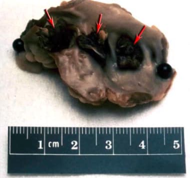
Cardioembolic stroke. Photo of left atrial thrombus.
-
Cardioembolic stroke. Streaming video: Mobile left atrial thrombus on echocardiography. Notice periodic bulging of the thrombus in the left atrium. ECG on this patient indicated atrial fibrillation.
-
Cardioembolic stroke. Streaming video: Patent foramen ovale. Notice the diversion of contrast from the right to the left atrium due to abnormal communication between the 2 chambers.
Tables
Frequency of Cardioembolic Stroke* |
|||
Study |
N |
Patient Age (Mean) |
Presumed Cardioembolic, % |
Oxfordshire, UK (1989) |
224 |
73 |
20† |
Melbourne, Australia (1989) |
353 |
-- |
19 |
Lausanne, Switzerland (1991) |
1311 |
65 |
18 |
Klosterneuburg, Austria (1992) |
365 |
68 |
19 |
Umea, Sweden (1992) |
953 |
72 |
31 |
Barcelona, Spain (1993) |
736 |
71 |
17 |
Guayaquil, Ecuador (1993) |
313 |
61 |
14 |
Giessen, Germany (1994) |
250†† |
-- |
17 |
Lund, Sweden (1994) |
166 |
73 |
28 |
Maastricht, Holland (1994) |
813 |
71 |
22 |
Paris, France (1995) |
250 |
-- |
29 |
Warsaw, Poland (1995) |
297 |
69 |
22 |
Barcelona, Spain (1997) |
1267 |
-- |
18 |
Taipei, Taiwan (1997) |
676 |
65 |
20 |
Riyadh, Saudi Arabia (1999) |
756 |
-- |
19 |
Athens, Greece (2000) |
885 |
70 |
38 |
Bensaçon, France (2000) |
1776 |
69 |
31 |
Santiago, Chile (2007) |
239 |
66 |
9.3 |
Mashhad, Iran (2007) |
1392 |
-- |
12 |
Aggregate |
13022 |
69 |
22 |
*Frequency of presumed cardioembolic stroke is a percentage of consecutive ischemic strokes, using each author's criteria. Criteria, design, and extent of evaluation varied substantially among studies. † 20% had a major embolic source. †† This study included transient ischemic attacks (TIAs). |
|||
Stroke Rates by CHADS2 Score |
||
CHADS2 Score |
Risk |
Stroke Rate Per Year, % |
0 |
Low |
1.9 |
1 |
Low |
2.8 |
2 |
Moderate |
4 |
3 |
High |
5.9 |
> 3 |
Very high |
> 8.5 |
Source: Gage BF, Waterman AD, Shannon W, et al. [13] |
||
Patient Features |
Antithrombotic Therapy |
Class of Recommendation |
Age < 60 y, no heart disease (lone AF) |
Aspirin (81-325 mg/d) or no therapy |
I |
Age < 60 y, heart disease but no risk factors* |
I |
|
Age 60-74 y, no risk factors* |
I |
|
Age 65-74 y with diabetes mellitus or CAD |
Oral anticoagulation (INR 2.0-3.0) |
I |
Age 75 y or older, women |
I |
|
Age 75 y or older, men, no other risk factors |
Oral anticoagulation (INR 2.0-3.0) or aspirin (81-325 mg/d) |
I |
Age 65 y or older, heart failure |
Oral anticoagulation (INR 2.0-3.0) |
I |
LV EF less than 35% or fractional shortening less than 25%, and hypertension |
I |
|
Rheumatic heart disease (mitral stenosis) |
I |
|
Prosthetic heart valves |
Oral anticoagulation (INR 2.0-3.0 or higher) |
I |
Previous thromboembolism |
I |
|
Persistent atrial thrombus on Tee |
IIA |
|
Source: ACC/AHA/ESC 2006 Guidelines for the management of patients with atrial fibrillation. [19] |
||
AF = atrial fibrillation; CAD = coronary artery disease; EF = ejection fraction; INR = international normalized ratio; LV = left ventricle; TEE = transesophageal echocardiography. |
||
* Risk factors for thromboembolism include heart failure (HF), (LV) ejection fraction less than 35%, and history of hypertension. |
||

What would you like to print?
- Overview Cardioembolic Stroke
- Overview of Cardiac Sources of Emboli
- Overview of the Disease Process
- Prevalence of Cardioembolic Strokes
- Evaluation in Suspected Cardioembolic Stroke
- Major Risk Sources
- Minor Risk Sources
- Diagnosis
- Recommended Tests
- Echocardiography
- Cardiac CT
- Cardiac MRI
- CT Scanning, MRI, Angiography
- Electrocardiography
- Medical Management in Cardioembolic Stroke
- Warfarin
- Direct thrombin inhibitors and Factor Xa inhibitors
- Surgical Care
- Cardioembolic Stroke Outcomes
- Special Considerations
- Show All
- Media Gallery
- Tables
- References


