Overview
The phrases "electroencephalogram (EEG) variant waves" or "normal EEG variants" refer to waves that are rare or unusual but not generally abnormal. They may be unusual in shape or in distribution. These variant waveforms include waveforms and patterns that are rare or unusual but are known to be generally benign. They can include complex wave mixtures that can appear unusual and can confuse the casual reader (for example, wave harmonics) as well as artifacts or electrical disturbances from structures that are not in or part of the brain and do not affect the brain or its function but appear in the EEG tracing. [1, 2]
Realizing that the EEG is fundamentally a recording of the electrical activity of a highly complex system is important. Moreover, this electrical activity is being recorded at the scalp and, although accurate, it is not the same as the recording of activity directly from the surface of brain or at the single cell level. Therefore, from person to person, a variation is expected of waveforms and occasional atypical waveforms that still fall within what would be considered normal. [3, 4, 5]
Odd-Looking Waveforms
The following waveforms are generally benign:
-
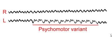
Psychomotor variant (rhythmic harmonic theta)
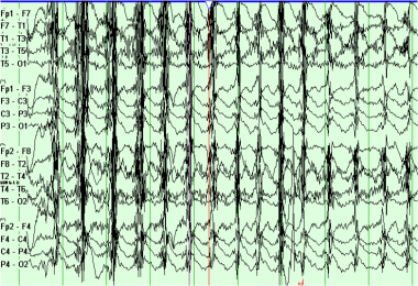
This is an unusual form that occurs as asymmetrical runs of theta or delta activity primarily in the temporal regions, lasting for a few seconds or as long as 30-45 seconds (see the following image).
This waveform is clearly a harmonic of 2 or more rhythms. The waves often have a bifid appearance.
It starts suddenly on 1 side and lasts for several seconds before terminating suddenly. This behavior resembles a seizure discharge, hence the name "psychomotor variant."
Generally considered benign, this waveform does not correlate with seizure disorder. It is best seen on a prolonged EEG and tends to be more common in children and young people. [6]
-
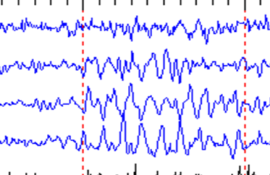
Mu (rhythm en arceau or wicket rhythm)
This waveform is recognized easily and has no pathological significance. The naive may not recognize it and assume it to be abnormal.
The mu waveform occurs in the central regions in the awake patient. It is seen best if a bone defect underlies the electrodes.
It can be markedly asymmetrical.
Often in the alpha range frequency, it has rounded positive aspects on 1 side and sharpened negative aspects on the other (see image shown below). [7]
It is not blocked by eye opening.
It becomes obvious when the alpha disappears (ie, alpha blocking).
Associated with fast activity, mu has a frequency about half that of fast activity.
The most classical feature of mu waveform is that it blocks with motor activity of the contralateral body (or the thought of such movement).
-
14- and 6-Hz waves
In these waves, the 2 frequencies are intimately intertwined and the complexes occur in bursts.
They generally are thought to be clinically insignificant.
They occur in healthy children and adolescents. Some claim that they are best seen in referential recordings during sleep. [6]
-
Small sharp spikes of sleep (SSS)
This waveform also is known as benign epileptiform transients of sleep (BETS).
These sharp, small waves occur on 1 or both sides (often asynchronously), especially in the temporal and frontal regions.
Rarely seen in children, they are seen most often in adults and the elderly. [7, 8] They can occur in epileptic patients but often are seen in healthy individuals. They can be regarded as a probable normal variant.
-
6-Hz spike and wave (phantom spike and wave)
These occur as bursts of miniature spike and wave complexes or runs of such complexes at 6 Hz rather than the usual 2-4 Hz.
Their significance is debated, but generally those occurring in the posterior head regions are regarded as benign.
Seen at all ages (but especially in adults), they often are confused with 14- and 6-Hz waves and may merge into them. [8]
The anterior variety are regarded by some as consistent with epilepsy, but further studies are needed to confirm this.
-
Wicket spikes
Wicket spikes are another waveform that is without pathological significance, occurring almost exclusively in adults. [8]
They are thought to be due to harmonics of 2 or more waveforms that combine to form pseudospikes.
Like wicket rhythm, they have rounded aspects to 1 side and sharp points to the other, giving the appearance of spikes or sharp waves.
When these are especially large, they may appear similar to pathological spikes. However, they generally can be distinguished by their morphology and at times by their defined background rhythms, which are harmonizing.
They can be seen either in wakefulness or sleep in the anterior or temporal head regions.
-
Subclinical rhythmic EEG discharges in adults
The typical pattern of subclinical rhythmic EEG discharges in adults (SREDA) consists of theta rhythm occurring in a widespread manner, maximal over the parietal and posterior temporal regions, and lasting for a few seconds to a minute without clinical signs or symptoms. It is described as "not evolving" and appears quite stable for its duration.
Another unusual variant is made up of predominantly delta frequencies as well as notched waveforms with a frontal distribution and a more prolonged duration that even includes sleep.
No significant clinical differences have been found between groups that had the atypical pattern and those with the typical pattern.
Although the mechanism of SREDA is not understood, it appears to represent a benign EEG phenomenon that should be distinguished from seizure discharges. [7]
-
Rhythmic midline theta
This is a focal rhythm maximal at the midline, most prominently at Cz, which occasionally may spread to adjacent electrodes. It has a frequency of 5-7 Hz and typically has an arciform, spiky, mulike appearance. It waxes and wanes, can appear during wakefulness or drowsiness, and is usually reactive to eye opening or limb movement. Although initially described in association with temporal lobe epilepsy, it probably represents a normal variant. [7]
Artifacts
These waveforms are produced by an electrical alteration of the recorded brain wave. It is important to realize that because the EEG is very sensitive to the recording of microvoltages, it is common to see the electrical effect of factors that occur outside of the skull. Although these disturbances do not represent brain abnormalities, their recognition is important because they can distort the recording of brain waves. [9]
Forehead, jaw, and eyelid muscle movements can cause artifacts by moving the electrodes. Movements in the surroundings produce disturbances by altering the ambient electrical fields. Moreover, the tongue and eyes have their own dipole electric charge. Therefore, their movement can get recorded by the electrodes. This, in turn, can affect the recorded brain wave.
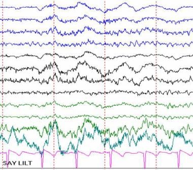 Artifact produced by tongue movement. The person was asked to say LILT, which caused the waves to slightly undulate.
ECG may contaminate the recording.
Sweating produces electrical disturbances by shorting electrode pairs.
Other sources of artifacts include ambient electrical waves from respirators, intravenous pump machines, televisions, and other electrical equipment.
-
Many are recognized by their characteristic appearance on the tracing, but others are identified by direct inspection and reported by the technologist or identified on the video tracing in video-EEG recording.
-
Artifacts show great variation because of their protean origin.
-
They may be single waves or recurrent waves (eg, intravenous infusion running), while others are prolonged disturbances (eg, sweating).
-
The specifics of each are not reviewed here because of their variability.
-
The following can be regarded as clinically insignificant:
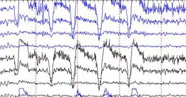
Chewing produces spurious spike and wave runs in the frontal and temporal regions from the temporalis muscles (see image below).
Sweating produces very slow waves, because the salt solution shorts out pairs of adjacent electrodes.
Eye movements occur with blinking and result from the electrical charge of the eye itself (see image below). They are frontal. Nystagmus also produces artifactual waves.
ECG and pulse motion produce unusual waveforms. ECG produces small spikes that are recurrent and are especially evident in the monopolar montages. Pulse artifacts occur with recurrent motion of electrodes sitting over a bounding pulse, which alters the contact with the skin. This causes a rhythmic disturbance that is synchronous with the heart but rounded rather than sharp in form.
Tremor and movement of the head or body may cause electrodes to move.
Electrode pops or movements can produce sudden, recurrent, or continuous electrical waves.
Electrical fields result from electrical devices and televisions.
ICU special waveforms may result from respirator-induced movements, intravenous drips and drip pumps, electrical fields, or cautery (eg, Bovie) in the operating room or emergency department.
Harmonics
EEG is a complex summation of many frequencies.
-
Different frequencies sometimes add to or cancel each other, creating odd waveforms or fluctuations of waveforms.
-
Pseudospikes or pseudoslow waves may be seen with intermixing of waves.
-
Many fascinating patterns have been generated by mixing artificially created computer-generated frequencies. These waveforms have the significance of the basic waveforms that underlie the patterns.
Resources
For related information on EEGs, see the following Medscape Reference articles:
Questions & Answers
Overview
What are psychomotor EEG variants?
What are 14- and 6-Hz EEG variant waves?
What are small sharp spikes of sleep (SSS) EEG variants?
What are 6-Hz spike and wave (phantom spike and wave) EEG variants?
What are wicket spikes EEG variants?
What are subclinical rhythmic EEG discharges in adults?
What are rhythmic midline theta EEG variants?
What are the harmonics of EEG?
-
Example of the psychomotor variant (rhythmic harmonic theta).
-
Example of mu waveforms.
-
Artifact produced by tongue movement. The person was asked to say LILT, which caused the waves to slightly undulate.
-
Example of EEG chewing artifact.
-
EEG artifact of eye blinking.