Continually Updated Clinical Reference
 
 
  All Sources     eMedicine     Medscape     Drug Reference     MEDLINE
 
eMedicine - Primary Torsion Dystonia : Article by

Quick Find
Authors & Editors
Introduction
Clinical
Differentials
Workup
Treatment
Medication
Follow-up
Multimedia
References

Related Articles
Apraxia and Related Syndromes

Chorea Gravidarum

Cortical Basal Ganglionic Degeneration

Dopamine-Responsive Dystonia

Hallervorden-Spatz Disease

Huntington Disease

Inherited Metabolic Disorders

Lysosomal Storage Disease

Neuroacanthocytosis

Neuroacanthocytosis Syndromes

Neuronal Ceroid Lipofuscinoses

Parkinson Disease

Parkinson Disease in Young Adults

Parkinson-Plus Syndromes

Prion-Related Diseases

Progressive Supranuclear Palsy

Striatonigral Degeneration

Systemic Lupus Erythematosus


Patient Education
Procedures Center

BOTOX® Injections Overview

BOTOX® Injections Preparation


Author: Rowena Emilia Tabamo, MD, Associate Director for Clinical Research, Institute for Neurodegenerative Disorders

Rowena Emilia Tabamo is a member of the following medical societies: American Academy of Neurology and Movement Disorders Society

Coauthor(s): Michele Tagliati, MD, Division Chief of Movement Disorders, Associate Professor, Department of Neurology, Mount Sinai School of Medicine; Susan B Bressman, MD, Chairperson, Department of Neurology,, Department of Neurology, St. Luke's-Roosevelt Hospitals; Professor, Beth Israel Deaconess Medical Center; Chairperson, Department of Neurology, Albert Einstein College of Medicine

Editors: Stephen T Gancher, MD, Adjunct Associate Professor, Department of Neurology, Oregon Health Sciences University; Francisco Talavera, PharmD, PhD, Senior Pharmacy Editor, eMedicine; Nestor Galvez-Jimenez, MD, Program Director of Movement Disorders, Department of Neurology, Division of Medicine, Director of Neurology Residency Training Program, Cleveland Clinic Florida; Selim R Benbadis, MD, Professor, Director of Comprehensive Epilepsy Program, Departments of Neurology and Neurosurgery, University of South Florida School of Medicine, Tampa General Hospital; Nicholas Y Lorenzo, MD, Chief Editor, eMedicine Neurology; Consulting Staff, Neurology Specialists and Consultants

Author and Editor Disclosure

Synonyms and related keywords: PTD, DMD, dystonia musculorum deformans, movement disorder, Oppenheim disease, Oppenheim's disease, primary torsion dystonia, idiopathic torsion dystonia, DYT1 gene

Background

Dystonia is a syndrome of sustained muscle contractions, usually producing twisting and repetitive movements or abnormal postures (Fahn, 1987).

In 1908, Schwalbe first described primary, or idiopathic, torsion dystonia in a Jewish family, and in 1911, Oppenheim termed this dystonia musculorum deformans (DMD). Initially believed to be a manifestation of hysteria, idiopathic torsion dystonia gradually became established as a neurologic entity with a genetic basis. DMD and Oppenheim disease are terms now used for childhood- and adolescent-onset dystonia due to the DYT1 gene.

With the recent mapping of genes for idiopathic torsion dystonia and identification of a gene for early onset dystonia, the description primary, or idiopathic, dystonia has evolved; it now may be viewed as secondary to or symptomatic of an identified cause. However, continuing to use primary torsion dystonia to classify a group of dystonias as a clinically and genetically heterogeneous group of movement disorders is justifiable because dystonia is the primary and sole abnormality attributable to the condition, and degeneration on pathologic examination is not clearly established.

Primary torsion dystonia may be focal, segmental, multifocal, or generalized, depending on which anatomic sites are involved (see Table 1).

Table 1. Anatomic Distribution of Primary Torsion Dystonia

Focal Single Body Site
Segmental Contiguous body regions
Multifocal Multiple, noncontiguous body sites
Generalized Leg involvement with other body sites
Hemidystonia Unilateral

Pathophysiology

Although secondary forms of dystonia are frequently associated with structural lesions of the basal ganglia and thalamus, no consistent histologic or biochemical findings are noted in primary torsion dystonia. However, perinuclear inclusion bodies have been described in the midbrain reticular formation and in the periaqueductal gray matter in 4 patients in whom DYT1 was clinically documented and genetically confirmed (McNaught, 2004).

No discernible abnormalities are seen on current structural neuroimaging studies. Abnormal brain networks have been described in different functional imaging studies; this is substantial evidence implicating dysfunction in dopaminergic pathways in the pathophysiology of primary torsion dystonia (Eidelberg, 1998; Perlmutter, 2004).

Besides motor control difficulties, defective sensory processing is described (Kaji, 2004).

Current models of basal ganglia circuitry have been adapted and suggesting dysfunction at the basal ganglia level. These aberrations involve the direct and indirect pathways and result in impaired inhibition at the cortical level with consequent loss of normal inhibitory reflexes at the level of the brainstem and spinal levels.

See Image 1 for a diagram of the basal ganglia circuitry dysfunction in dystonia.

Frequency

United States

The relative frequencies of primary and secondary forms of dystonia are not known.

In the Movement Disorder Center at Columbia Presbyterian Medical Center, 71% of more than 3000 patients with the disorder had primary torsion dystonia; the remaining 29% had secondary etiologies, with tardive dystonia being the leading acquired cause (unpublished data).

The prevalence of primary torsion dystonia is difficult to estimate because of the variation in its expression and the tendency for mild cases to go undiagnosed. In Rochester, Minnesota, the prevalence was calculated to be approximately 330 cases per million population. Late-onset focal primary dystonia was 10 times more common than early-onset generalized primary torsion dystonia (Nutt, 1998). Several large studies showed that early-onset primary torsion dystonia is 5-10 times more common in Ashkenazi Jews than in people who were not Jewish or in Jewish individuals not of Ashkenazi heritage (Zibler, 1984).

International

In a recent European collaborative study (the Epidemiological Study of Dystonia in Europe [ESDE]), investigators found a crude annual prevalence of 15.2 cases per 100,000 individuals, the majority of whom had focal dystonia at a rate of 11.7 cases per 100,000 individuals (ESDE Collaborative Group, 1999).

Race

Childhood- and adolescent-onset primary dystonia is more common in Jews of Eastern European or Ashkenazi ancestry than in other groups.

  • Many cases of early primary torsion dystonia, especially those among non-Jewish populations, are not due to the TOR1A GAG deletion in DYT1. The DYT6 locus was identified by means of linkage analysis in 15 affected members from 2 Swiss Mennonite families (Almasy, 1997).
  • A genomewide search for primary torsion dystonia in a large family from central Italy in whom 11 members were definitely affected revealed a novel locus, namely, DYT13 (Valente, 2001).

Sex

In a large study of 957 cases of primary dystonia from Europe, segmental and focal dystonias had notable female predilections. This finding suggested that patients with focal dystonia should not be treated as a homogenous group and that sex-linked factors may play a role (ESDE Collaborative Group, 1999).


History

The following history should be elicited:

  • Age of onset
  • Initial site of involvement and progression to other body sites and time course of progression
  • Occurrence of dystonia at rest, with any specific voluntary action or, posture maintenance
  • Presence or absence of tremor or other movement disorders.
  • Presence or absence of a sensory trick, or geste antagoniste
  • A family history of similar symptoms or other involuntary movements, the age of onset of similar symptoms, and body part predominantly affected
  • Inquiry about other imaging or laboratory abnormalities (ie, MRI findings, serum ceruloplasmin concentrations) that suggest another cause of dystonia
  • Previous therapeutic trial and response to low-dose levodopa to exclude dopamine-responsive dystonia
  • Any secondary etiologies, such as trauma, infectious process, birth injury, or developmental delay
  • Use of any medications reported to cause dystonia, such as levodopa, dopamine agonists, antipsychotics, neuroleptics, dopamine-blocking agents, metoclopramide, fenfluramine, flecainide, ergot agents, anticonvulsive agents, and certain calcium channel blockers
  • Other neurologic complaints associated with the dystonic symptoms
  • Pain, which is not usually a prominent feature except in some cases of cervical dystonia and other forms of secondary dystonia (eg, reflex sympathetic dystrophy and foot dystonia occurring with Parkinson disease)
  • Aggravating or attenuating factors
  • Degree of functional impairment resulting from the dystonia
  • Medication trials, benefits, and adverse effects
  • Additional questions about the following may help in determining if dystonia is affecting other body parts (Such involvement might not be otherwise volunteered.):
    • Increased blinking
    • Intermittent puckering of the mouth
    • Chewing movements
    • Tongue popping
    • Stuttering
    • Difficulty speaking
    • Becoming breathless when speaking with a soft voice
    • Turning, tilting, or shifting of the head in any direction
    • Jerking of the head
    • Twisting of the body
    • Tremors of the hands or feet, arms, or legs
    • Twisting or moving involuntarily when using hands or walking
    • Difficulty with writing
    • History of clumsiness
    • Cramps when using the hands or legs
    • Toes going up or down involuntarily or being pigeon toed

Physical

It is important to note the distribution of body parts affected. Although classification of the distribution is arbitrary, it may serve as a useful guide in clinical practice and may help in grouping families and patients for clinical trials and genetic studies.

  • Distributions are classified as follows:
    • Focal (single body region)
    • Segmental (contiguous regions)
    • Multifocal (noncontiguous regions)
    • Hemidystonia (a type of multifocal distribution involving an ipsilateral arm and leg)
    • Generalized (leg, trunk, and 1 other region or both legs with or without trunk involvement plus 1 other region)
  • The central features that distinguish dystonia from other involuntary movement disorders are the posture-assuming features or directional quality and patterned predictable involvement of a specific set of muscles involved.
  • Although the pattern of muscle contractions in dystonia is consistent and predictable, involuntary movements vary with changing postures or tasks.
  • The site of involvement may remain focal or progress to involve other parts of the body over time.
  • The speed of dystonic contractions may be rapid or slow.
  • Various sensory tricks may be performed that diminish the dystonic movements, termed geste antagoniste.
  • Dystonic movements intensify with voluntary action. Movements of primary dystonia commonly occur with specific actions and are not present at rest. As the dystonic condition progresses, relatively nonspecific voluntary actions can bring out the dystonic movements. With still further worsening, the affected limb can develop dystonic movements while at rest, and the patient eventually develops sustained posturing.
  • Irregular, rhythmic contractions termed dystonia tremors may be observed. The tremor is less regular than essential tremor.
  • Facial muscles are affected, as manifested by patterned and sustained contractions of the forehead, eyelids, and lower face. Limbs may be affected as well, and specific voluntary tasks may intensify such contractions. Examples are writing when the upper extremities are affected and walking forward but not backward when lower extremities are affected.
  • It is important to note other physical and abnormal neurologic findings in addition to the dystonia.

Causes

Causes of dystonia have historically consisted of 2 main groups: idiopathic (or primary) and symptomatic (or secondary) (Fahn, 1998). Idiopathic dystonia was distinguished from the symptomatic dystonias both by its lack of known cause and the absence of consistent brain pathology. However, it has become clearer that idiopathic dystonia consists of a group of clinical syndromes that are likely to have a genetic basis.

Table 2 summarizes the clinical characteristics of primary torsion dystonia associated with different genes. Table 3 lists the genetic loci for dystonia.

Table 2. Clinical Characteristics of Primary Torsion Dystonia Associated with Different Genes

Characteristic DYT1 DYT6 DYT7 DYT13
Age of onset Early (<26 y); rare cases of late onset Childhood or adulthood Adult 5-40 y (mean, 15.6 y)
Site of involvement Limb onset (>95% of patients have arm involvement), trunk, neck, cranial ( <15%) Limb, neck, or cranial muscles; cranial involvement with dysarthria and dysphagia Cervicocranial Prominent cervicocranial and upper-limb involvement
Mode of transmission Autosomal dominant with reduced penetrance (30-40%) Autosomal dominant with reduced penetrance Autosomal dominant with reduced penetrance (12-15%) Autosomal dominant
Locus 9q32 8p 18p 1p36.13-p36.32
Pathophysiology Mutation in gene TOR1A coding for an adenosine triphosphate–binding protein, resulting from a GAG deletion No data No data No data
Families described Ashkenazi and non-Ashkenazi groups Mennonite or Amish German Italian

Table 3. Genetic Loci for Dystonia

Gene Locus Features
DYT1* 9q34 Early, limb-onset primary torsion dystonia; autosomal dominant with 30% penetrance; gene encodes torsin A; all mutations except 1 are GAG deletions
DYT2 None Autosomal recessive in Gypsy populations; early onset
DYT3 Xq13.1 X-linked (ie, Lubag) dystonia parkinsonism; almost all due to a founder Filipino mutation; young adult-onset, cranial (including larynx and/or stridor) and limb dystonia, parkinsonism develops (or is present at onset) with shuffling, drooling
DYT4 None Whispering dysphonia in Australian family (autosomal dominant)
DYT5 14q22.1 Childhood-onset dopa-responsive dystonia (DRD) and parkinsonism; autosomal dominant, sex influenced, reduced penetrance (higher in girls than in boys); gene encodes guanosine triphosphate cyclohydrolase I, with many different mutations
DYT6* 8p Adolescent and early-adult onset, mixed phenotype with limb, cervical, and cranial onset and limited and generalized spread; so far, found only in Mennonite families; autosomal dominant with reduced penetrance
DYT7* 18p Late-onset primary cervical dystonia in North German families; autosomal dominant with reduced penetrance
DYT8 2q33-35 Paroxysmal nonkinesiogenic dyskinesia or chorea, autosomal dominant
DYT9 1p21 Episodic choreoathetosis/spasticity (CSE), episodic choreoathetosis with spasticity, autosomal dominant
DYT10 16p11.2-q12.1 Paroxysmal kinesiogenic dyskinesia or chorea, autosomal dominant
DYT11 7q21 Myoclonus-dystonia, autosomal dominant, childhood-onset dystonia (especially limbs and neck) and myoclonus (especially neck, shoulders, face); often improves with alcohol
DYT12 19q13 Rapid-onset dystonia parkinsonism
DYT13* 1p36.13-35.32 Prominent craniocervical and upper-limb involvement and mild severity in a large Italian family

Note.—Although the etiologies for these dystonic syndromes are attributed mainly to genetic causes and to no other secondary causes, only some these conditions have dystonia as the sole clinical finding to fulfill the criteria for a diagnosis of primary torsion dystonia.

*Adapted from Bressman et al, 1989.

  • Types of dystonia
    • Idiopathic or primary torsion dystonia: Despite a negative family history, a genetic basis for dystonia is not ruled out completely, as its mode of inheritance usually is autosomal dominant with reduced penetrance.
    • Sporadic and familial torsion dystonia
    • Inherited (ie, hereditary) dystonia
  • Secondary etiologies of dystonia
    • Vascular
      • Cerebrovascular, or ischemic injury
      • Arteriovenous malformation
      • Perinatal cerebral injury
    • Infectious
      • Viral encephalitis
      • Subacute sclerosing panencephalitis
      • AIDS
      • Creutzfeldt-Jakob disease
    • Trauma
      • Head trauma
      • Peripheral trauma
    • Brain tumor
    • Toxins - Manganese, carbon monoxide, carbon disulfide, methanol, disulfiram, wasp sting
  • Drugs - Levodopa, dopamine agonists, antipsychotics, metoclopramide, fenfluramine, flecainide, ergot agents, anticonvulsant agents, certain calcium channel blockers
  • Metabolic conditions
    • Kernicterus
    • Wilson disease
    • Amino acid disorders
    • Glutaric acidemia
    • Methylmalonic acidemia
    • Homocystinuria
    • Hartnup disease
    • Tyrosinosis
    • Lipid disorders
    • Metachromatic leukodystrophy
    • Neuronal ceroid lipofuscinosis
    • Dystonic lipidoses - Niemann-Pick disease, type C (ie, sea blue histiocytosis)
    • Primary antiphospholipid antibody syndrome
    • Gangliosidoses (ie, GM1, GM2)
    • Mitochondrial encephalopathies (eg, Leigh disease, Leber disease)
    • Lesch-Nyhan syndrome
    • Triosephosphate isomerase deficiency
    • Vitamin E deficiency
    • Biopterin deficiency
  • Genetic factors
    • Dystonia plus syndromes
    • Myoclonus dystonia
    • Dopa-responsive dystonia (DRD)
    • Rapid-onset dystonia parkinsonism
    • Lubag or X-linked dystonia parkinsonism
  • Neurodegenerative conditions
    • Progressive supranuclear palsy
    • Multiple systems atrophy
    • Corticobasal-ganglionic degeneration
    • Hallervorden-Spatz disease
    • Hypobetalipoproteinemia, acanthocytosis, retinitis pigmentosa, pallidal degeneration (HARP) syndrome
    • Neuroacanthocytosis
    • Spinocerebellar ataxia (SCA), types 1, 2, or 3
    • Ataxia telangiectasia
    • Huntington disease
    • Dentatorubropalidoluysian atrophy
  • Demyelination - Multiple sclerosis
  • Structural conditions
    • Atlantoaxial subluxation
    • Syringomyelia
    • Arnold-Chiari malformation
    • Congenital Klippel-Feil syndrome


Apraxia and Related Syndromes
Chorea Gravidarum
Cortical Basal Ganglionic Degeneration
Dopamine-Responsive Dystonia
Hallervorden-Spatz Disease
Huntington Disease
Inherited Metabolic Disorders
Lysosomal Storage Disease
Neuroacanthocytosis
Neuroacanthocytosis Syndromes
Neuronal Ceroid Lipofuscinoses
Parkinson Disease
Parkinson Disease in Young Adults
Parkinson-Plus Syndromes
Prion-Related Diseases
Progressive Supranuclear Palsy
Striatonigral Degeneration
Systemic Lupus Erythematosus

Other Problems to be Considered

If dystonia clinically manifests with another syndrome complex, consider the following differential diagnoses:

Parkinsonism - Dopamine-responsive dystonia, juvenile Parkinson disease, Huntington disease, X-linked dystonia parkinsonism, rapid-onset dystonia parkinsonism, dystonic lipidoses, Hallervorden-Spatz disease, corticobasal ganglionic degeneration, basal ganglia calcification, progressive supranuclear palsy, Parkinson disease, Machado-Joseph disease (SCA, type 3), Wilson disease, gangliosidoses, neuroacanthocytosis

Neuropathy - Metachromatic leukodystrophy, acanthocytosis, SCA types 2 and 3, GM2 gangliosidosis

Ataxia - Ataxia-telangiectasia, neuronal ceroid lipofuscinosis, metachromatic leukodystrophy, Hartnup disease

Optic and/or retinal conditions - GM2 gangliosidosis, Hallervorden-Spatz disease, metachromatic leukodystrophy

Oculomotor findings - Dystonic lipidoses, SCA types 1-3, ataxia-telangiectasia, corticobasal ganglionic degeneration, progressive supranuclear palsy, Huntington disease

Tics - Neuroacanthocytosis


Lab Studies

  • Algorithm for dystonia workup when history and when physical findings suggest only dystonia
    • Age of onset younger than 26 years (or a relative with early-onset dystonia)
      • DYT1 testing (with genetic counseling)
      • If negative for DYT1 testing, consider trial of levodopa
      • If no response to levodopa trial, perform MRI, ceruloplasmin test, and slitlamp examination
    • Age of onset older than 26 years
      • MRI
      • Serum ceruloplasmin
  • Algorithm for dystonia workup when the history and physical findings show dystonia and other deficits (see Physical, Causes, and Differentials)
    • Possibly tardive condition: Assess for a history of exposure to a dopamine blocking agent
    • Possibly structural condition: Perform MRI and CT angiography
    • Possibly metabolic and/or neurodegenerative: Perform the following:
      • Trial of levodopa
      • Measurement of ceruloplasmin levels
      • Slitlamp examination
      • MRI of brain
      • Smear for acanthocytes
      • Antiphospholipid antibody testing
      • Genetic testing for Huntington disease, SCA, mitochondrial diseases
      • Lysosomal screening (GM1, GM2 gangliosidoses)
      • Test for serum and urine organic acids and amino acids
      • Chromosomal analysis
      • Alpha-fetoprotein test
      • Determination of lactate and pyruvate in serum and CSF with the lactate-pyruvate ratio
      • Skin, muscle, and nerve biopsy
      • CSF analysis
      • Electromyography (EMG), nerve conduction velocity (NCV) test
      • EEG


Medical Care

Therapy for most people with dystonia is symptomatic, directed at controlling the intensity of the dystonic contractions.

  • Although no curative treatment for dystonia is available, secondary forms of dystonia, such as Wilson disease and DRD, can be treated with specific therapy, which may help reverse symptoms.
  • Early diagnosis and start of treatment for dystonia, though not proven to alter its course or increase the likelihood for remission, may improve quality of life and alleviate the disability of patients with dystonia.
  • Available therapies for dystonia include oral medications, intramuscular or subcutaneous botulinum toxin injections, surgical procedures, and physical and/or rehabilitation therapies.
  • Overall, about 40% of patients improve with oral therapy. Adverse effects of the particular agents used can limit the benefits.
  • Overall, the goals of therapy should be directed at increasing movement, alleviating pain, preventing contractures, restoring functional abilities, and minimizing adverse effects from medical therapy.

Surgical Care

Surgical care is reserved for patients with severe symptoms in whom drug therapy fails. In general, it should be considered in patients with generalized dystonia because these patients are severely affected, because their condition is most likely to be refractory to therapy, or because they have unfavorable responses to medical therapy primarily due to adverse effects related to their need for increasing doses or to drug interactions from polypharmacy. Careful patient selection is one of the most important aspects of ensuring a successful surgical outcome.

  • Thalamotomy was originally the preferred surgery for dystonia. However, dystonic symptoms associated with Parkinson disease have remarkably improved after pallidotomy or pallidal deep brain stimulation (DBS). Bilateral pallidotomy surgery may be associated with uncontrollable adverse effects, and initial improvement of symptoms may not be sustained.
  • Given the development of high-frequency stimulation instead of inducing lesions, surgical procedures have become safer and adverse effects are easier to control than before. As the disease progresses, stimulation may be varied (Krause, 2004).
  • Over the past few years, DBS of the globus pallidus interna (GPI) has gained widespread acceptance as an effective treatment for primary generalized dystonia (Tronnier, 2000; Vercueil, 2001; Coubes, 2004)
    • In a 2-year follow-up study, Coubes et al (2004) found that GPI DBS was efficient in most cases of primary dystonia, whatever the topography of the symptoms (ranging from spasmodic torticollis to generalized dystonia).
    • In a 3-year follow up study by Krause et al (2004), patients with primary generalized dystonia benefited from GPI stimulation, though in 1 patient had secondary worsening of symptoms approximately 3 years after DBS implantation.
  • GPI DBS is becoming popular in patients with primary dystonia because of its effectiveness and safety. It can be proposed at the initial phase of the disease to limit the functional consequences and to improve the prognosis for functional recovery. The consensus is that the secondary forms are less responsive than primary forms, yet responses in secondary forms do occur (Bakay, 2004).
  • At present, although the GPI is the most common target for dystonia, other targets used in the past, included pallidal and nigral outflow or the thalamus, should also be considered (Lozano, 2004).
  • Selective peripheral denervation with partial rhizotomy performed by an experienced surgeon may have a role in cervical dystonia that does not respond to other therapies (Bertrand, 1988).
  • Myectomy may be beneficial for blepharospasm and minimally effective for cervical dystonia. Problems include weakness and disfigurement.


The goals of pharmacotherapy are to reduce morbidity and prevent complications.

Drug Category: Anticholinergics

In general, these are the most successful medications for oral therapy for most forms of dystonia. This family of drugs includes trihexyphenidyl (Artane), benztropine (Cogentin), procyclidine (Kemadrin), diphenhydramine (Benadryl), and ethopropazine (Parsidol). Approximately 40% of patients improve, though adverse effects often limit the benefits. Slow uptitration helps to reduce the occurrence of early adverse effects.

High doses of up to 120 mg/day have been used to achieve maximal benefit. In general, the dose is increased slowly in 3 or 4 divided doses until adverse effects limit further increases.

Drug NameTrihexyphenidyl (Artane, Benzhexol hydrochloride)
DescriptionBenefits often delayed by several wk; patients must take for several wk before full benefits appear. Trial may take as long as 3 mo.
Adult DoseStarting dose: 2.5 mg/d PO; increase weekly to tid/qid until benefit achieved or adverse effects appear; adults rarely tolerate high doses
Maintenance dose: 5-15 mg/d PO divided tid/qid
Pediatric Dose100-120 mg/d PO is maximum tolerated dose
ContraindicationsDocumented hypersensitivity; acute narrow-angle glaucoma; pyloric or duodenal obstruction; stenosing peptic ulcers; bladder-neck obstruction; achalasia; myasthenia gravis; relative contraindications are dementia, memory impairment, and urinary hesitancy
InteractionsAmantadine may increase anticholinergic adverse effects (disappear when dose reduced); may decrease serum concentrations of haloperidol, worsening schizophrenic symptoms; may reduce pharmacologic or therapeutic actions of phenothiazines
PregnancyC - Safety for use during pregnancy has not been established.
PrecautionsAdverse effects include blurred vision, constipation, dry mouth, urinary retention, short-term memory loss, confusion, psychosis, restlessness, insomnia, nightmares, hallucinations, and heat intolerance; rapid decrease in dose may precipitate cholinergic symptoms, including nausea, diarrhea, and bradycardia; dose adjustment may be required in elderly patients; caution in tachycardia, cardiac hypotension, prostatic hypertrophy, arrhythmias, hypertension; caution in any tendency to urinary retention, liver or kidney disorders, or obstructive disease of GI or GU tract; if dry mouth severe and impairs swallowing or speaking or if loss of appetite and weight occurs, reduce dosage or temporarily discontinue

Drug Category: Muscle relaxants

The most commonly used muscle relaxant in dystonia is baclofen, but other muscle relaxants include tizanidine (Zanaflex) and cyclobenzaprine (Flexeril), with limited benefits reported in some patients. Adverse effects are common and include sedation and dysphoria.

Drug NameBaclofen (Lioresal)
DescriptionDerivative of gamma-aminobutyric acid (GABA) that reduces spinal-cord interneuron and motor neuron excitability, possibly by activating presynaptic GABA-B receptor by L-isomer. Effective in about 20% of patients. Appears to offer dramatic benefit in as many as 30% of children with dystonia, though not always sustained. Adults less likely than children to benefit.

Intrathecal baclofen infusion given with implanted refillable pump of some benefit in secondary dystonia, especially with spasticity (Ford, 1996). Patients with primary dystonia also may benefit. Before implantation, trial of intrathecal series of bolus infusions during lumbar puncture (LP) usually performed.

Adult DoseStandard dose varies, successful therapeutic range 40-120 mg/d PO divided tid/qid; to achieve therapeutic levels without adverse effects, gradually increase by 2.5-5 mg/wk
Intrathecal administration: Test dosing during LP usually carried out on 3 consecutive days by using infusions of 50, 75, and 100 mcg; some patients with dystonia respond to high doses, but risk of CNS depression and respiratory arrest increases with dose; after pump implantation, pump rate usually adjusted to deliver successful trial dose over 24 h and increased by 10-15% q2d until maximum response achieved (Bressman, 2000)
Pediatric Dose10-20 mg/d PO
ContraindicationsDocumented hypersensitivity
InteractionsMay exacerbate lethargy produced by CNS depressants or dry mouth produced by anticholinergics; may increase blood glucose levels, requiring adjustments of antidiabetic medications; may cause additive sedation with psychotropics; may potentiate hypotensive effects of monoamine oxidase inhibitors (MAOIs)
PregnancyC - Safety for use during pregnancy has not been established.
PrecautionsMain adverse effects are lethargy, upset stomach, dizziness, dry mouth, urinary urgency or hesitation; confusion, hallucinosis, and paranoia rare; rapid decrease in dose may precipitate psychosis or seizures; adjust dose in renal impairment; avoid abrupt withdrawal in elderly patients; caution in patients with history of autonomic dysreflexia and when spasticity used to increase function; withdrawal can cause autonomic dysreflexia

Drug Category: Benzodiazepines

Lorazepam and clonazepam (Klonopin) may be used. They should be uptitrated slowly and decreased gradually, as abrupt cessation may lead to withdrawal symptoms.

Drug NameClonazepam (Klonopin)
DescriptionSuppresses muscle contractions by facilitating inhibitory GABA neurotransmission and other inhibitory transmitters.
Adult Dose1-8 mg/d PO
Pediatric DoseNot established
ContraindicationsDocumented hypersensitivity; severe liver disease; acute narrow-angle glaucoma
InteractionsPhenytoin and barbiturates may reduce effects; CNS depressants increase toxicity
PregnancyC - Safety for use during pregnancy has not been established.
PrecautionsSedation and ataxia are limiting adverse effects for most patients; some patients have irritability, confusion, psychosis, or depression at high doses; withdrawal symptoms, including worsening of dystonia, if doses lowered suddenly

Drug Category: Dopaminergic medications

Levodopa is the first drug that many specialists in dystonia prescribe. The dopa-responsive form of dystonia shows a dramatic response to levodopa. Levodopa has minimal adverse effects (eg, nausea) and can be administered for an indefinite time. Rapid discontinuation is possible. Other dopamine agonists, such as pramipexole (Mirapex) may also be tried.

Carbidopa/levodopa is a valuable diagnostic and therapeutic tool for DRD; when administered in gradually increasing doses, it is well tolerated in children.

Drug NameCarbidopa/levodopa (Sinemet)
DescriptionLarge neutral amino acid absorbed in proximal small intestine by saturable carrier-mediated transport system. Meals that include other large neutral amino acids decrease absorption. Only patients with meaningful motor fluctuations need consider low-protein or protein-redistributed diet. Increased consistency of absorption achieved when levodopa taken 1 h after meals. Nausea often reduced if levodopa taken immediately after meals; some patients with nausea benefit from additional carbidopa in doses up to 200 mg/d.

Half-life of levodopa/carbidopa approximately 2 h.

Provide at least 70-100 mg/d of carbidopa. When more carbidopa required, substitute 1 25-mg/100-mg tab for each 10-mg/100-mg tab. When more levodopa required, substitute 25-mg/250-mg tab for 25-mg/100-mg or 10-mg/100-mg tab.

Slow-release (SR) formulation absorbed more slowly and provides more sustained levodopa levels than immediate-release (IR) form. SR form as effective as IR form when levodopa initially required and may be more convenient when fewer intakes desired.

Adult DoseStarting dose: Half of 25-mg/100-mg tab PO qd; increase q5-7d by half tab in bid/tid schedule
DRD: Half to 1 tab PO bid/tid
Non-DRD dystonia: 25-mg/250-mg PO tid
Pediatric DoseNot established
ContraindicationsDocumented hypersensitivity; narrow-angle glaucoma; malignant melanoma; undiagnosed skin lesions
InteractionsHydantoins, pyridoxine, phenothiazine, and hypotensive agents may decrease effects; antacids and MAOIs increase toxicity
PregnancyC - Safety for use during pregnancy has not been established.
PrecautionsMost common acute adverse effects are nausea, hypotension, and hallucinations. Long-term adverse effects include motor fluctuations and dyskinesia (eg, chorea); certain adverse CNS effects (eg, dyskinesias) may occur at low dosages and early in therapy with SR form; caution in patients with history of myocardial infarction, arrhythmias, asthma, or peptic ulcer disease; sudden discontinuation may cause worsening of Parkinson disease; high-protein diets should be distributed throughout day to avoid fluctuations in levodopa absorption

Drug Category: Antidopaminergic medications

The usefulness of these agents in primary dystonia is controversial. Some small controlled studies have shown a benefit, whereas others have not. Percentages of patients who benefitted in large, open-label studies were 11-30%.

The risk of developing permanent involuntary movements (ie, tardive syndromes) superimposed on preexisting dystonia limits the long-term use of most dopamine receptor blockers. Because of the risk of permanent tardive syndromes, typical neuroleptics should not be used to treat dystonia except in extremely severe cases.

Dopamine depleters, such as reserpine and tetrabenazine, are especially useful in the treatment of tardive dystonia. Neither tetrabenazine nor reserpine is convincingly implicated as the cause of tardive syndromes.

Atypical neuroleptics, such as clozapine, have been used to treat tardive dystonia. Initial data on the use of these agents in treating primary dystonia are not promising.

For severe dystonia in children, a combination of an anticholinergic, a dopamine depleter, and a dopamine receptor blocker called the Marsden cocktail, is reported to be of benefit. However, treatment with dopamine receptor blocker may cause involuntary movements (eg, dyskinesia, akathisia, dystonia) that may persist after the agent is stopped and may be permanent.

Drug NameTetrabenazine
DescriptionDopamine depleter/receptor blocker not available in United States but preferred over reserpine because, unlike reserpine, adverse effects and maximal benefits usually seen in <2 wk.
Adult DoseStarting dose: 12.5 mg PO qd/bid; increase slowly
Maintenance dose: 25-400 mg/d; mean effective dose in author's center is 100 mg/d
Pediatric DoseNot established
ContraindicationsDocumented hypersensitivity; Parkinson symptoms and depression, which may be exacerbated
InteractionsInhibits actions of most dopaminergic medications, including vasoconstrictive effects of high-dose dopamine; may potentiate hypotensive effects of antihypertensive medications
Pregnancy
PrecautionsAdverse effects include sedation, apathy, nausea, orthostatic hypotension, insomnia, acute (reversible) dystonic reactions, acute (reversible) restlessness (known as akathisia), and confusion (can be reversed with dose reduction or discontinuation); depression uncommon but can be severe and life threatening if not recognized and treated (usually with dose reduction); drug-induced parkinsonism often limiting factor in treating patients who seem to benefit from antidopaminergic agents; parkinsonism reversible and dose dependent and can be controlled with dose reduction

Drug Category: Toxoids

Botulinum toxins are the most effective way to treat focal dystonia. The benefit from botulinum toxin A was proven in controlled trials for several focal dystonias: blepharospasm, torticollis, spasmodic dysphonia, and brachial dystonia.

Botulinum toxin B (Myobloc) is a sterile liquid formulation of purified neurotoxin that acts at neuromuscular junctions to produce flaccid paralysis by inhibiting acetylcholine release. It specifically cleaves synaptic vesicle-associated membrane protein (VAMP, also known as synaptobrevin), a component of the protein complex responsible for docking and fusion of synaptic vesicles to presynaptic membranes, a necessary step for neurotransmitter release. The most commonly reported adverse events are dry mouth, dysphagia, dyspepsia, and pain at the injection site.

Drug NameBotulinum toxin A (Botox)
DescriptionPotent neurotoxin that prevents release of acetylcholine at neuromuscular junction by specific action on proteins responsible for fusion of acetylcholine-containing vesicles with presynaptic membrane. Injected into affected muscle, producing temporary muscle weakness and atrophy. Seven serotypes; at present, only serotypes A and B are commercially available. Effect not permanent. Onset of benefit usually within 3-7 d. Duration of benefit may be 3-6 mo.
Adult DoseVaries according to muscles involved and individual patient; use small doses initially and increase prn; mean doses for common dystonias as follows:
Spasmodic dysphonia: 1.5 U
Blepharospasm: 50 U
Oromandibular dystonia: 50 U
Cervical dystonia: 200 U
Limb injection doses vary from <5 U when small muscles involved to high doses when large muscles involved
Pediatric DoseNot established
ContraindicationsDocumented hypersensitivity
InteractionsAminoglycosides or drugs that interfere with neuromuscular transmission may potentiate effects
PregnancyC - Safety for use during pregnancy has not been established.
PrecautionsMajor adverse effects are weakness of noninjected muscles or weakness of noninjected muscles due to local spread of toxin (symptoms due depend on site of injection); with eyelid injection, ptosis and diplopia may occur, whereas dysphagia may occur after cervical or intraoral injections; systemic symptoms of malaise, upset stomach, muscle aches, and low-grade fever uncommon; may worsen symptoms of myasthenia gravis, Eaton-Lambert syndrome, and amyotrophic lateral sclerosis

Limitations of injections include inability to treat many muscles because of dose considerations; involvement of muscles inaccessible or unsafe to inject, eg, prevertebral muscles involved in anterocollis; adverse effects include excessive weakness and diffusion of toxin to uninvolved muscles

Drug NameBotulinum Toxin Type B (Myobloc)
DescriptionParalyzes muscle by blocking neurotransmitter release. Cleaves synaptic vesicle association membrane protein (VAMP, synaptobrevin), component of protein complex responsible for docking and fusion of synaptic vesicle to presynaptic membrane (necessary step for neurotransmitter release).
Adult DoseCervical dystonia: 2500-5000 U IM divided among affected muscles in patients treated previously with any type of botulinum toxin; use decreased dose in untreated patients
Pediatric DoseNot established
ContraindicationsDocumented hypersensitivity; coadministration of neuromuscular blockers; diseases of neuromuscular transmission; coagulopathy; uncooperative patient
InteractionsAminoglycosides or drugs that interfere with neuromuscular transmission may potentiate effects
PregnancyC - Safety for use during pregnancy has not been established.
PrecautionsCaution if inflammation, excessive weakness, or atrophy at proposed injection site; may increase risk of dysphagia and respiratory complications; concurrent use with botulinum toxin type A or within 4 mo of type B administration not recommended; presence of antibodies to botulinum toxin type B may reduce effects of therapy (avoid high doses or frequent administration)


Further Outpatient Care

  • Advise physical therapy and relaxation techniques with gentle stretching and range-of-motion exercises.
  • Braces and assistive devices may help increase mobility, improve strength, and prevent contractures.

Patient Education


Media file 1:  
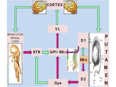
Idiopathic torsion dystonia. Major nuclear complex of the basal ganglia is the striatum, which is composed of the caudate and putamen. The striatum receives glutamatergic input from the cerebral cortex and dopaminergic input from the substantia nigra pars compacta (SNc). Two types of spiny projection neurons receive cortical and nigral inputs: those that project directly and those that project indirectly to the internal segment of the globus pallidus (GPI), which is the major output site of the basal ganglia. Complementary action of both of these pathways regulates the overall function of the GPI. The GPI, which, in turn, provides tonic inhibitory (ie, gamma-aminobutyric acid [GABA]–ergic) discharges downstream into the thalamic nuclei that project to the frontal cortical and other CNS areas.Direct pathway (D1) inhibits the substantia nigra pars reticulata (SNr) and the GPI, which are the major output sites, resulting in a net disinhibition and facilitation of thalamocortical circuits. Indirect pathway (D2), through serial connections with the globus pallidus pars externa (GPe) and the subthalamic nucleus (STN), is excitatory to the GPI, resulting in further inhibitory action on thalamocortical pathways. In this model, the mean discharge rate of the GPI is the key factor that determines a hypokinetic or hyperkinetic movement disorder. Increased inhibitory influences of the GPI on the thalamocortical circuitry result in hypokinetic disorders, such as Parkinson disease, whereas decreased GPI activity results in hyperkinetic disorders, such as hemiballismus. VL = ventrolateral thalamus.
Click to see larger pictureClick to see detailView Full Size Image
Media type:  Photo


  • Almasy L, Bressman SB, Raymond D, et al. Idiopathic torsion dystonia linked to chromosome 8 in two Mennonite families. Ann Neurol. Oct 1997;42(4):670-3. [Medline].
  • Augood SJ, Martin DM, Ozelius LJ, et al. Distribution of the mRNAs encoding torsinA and torsinB in the normal adult human brain. Ann Neurol. Nov 1999;46(5):761-9. [Medline].
  • Balash Y, Giladi N. Efficacy of pharmacological treatment of dystonia: evidence-based review including meta-analysis of the effect of botulinum toxin and other cure options. Eur J Neurol. Jun 2004;11(6):361-70. [Medline].
  • Bara-Jimenez W, Catalan MJ, Hallett M, Gerloff C. Abnormal somatosensory homunculus in dystonia of the hand. Ann Neurol. Nov 1998;44(5):828-31. [Medline].
  • Bertrand C, Molina-Negro P, Bouvier G, Gorczyca W. Observations and analysis of results in 131 cases of spasmodic torticollis after selective denervation. Appl Neurophysiol. 1987;50(1-6):319-23. [Medline].
  • Bertrand CM, Molina-Negro P. Selective peripheral denervation in 111 cases of spasmodic torticollis: rationale and results. In: Fahn S, Marsden CD, Calne DB, eds. Dystonia. In: Advances in Neurology. Vol 50. New York, NY: Raven;. 1988: 637-43.
  • Bhidayasiri R. An Overview of dystonia, II: treatment. CNS News. 2005;September:38-40.
  • Black KJ, Gado MH, Perlmutter JS. PET measurement of dopamine D2 receptor-mediated changes in striatopallidal function. J Neurosci. May 1 1997;17(9):3168-77. [Medline].
  • Bradley EA, Bartley GB, Chapman KL, Waller RR. Surgical correction of blepharoptosis in patients with myasthenia gravis. Ophthal Plast Reconstr Surg. Mar 2001;17(2):103-10. [Medline].
  • Bressman SB, de Leon D, Brin MF, et al. Idiopathic dystonia among Ashkenazi Jews: evidence for autosomal dominant inheritance. Ann Neurol. Nov 1989;26(5):612-20. [Medline].
  • Bressman SB, Greene PE. Dystonia. Curr Treat Options Neurol. May 2000;2(3):275-285. [Medline].
  • Brin MF, Comella C, Jankovic J. Dystonia: Etiology, Clinical Features, and Treatment. Philadelphia, Pa: Lippincott, Williams, and Wilkins;. 2004.
  • Burke RE, Fahn S, Marsden CD. Torsion dystonia: a double-blind, prospective trial of high-dosage trihexyphenidyl. Neurology. Feb 1986;36(2):160-4. [Medline].
  • Byl NN, Merzenich MM, Jenkins WM. A primate genesis model of focal dystonia and repetitive strain injury: I. Learning-induced dedifferentiation of the representation of the hand in the primary somatosensory cortex in adult monkeys. Neurology. Aug 1996;47(2):508-20. [Medline].
  • Cohen LG, Hallett M. Hand cramps: clinical features and electromyographic patterns in a focal dystonia. Neurology. Jul 1988;38(7):1005-12. [Medline].
  • Cooper IS. 20-year followup study of the neurosurgical treatment of dystonia musculorum deformans. Adv Neurol. 1976;14:423-52. [Medline].
  • Cooper IS. 20-year followup study of the neurosurgical treatment of dystonia musculorum deformans. Adv Neurol. 1976;14:423-52. [Medline].
  • Coubes P, Echenne B, Roubertie A, et al. Treatment of early-onset generalized dystonia by chronic bilateral stimulation of the internal globus pallidus. Apropos of a case [in French]. Neurochirurgie. May 1999;45(2):139-44. [Medline].
  • Coubes P, Cif L, El Fertit H, et al. Electrical stimulation of the globus pallidus internus in patients with primary generalized dystonia: long-term results. J Neurosurg. Aug 2004;101(2):189-94. [Medline].
  • Defazio G, Livrea P, Guanti G, et al. Genetic contribution to idiopathic adult-onset blepharospasm and cranial-cervical dystonia. Eur Neurol. 1993;33(5):345-50. [Medline].
  • Eidelberg D, Moeller JR, Ishikawa T, et al. The metabolic topography of idiopathic torsion dystonia. Brain. Dec 1995;118 ( Pt 6):1473-84. [Medline].
  • Eidelberg D, Moeller JR, Antonini A, et al. Functional brain networks in DYT1 dystonia. Ann Neurol. Sep 1998;44(3):303-12. [Medline].
  • Epidemiologic Study of Dystonia in Europe (ESDE) Collaborative Group. Sex-related influences on the frequency and age of onset of primary dystonia. Neurology. Nov 10 1999;53(8):1871-3. [Medline].
  • Fahn S, Marsden CD, Calne DB. Classification and investigation of dystonia. Mov Disord. 1987;332-58.
  • Fahn S, Hallet M, De Long M, eds. Advances in Neurology: Dystonia 4. Vol 94. Philadelphia, Pa: Lippincott, Williams and Wilkins; 2004.
  • Fish DR, Sawyers D, Allen PJ, et al. The effect of sleep on the dyskinetic movements of Parkinson''s disease, Gilles de la Tourette syndrome, Huntington''s disease, and torsion dystonia. Arch Neurol. Feb 1991;48(2):210-4. [Medline].
  • Fletcher NA, Harding AE, Marsden CD. A genetic study of idiopathic torsion dystonia in the United Kingdom. Brain. Apr 1990;113 ( Pt 2):379-95. [Medline].
  • Ford B, Greene P, Louis ED, et al. Use of intrathecal baclofen in the treatment of patients with dystonia. Arch Neurol. Dec 1996;53(12):1241-6. [Medline].
  • Greene P. Baclofen in the treatment of dystonia. Clin Neuropharmacol. Aug 1992;15(4):276-88. [Medline].
  • Grundman K. Primary torsion dystonia. Arch Neurol. 2005;62(4):682-5.
  • Herz E. Dystonia. Historical review: analysis of dystonic symptoms and physiologic mechanisms involved. Arch Neurol Psychiatry. 1944;51:305-18.
  • Kaji R, Rothwell JC, Katayama M, et al. Tonic vibration reflex and muscle afferent block in writer''s cramp. Ann Neurol. Aug 1995;38(2):155-62. [Medline].
  • Kaji R, Nagako M, Urushihara R. Sensory deficits in dystonia and their significance In: Fahn S, Hallet M, DeLong M, eds. Advances in Neurology: Dystonia 4. Vol 94. Philadelphia, Pa: Lippincott, Williams and Wilkins;. 2004: 11-7.
  • Krause M, Fogel W, Kloss M, et al. Pallidal stimulation for dystonia. Neurosurgery. Dec 2004;55(6):1361-8; discussion 1368-70. [Medline].
  • Krauss JK, Yianni J, Loher TJ, Aziz TZ. Deep brain stimulation for dystonia. J Clin Neurophysiol. Jan-Feb 2004;21(1):18-30. [Medline].
  • Leube B, Kessler KR, Goecke T, et al. Frequency of familial inheritance among 488 index patients with idiopathic focal dystonia and clinical variability in a large family. Mov Disord. Nov 1997;12(6):1000-6. [Medline].
  • Leube B, Hendgen T, Kessler KR, et al. Evidence for DYT7 being a common cause of cervical dystonia (torticollis) in Central Europe. Am J Med Genet. Sep 19 1997;74(5):529-32. [Medline].
  • Leube B, Rudnicki D, Ratzlaff T, et al. Idiopathic torsion dystonia: assignment of a gene to chromosome 18p in a German family with adult onset, autosomal dominant inheritance and purely focal distribution. Hum Mol Genet. Oct 1996;5(10):1673-7. [Medline].
  • Lozano A, Abosch A. Pallidal stimulation for dystonia. In: Fahn S, Hallet M, De Long M, eds. Advances in Neurology: Dystonia 4. Vol 94. Philadelphia, Pa: Lippincott, Williams and Wilkins;. 2004: 301-8.
  • Magyar-Lehmann S, Antonini A, Roelcke U, et al. Cerebral glucose metabolism in patients with spasmodic torticollis. Mov Disord. Sep 1997;12(5):704-8. [Medline].
  • McNaught KS, Kapustin A, Jackson T, et al. Brainstem pathology in DYT1 primary torsion dystonia. Ann Neurol. Oct 2004;56(4):540-7. [Medline].
  • Misbahuddin A, Warner TT. Dystonia: an update on genetics and treatment. Curr Opin Neurol. Aug 2001;14(4):471-5. [Medline].
  • Misbahuddin A, Placzek MR, Taanman JW, et al. Mutant torsin A, which causes early-onset primary torsion dystonia, is redistributed to membranous structures enriched in vesicular monoamine transporter in cultured human SH-SY5Y cells. Mov Disord. Apr 2005;20(4):432-40. [Medline].
  • Muller U, Steinberger D, Nemeth AH. Clinical and molecular genetics of primary dystonias. Neurogenetics. Mar 1998;1(3):165-77. [Medline].
  • Nakashima K, Rothwell JC, Thompson PD, et al. The blink reflex in patients with idiopathic torsion dystonia. Arch Neurol. Apr 1990;47(4):413-6. [Medline].
  • Nemeth AH. The genetics of primary dystonias and related disorders. Brain. Apr 2002;125(Pt 4):695-721. [Medline].
  • Nutt JG, Muenter MD, Aronson A, et al. Epidemiology of focal and generalized dystonia in Rochester, Minnesota. Mov Disord. 1988;3(3):188-94. [Medline].
  • Ondo WG, Desaloms JM, Jankovic J, Grossman RG. Pallidotomy for generalized dystonia. Mov Disord. Jul 1998;13(4):693-8. [Medline].
  • Ozelius L, Kramer PL, Moskowitz CB, et al. Human gene for torsion dystonia located on chromosome 9q32-q34. Neuron. May 1989;2(5):1427-34. [Medline].
  • Panizza M, Lelli S, Nilsson J, Hallett M. H-reflex recovery curve and reciprocal inhibition of H-reflex in different kinds of dystonia. Neurology. May 1990;40(5):824-8. [Medline].
  • Pauls DL, Korczyn AD. Complex segregation analysis of dystonia pedigrees suggests autosomal dominant inheritance. Neurology. Jul 1990;40(7):1107-10. [Medline].
  • Risch N, de Leon D, Ozelius L, et al. Genetic analysis of idiopathic torsion dystonia in Ashkenazi Jews and their recent descent from a small founder population. Nat Genet. Feb 1995;9(2):152-9. [Medline].
  • Rothwell JC, Obeso JA, Day BL, Marsden CD. Pathophysiology of dystonias. Adv Neurol. 1983;39:851-63. [Medline].
  • Shang H, Clerc N, Lang D, et al. Clinical and molecular genetic evaluation of patients with primary dystonia. Eur J Neurol. Feb 2005;12(2):131-8. [Medline].
  • Stojanovic M, Cvetkovic D, Kostic VS. A genetic study of idiopathic focal dystonias. J Neurol. Aug 1995;242(8):508-11. [Medline].
  • Tasker RR, Doorly T, Yamashiro K. Thalamotomy in generalized dystonia. Adv Neurol. 1988;50:615-31. [Medline].
  • Tinazzi M, Frasson E, Bertolasi L, et al. Temporal discrimination of somesthetic stimuli is impaired in dystonic patients. Neuroreport. May 14 1999;10(7):1547-50. [Medline].
  • Tronnier VM, Fogel W. Pallidal stimulation for generalized dystonia: report of three cases. J Neurosurg. Mar 2000;92(3):453-6. [Medline].
  • Trost M. Dystonia update. Curr Opin Neurol. Aug 2003;16(4):495-500. [Medline].
  • Valente EM, Bentivoglio AR, Cassetta E, et al. DYT13, a novel primary torsion dystonia locus, maps to chromosome 1p36.13--36.32 in an Italian family with cranial-cervical or upper limb onset. Ann Neurol. Mar 2001;49(3):362-6. [Medline].
  • Vayssiere N, van der Gaag N, Cif L, et al. Deep brain stimulation for dystonia confirming a somatotopic organization in the globus pallidus internus. J Neurosurg. Aug 2004;101(2):181-8. [Medline].
  • Vercueil L, Pollak P, Fraix V, et al. Deep brain stimulation in the treatment of severe dystonia. J Neurol. Aug 2001;248(8):695-700. [Medline].
  • Vitek JL, Chockkan V, Zhang JY, et al. Neuronal activity in the basal ganglia in patients with generalized dystonia and hemiballismus. Ann Neurol. Jul 1999;46(1):22-35. [Medline].
  • Whone AL, Von Spiczak S, Edwards M, et al. Opioid binding in DYT1 primary torsion dystonia: an 11C-diprenorphine PET study. Mov Disord. Dec 2004;19(12):1498-503. [Medline].
  • van der Kamp W, Berardelli A, Rothwell JC, et al. Rapid elbow movements in patients with torsion dystonia. J Neurol Neurosurg Psychiatry. Sep 1989;52(9):1043-9. [Medline].

Primary Torsion Dystonia excerpt

Article Last Updated: Mar 30, 2007