Practice Essentials
Hydrocephalus can be defined broadly as a disturbance of cerebrospinal fluid (CSF) formation, flow, or absorption, leading to an increase in volume occupied by this fluid in the central nervous system (CNS). [1] This condition could also be termed a hydrodynamic CSF disorder. See the image below.
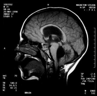 Noncommunicating obstructive hydrocephalus caused by obstruction of the foramina of Luschka and Magendie. This MRI sagittal image demonstrates dilatation of lateral ventricles with stretching of corpus callosum and dilatation of the fourth ventricle.
Signs and symptoms
Clinical features of hydrocephalus are influenced by the patient's age, the cause of the hydrocephalus, the location of the obstruction, its duration, and its rapidity of onset.
Symptoms in infants include poor feeding, irritability, reduced activity, and vomiting.
Symptoms in children and adults include the following:
-
Slowing of mental capacity, cognitive deterioration
-
Headaches (initially in the morning)
-
Neck pain, suggesting tonsillar herniation
-
Vomiting, more significant in the morning
-
Blurred vision: A consequence of papilledema and, later, of optic atrophy
-
Double vision: Related to unilateral or bilateral sixth nerve palsy
-
Difficulty in walking secondary to spasticity: Preferentially affects the lower limbs because the periventricular pyramidal tract is stretched by the hydrocephalus
-
Drowsiness
Children may also exhibit stunted growth and sexual maturation from third ventricle dilatation. Adults may also have nausea that is not exacerbated by head movements; incontinence (urinary first, fecal later if condition remains untreated) indicates significant destruction of the frontal lobes and advanced disease.
Symptoms of normal pressure hydrocephalus (NPH) include the following:
-
Gait disturbance: Usually the first symptom and may precede other symptoms by months or years; magnetic gait is used to emphasize the tendency of the feet to remain "stuck to the floor" despite patients’ best efforts to move them
-
Dementia (of varying degrees): Should be a late finding in pure (shunt-responsive) NPH; presents as an impairment of recent memory or as a "slowing of thinking"; spontaneity and initiative are decreased
-
Urinary incontinence: May present as urgency, frequency, or a diminished awareness of the need to urinate
-
Other symptoms that can occur: Personality changes and Parkinsonism
-
Rarely: Headaches; seizures are extremely rare—consider an alternative diagnosis
See Clinical Presentation for more detail.
Diagnosis
Examination in infants may reveal the following findings:
-
Head enlargement (head circumference ≥98th percentile for age), especially crossing percentiles on the growth chart
-
Dysjunction/splaying of sutures
-
Dilated scalp veins
-
Tense/bulging fontanelle
-
Setting-sun sign: Characteristic of increased intracranial pressure (ICP); downward deviation of the ocular globes, retracted upper lids, visible white sclerae above irises
-
Increased limb tone (spasticity preferentially affecting the lower limbs)
Children and adults may demonstrate the following findings on physical examination:
-
Papilledema (optic nerve swelling), although this does not develop acutely
-
Failure of upward gaze: Due to pressure on the tectal plate through the suprapineal recess; the limitation of upward gaze is of supranuclear origin
-
Unsteady gait
-
Large head
-
Unilateral or bilateral sixth nerve palsy (secondary to increased ICP)
Children may also exhibit the Macewen sign, in which a "cracked pot" sound is noted on percussion of the head.
Patients with NPH may exhibit the following findings on examination:
-
Normal muscle strength; no sensory loss
-
Increased reflexes and Babinski response in one or both feet: Search for vascular risk factors (causing associated brain microangiopathy or vascular Parkinsonism), which are common in NPH patients
-
Variable difficulty in walking: May have mild imbalance to inability to walk or to stand; the classic gait impairment consists of short steps, wide base, externally rotated feet, and lack of festination (hastening of cadence with progressively shortening stride length, a hallmark of the gait impairment of Parkinson disease)
-
Frontal release signs (in late stages): Appearance of sucking and grasping reflexes
Testing
No specific blood tests are recommended in the workup for hydrocephalus. However, consider genetic testing and counseling when X-linked hydrocephalus is suspected, and evaluate the CSF in posthemorrhagic and postmeningitic hydrocephalus for protein concentration and to exclude residual infection.
Obtain electroencephalography in patients with seizures.
Imaging studies
The following imaging studies may be used to evaluate patients with suspected hydrocephalus:
-
Computed tomography (CT) scanning: To assess size of ventricles and other structures
-
Magnetic resonance imaging (MRI): To assess for Chiari malformation or cerebellar or periaqueductal tumors
-
Ultrasonography through anterior fontanelle in infants: To assess for subependymal and intraventricular hemorrhage; to follow infants for possible progressive hydrocephalus
-
Skull radiography: To detect erosion of sella turcica, or "beaten copper cranium" (or "beaten silver cranium")—the latter can also be seen in craniosynostosis; (after shunt insertion) to confirm correct positioning of installed hardware
-
MRI cine: To measure CSF stroke volume (SV) in the cerebral aqueduct; however, such measurements don’t appear to be useful in predicting response to shunting [2]
-
Diffusion tensor imaging (DTI): To detect differences in fractional anisotropy and mean diffusivity of the brain parenchyma surrounding the ventricles; allows recognition of microstructural changes in periventricular white matter region that may be too subtle on conventional MRI [3]
-
Radionuclide cisternography (in NPH): To assess the prognosis with regard to possible shunting—however, due to its poor sensitivity in predicting shunt response when the ventricular to total intracranial activity (V/T) ratio is less than 32%, this test is no longer commonly used
See Workup for more detail.
Management
Surgery
Surgical treatment is the preferred therapeutic option in patients with hydrocephalus. [4] Most patients eventually undergo shunt placements, such as the following:
-
Ventriculoperitoneal (VP) shunt (most common)
-
Ventriculoatrial (VA) shunt (or "vascular shunt")
-
Lumboperitoneal shunt: Only used for communicating hydrocephalus, CSF fistula, or pseudotumor cerebri)
-
Torkildsen shunt (rarely): Effective only in acquired obstructive hydrocephalus (ventriculocisternostomy)
-
Ventriculopleural shunt (second-line therapy): Used if other shunt types contraindicated
Rapid-onset hydrocephalus with ICP is an emergency. The following procedures can be done, depending on each specific case:
-
Ventricular tap in infants
-
Open ventricular drainage in children and adults (EVD, external ventricular drain)
-
Lumbar puncture (LP) in posthemorrhagic and postmeningitic hydrocephalus
-
VP or VA shunt
Repeat LPs can be performed for cases of hydrocephalus after intraventricular hemorrhage (which can resolve spontaneously). If reabsorption does not resume when the CSF protein content is less than 100 mg/dL, spontaneous resorption is unlikely to occur. LPs can be performed only in cases of communicating hydrocephalus.
Alternatives to shunting include the following:
-
Choroid plexectomy or choroid plexus coagulation
-
Opening of a stenosed aqueduct
-
Endoscopic fenestration of the floor of the third ventricle (however, contraindicated in communicating hydrocephalus)
Conservative management
Medical treatment is not effective in long-term treatment of chronic hydrocephalus; it is used as a temporizing measure to delay surgical intervention. Medical therapy may be tried in premature infants with posthemorrhagic hydrocephalus (in the absence of acute hydrocephalus, repeated taps are done not only to allow for potential resolution, but also to allow the protein level to reduce low enough that it will not clog any placed shunt). Normal CSF absorption may resume spontaneously during this interim period. Medical agents include carbonic anhydrase inhibitors (eg, acetazolamide) and loop diuretics (eg, furosemide) for the treatment of hydrocephalus are controversial and should be used only as temporary measures (such as patients who already have non-programmable shunts, or when shunt placement is not able to be done at that time).
See Treatment and Medication for more detail.
Background
Hydrocephalus can be defined broadly as a disturbance of formation, flow, or absorption of cerebrospinal fluid (CSF) that leads to an increase in volume occupied by this fluid in the CNS. [1] This condition also could be termed a hydrodynamic disorder of CSF. Acute hydrocephalus occurs over days, subacute hydrocephalus occurs over weeks, and chronic hydrocephalus occurs over months or years. Conditions such as cerebral atrophy and focal destructive lesions also lead to an abnormal increase of CSF in CNS. In these situations, loss of cerebral tissue leaves a vacant space that is filled passively with CSF. Such conditions are not the result of a hydrodynamic disorder and therefore are not classified as hydrocephalus. An older misnomer used to describe these conditions was hydrocephalus ex vacuo.
Benign external hydrocephalus (benign enlargement of the subarrachnoid spaces of infancy) is a self-limiting absorption deficiency of infancy and early childhood with mildly raised intracranial pressure (ICP) and enlarged subarachnoid spaces. The ventricles usually are not enlarged significantly, and resolution within 1 year is the rule. [5]
Normal pressure hydrocephalus (NPH) describes a condition that rarely occurs in patients younger than 60 years. [6] Enlarged ventricles and normal CSF pressure at lumbar puncture (LP) in the absence of papilledema led to the term NPH. However, intermittent intracranial hypertension has been noted during monitoring of patients in whom NPH is suspected, usually at night. The classic Hakim triad of symptoms includes gait apraxia, incontinence, and dementia. Headache is not a typical symptom in NPH.
Communicating hydrocephalus occurs when full communication occurs between the ventricles and subarachnoid space. It is caused by overproduction of CSF (rarely), defective absorption of CSF (most often, includes conditions such as intracranial hemorrhage or meningitis resulting in damage to the arachnoid granulations, where CSF is reabsorbed), or venous drainage insufficiency (occasionally). See the image below.
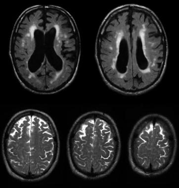 Communicating hydrocephalus with surrounding "atrophy" and increased periventricular and deep white matter signal on fluid-attenuated inversion recovery (FLAIR) sequences. Note that apical cuts (lower row) do not show enlargement of the sulci, as is expected in generalized atrophy. Pathological evaluation of this brain demonstrated hydrocephalus with no microvascular pathology corresponding with the signal abnormality (which likely reflects transependymal exudate) and normal brain weight (indicating that the sulci enlargement was due to increased subarachnoid cerebrospinal fluid [CSF] conveying a pseudoatrophic brain pattern).
Noncommunicating hydrocephalus occurs when CSF flow is obstructed within the ventricular system or in its outlets to the arachnoid space, resulting in impairment of the CSF from the ventricular to the subarachnoid space. The most common form of noncommunicating hydrocephalus is obstructive and is caused by intraventricular or extraventricular mass-occupying lesions that disrupt the ventricular anatomy. [7] See the images below.
Noncommunicating obstructive hydrocephalus caused by obstruction of the foramina of Luschka and Magendie. This MRI sagittal image demonstrates dilatation of lateral ventricles with stretching of corpus callosum and dilatation of the fourth ventricle.
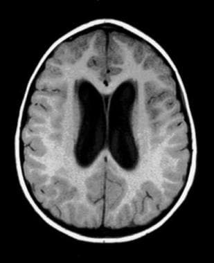 Noncommunicating obstructive hydrocephalus caused by obstruction of foramina of Luschka and Magendie. This MRI axial image demonstrates dilatation of the lateral ventricles.
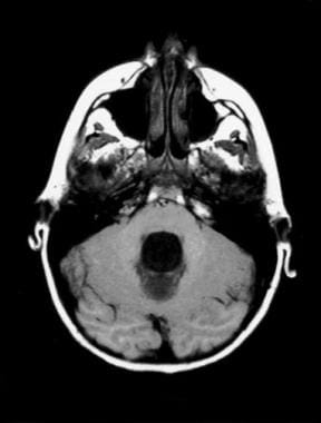 Noncommunicating obstructive hydrocephalus caused by obstruction of foramina of Luschka and Magendie. This MRI axial image demonstrates fourth ventricle dilatation.
Congenital hydrocephalus applies to the ventriculomegaly that develops in the fetal and infancy periods, often associated with macrocephaly. [8] The most common causes of congenital hydrocephalus are obstruction of the cerebral aqueduct flow, Arnold-Chiari malformation or Dandy–Walker malformation. [9] These patients may stabilize in later years due to compensatory mechanisms but may decompensate, especially following minor head injuries. During these decompensations, determining the extent to which any new neurological deficits may be due to the new acute event, compared with hydrocephalus that may have gone unnoticed for many years, is difficult. An extremely severe variant of congenital hydrocephalus is hydranencephaly, where the brain's cerebral hemispheres are absent to varying degrees and the remaining cranial cavity is filled with cerebrospinal fluid.
Pathophysiology
Normal CSF production is 0.20-0.35 mL/min; most CSF is produced by the choroid plexus, which is located within the ventricular system, mainly the lateral and fourth ventricles. The capacity of the lateral and third ventricles in a healthy person is 20 mL. Total volume of CSF in an adult is 120 mL.
Normal route of CSF from production to clearance is the following: From the choroid plexus, the CSF flows to the lateral ventricle, then to the interventricular foramen of Monro, the third ventricle, the cerebral aqueduct of Sylvius, the fourth ventricle, the two lateral foramina of Luschka and one medial foramen of Magendie, the subarachnoid space, the arachnoid granulations, the dural sinus, and finally into the venous drainage.
ICP rises if production of CSF exceeds absorption. This occurs if CSF is overproduced, resistance to CSF flow is increased, CSF resorption is decreased, or venous sinus pressure is increased. CSF production falls as ICP rises. Compensation may occur through transventricular absorption (subependymal flow) of CSF and also by absorption along nerve root sleeves (which may result in enlarged optic nerve sheaths). The temporal and frontal horns dilate first, often asymmetrically. This may result in elevation of the corpus callosum, stretching or perforation of the septum pellucidum, thinning of the cerebral mantle, or enlargement of the third ventricle downward into the pituitary fossa (which may cause pituitary dysfunction) as well as dorsal midbrain compression resulting in Parinaud's syndrome (aralysis of upgaze, Pseudo-Argyll Roberson pupils, convergence-retraction nystagmus, eyelide retraction, and setting sun sign).
The mechanism of NPH has not been elucidated completely. Current theories include increased resistance to flow of CSF within the ventricular system or subarachnoid villi; intermittently elevated CSF pressure, usually at night; and ventricular enlargement caused by an initial rise in CSF pressure. The enlargement is maintained despite normal pressure because of the Laplace law. Although pressure is normal, the enlarged ventricular area reflects increased force on the ventricular wall.
Frequency
United States
The incidence of congenital hydrocephalus is 3 per 1,000 live births; the incidence of acquired hydrocephalus is not known exactly due to the variety of disorders that may cause it.
International
Incidence of acquired hydrocephalus is unknown. About 100,000 shunts are implanted each year in the developed countries, but little information is available for other countries.
Mortality/Morbidity
In untreated hydrocephalus, death may occur by tonsillar herniation secondary to raised ICP with compression of the brain stem and subsequent respiratory arrest.
Shunt dependence occurs in 75% of all cases of treated hydrocephalus and in 50% of children with communicating hydrocephalus. Patients are hospitalized for scheduled shunt revisions or for treatment of shunt complications or shunt failure. Poor development of cognitive function in infants and children, or loss of cognitive function in adults, can complicate untreated hydrocephalus. It may persist after treatment. Visual loss can complicate untreated hydrocephalus and may persist after treatment.
Epidemiology
Sex
Generally, incidence is equal in males and females. The exception is Bickers-Adams syndrome (X-linked hydrocephalus with stenosis of aqueduct of Sylvius), transmitted by females and manifested in males. NPH has a slight male preponderance.
Age
Incidence of human hydrocephalus presents a bimodal age curve. One peak occurs in infancy and is related to the various forms of congenital malformations and premature birth. Another peak occurs in adulthood, mostly resulting from NPH. Adult hydrocephalus represents approximately 40% of total cases of hydrocephalus.
The outcome of pediatric hydrocephalus has been studied frequently, but much remains unresolved about long-term and social outcomes. [10]
Prognosis
Long-term outcome is related directly to the cause of hydrocephalus.
Up to 50% of patients with large intraventricular hemorrhage develop permanent hydrocephalus requiring shunt.
Following removal of a posterior fossa tumor in children, 20% develop permanent hydrocephalus requiring a shunt. The overall prognosis is related to type, location, and extent of surgical resection of the tumor.
Satisfactory control was reported for medical treatment in 50% of hydrocephalic patients younger than 1 year who had stable vital signs, normal renal function, and no symptoms of elevated ICP.
Criteria exist for predicting improvement with shunting in NPH, but they are controversial.
-
If gait disturbance precedes mental deterioration, the chance of improvement is 77%. Patients with dementia and no gait disturbance rarely respond to shunting.
-
Focal impingement of corpus callosum on MRI indicates unstable ICP and is associated with a good response to shunting.
-
Initial OP of CSF greater than 100 mm H2 O predicts better response.
-
Response to a single LP or to controlled CSF drainage via lumbar subarachnoid catheter (ELD) has some value in predicting outcome.
-
Cerebral blood flow of 32 mL/100 g per minute or greater predicts clinical improvement after shunt.
-
CSF pressure of 180 mm H2 O with frequent Lundberg B waves on continuous CSF pressure monitoring is associated with good prognosis after shunting. Lundberg B waves represent an accentuation of physiological phenomena, reflecting arterial waves. They represent fluctuating ICP waves of 4-8 per minute frequency and 20-30 mm Hg (260-400 mm H2 O) amplitude. Occasionally they can occur in normal sleep.
-
Large ventricles with flattened or invaginated sulci (entrapped sulci) suggest that hydrocephalus is not due to atrophy alone. These patients have good prognosis with shunting.
-
If isotopic cisternography shows persistent ventricular activity on a late scan (42-72 h), the probability of improving with shunting is 75%.
Patient Education
Knowledge of the signs and symptoms of shunt malfunction or infection and the necessity for emergent medical evaluation in these instances is mandatory in patients, family members, and caregivers.
The patient, family, and caregivers should know that periodic re-evaluation is necessary.
Pumping the shunt is contraindicated in most cases.
Patients with vascular shunts, and some patients with other types of shunts, should receive prophylactic antibiotics before dental procedures or instrumentation of the bladder.
-
Noncommunicating obstructive hydrocephalus caused by obstruction of the foramina of Luschka and Magendie. This MRI sagittal image demonstrates dilatation of lateral ventricles with stretching of corpus callosum and dilatation of the fourth ventricle.
-
Noncommunicating obstructive hydrocephalus caused by obstruction of foramina of Luschka and Magendie. This MRI axial image demonstrates dilatation of the lateral ventricles.
-
Noncommunicating obstructive hydrocephalus caused by obstruction of foramina of Luschka and Magendie. This MRI axial image demonstrates fourth ventricle dilatation.
-
Communicating hydrocephalus with surrounding "atrophy" and increased periventricular and deep white matter signal on fluid-attenuated inversion recovery (FLAIR) sequences. Note that apical cuts (lower row) do not show enlargement of the sulci, as is expected in generalized atrophy. Pathological evaluation of this brain demonstrated hydrocephalus with no microvascular pathology corresponding with the signal abnormality (which likely reflects transependymal exudate) and normal brain weight (indicating that the sulci enlargement was due to increased subarachnoid cerebrospinal fluid [CSF] conveying a pseudoatrophic brain pattern).


