Continually Updated Clinical Reference
 
 
  All Sources     eMedicine     Medscape     Drug Reference     MEDLINE
 
eMedicine - Ependymoma : Article by

Quick Find
Authors & Editors
Introduction
Clinical
Differentials
Workup
Treatment
Medication
Follow-up
Miscellaneous
Multimedia
References

Related Articles
Acute Disseminated Encephalomyelitis

Aseptic Meningitis

Brainstem Gliomas

Cavernous Sinus Syndromes

Cerebral Aneurysms

Craniopharyngioma

Glioblastoma Multiforme

Haemophilus Meningitis

HIV-1 Associated CNS Conditions: Meningitis

HIV-1 Associated Opportunistic Infections: CNS Cryptococcosis

HIV-1 Associated Opportunistic Infections: CNS Toxoplasmosis

HIV-1 Associated Opportunistic Infections: Cytomegalovirus Encephalitis

HIV-1 Associated Vacuolar Myelopathy

Intracranial Epidural Abscess

Leptomeningeal Carcinomatosis

Low-Grade Astrocytoma

Medulloblastoma

Meningococcal Meningitis

Oligodendroglioma

Spinal Epidural Abscess

Staphylococcal Meningitis

Subdural Empyema

Tropical Myeloneuropathies

Tuberculous Meningitis

Viral Encephalitis

Viral Meningitis


Patient Education
Click here for patient education.


Author: Subrata Ghosh, MD, MBBS, MS, Staff Physician, Assistant Professor of Neurosurgery, Baylor College of Medicine, Houston, Division of Neurosurgery, St. Luke's Episcopal Hospital, Texas Medical Center, Houston, TX

Coauthor(s): Draga Jichici, HBSc, MD, FRCP(C), FAHA, Assistant Professor, Department of Medicine, Division of Critical Care Medicine, McMaster University Medical School, Canada

Editors: Rodrigo O Kuljis, MD, Esther Lichtenstein Professor of Psychiatry and Neurology, Director, Division of Cognitive and Behavioral Neurology, Department of Neurology, University of Miami School of Medicine; Francisco Talavera, PharmD, PhD, Senior Pharmacy Editor, eMedicine; Jorge Kattah, MD, Head, Program Director, Professor, Department of Neurology, University of Illinois College of Medicine at Peoria; Matthew J Baker, MD, Consulting Staff, Collier Neurologic Specialists, Naples Community Hospital; Nicholas Y Lorenzo, MD, Chief Editor, eMedicine Neurology; Consulting Staff, Neurology Specialists and Consultants

Author and Editor Disclosure

Synonyms and related keywords: ependymal cells neoplasms, hydrocephalus, neuroepithelial tumor of the spinal cord, hyperdiploid tumor, tetraploid tumor, meningitis, encephalitis, astrocytoma, medulloblastoma, oligodendroglioma, brain tumor

Background

Ependymomas are neoplasms of ependymal cells that occur throughout the entire neuraxis in association with the lining of the cerebral ventricles and central canal of the spinal cord.

Pathophysiology

Ependymomas occur most commonly in the intracranial and intraspinal areas, with lesions rarely occurring in the sacral area. Other unusual ectopic sites of ependymoma are the mediastinum, ovary, and broad ligament. In general, the anatomic location determines the pathophysiological manifestations of the tumor. Supratentorial tumors present with mass effect, focal neurological signs, and occasional obstruction of ventricular outflow. The relationship with the ventricular system is more apparent in tumors of the posterior fossa (mostly of the fourth ventricle), which usually present with obstructive hydrocephalus with or without signs of brain stem compression.

Frequency

United States

Ependymomas are infrequent tumors, representing 2-8% of all brain tumors. However, ependymomas are the third most common brain tumor in children (8-12%) with up to 30% occurring in children younger than 3 years. Half of the ependymomas occur in the first 2 decades of life; two-thirds are located in the posterior fossa (>than 90% are in the fourth ventricle). Interestingly, despite their overall low frequency, ependymomas are the most frequent neuroepithelial tumors of the spinal cord.

Mortality/Morbidity

From the biological perspective, ependymomas do not usually proliferate rapidly, are not invasive, and usually do not metastasize. The associated morbidity can mainly be accounted for by the local space-occupying effects of the tumor. In unusual cases, the risk of sudden death from large intracranial ependymomas results from increased intracranial pressure secondary to obstructive hydrocephalus.

Race

No race predilection is reported.

Sex

No sex predilection is reported.

Age

Peak age at presentation ranges from 7 weeks to 16 years with a mean of 3.7 years. A second, lower peak age of presentation occurs in the third decade of life.


History

Presenting features are insidious and progressive in nature.

  • Nausea and vomiting (80%) is the most common presenting symptom, secondary to increased intracranial pressure.
  • Headache (50%), due to the local effect of pressure or increased intracranial pressure, is usually worse in the morning.
  • Change in behavior (50%) includes lethargy, irritability, diminished social interaction, and loss of appetite (prevalent in younger children).
  • Difficulty with balance (30%) reflects cerebellar involvement or mass effect.

Physical

  • Papilledema (60%)
  • Ataxia (45%)
  • Nystagmus with or without gaze palsy (40%)
  • Lower cranial nerve palsies (10%)
  • Apraxia or hemiparesis (20%)
  • Increase in head circumference in children younger than 2 years (10%)

Causes

No particular genetic or molecular marker or familial predisposition has been identified for this tumor type. In one series, only a few ependymomas were reported to be hyperdiploid or tetraploid.

  • Other diagnostic considerations:
    • Meningitis
    • Encephalitis
    • Other brain tumors (astrocytoma, medulloblastoma, oligodendroglioma)
    • Meningitis and encephalitis can be readily differentiated by their more abrupt onset, associated fever, or signs of meningeal irritation.
    • Differentiation from other types of brain tumors (astrocytoma, medulloblastoma, oligodendroglioma) is radiological and pathological.


Acute Disseminated Encephalomyelitis
Aseptic Meningitis
Brainstem Gliomas
Cavernous Sinus Syndromes
Cerebral Aneurysms
Craniopharyngioma
Glioblastoma Multiforme
Haemophilus Meningitis
HIV-1 Associated CNS Conditions: Meningitis
HIV-1 Associated Opportunistic Infections: CNS Cryptococcosis
HIV-1 Associated Opportunistic Infections: CNS Toxoplasmosis
HIV-1 Associated Opportunistic Infections: Cytomegalovirus Encephalitis
HIV-1 Associated Vacuolar Myelopathy
Intracranial Epidural Abscess
Leptomeningeal Carcinomatosis
Low-Grade Astrocytoma
Medulloblastoma
Meningococcal Meningitis
Oligodendroglioma
Spinal Epidural Abscess
Staphylococcal Meningitis
Subdural Empyema
Tropical Myeloneuropathies
Tuberculous Meningitis
Viral Encephalitis
Viral Meningitis

Other Problems to be Considered

Brainstem syndromes


Lab Studies

  • Laboratory studies are not helpful for diagnosis.

Imaging Studies

  • MRI is the diagnostic tool of choice. It reveals discrete, heterogeneous masses with variable enhancement.
    • Evidence of calcification, necrosis, and cystic change are occasionally seen; hemorrhage is rare.
    • The tumor characteristically displaces rather than infiltrates brain parenchyma with minimal peritumoral edema.
  • CT scan is used in emergency situations, although its resolution is inferior to that of MRI. The CT appearance of ependymoma varies, but calcification is more evident on CT scan.

Procedures

  • Ventriculostomy is not required preoperatively because patients are usually stable. In fact, it should be avoided if possible because of the potentially fatal risk of upward herniation or hemorrhage within the tumor with brain stem compression.
  • Lumbar puncture is also contraindicated because of a similar risk of downward (tonsillar) herniation preoperatively.

Histologic Findings

The presence of a classic, well-circumscribed lesion with moderate cellularity, punctuated by areas of an acellular, fibrillary zone (perivascular pseudorosettes) is common. Variants include the following:

  • Clear cell type with perinuclear halo
  • Papillary type with an extensive epithelial component
  • True rosette formation
  • Myxopapillary type with prominent perivascular and intercellular mucin
  • Rare melanotic type containing lipofuscin and no melanin pigment

No consensus exists about histological grading and its relationship to survival or clinical behavior. Most centers accept the World Health Organization (WHO) revised classification of grade I, benign, and grade II, anaplastic types, the former being less invasive and showing less incidence of metastasis. Only 10% of ependymomas metastasize to other areas of the neuraxis; these metastases are almost always associated with tumor recurrence at the primary site, which emphasizes the importance of local control.


Medical Care

Preoperative and perioperative steroids are recommended to help limit edema and alleviate some symptoms.

Surgical Care

Surgery remains the most effective therapy for this tumor. It establishes tissue diagnosis, restores normal cerebrospinal fluid flow, and can be used to attempt total removal of the tumor.

  • A second-look surgery for unexpected residual lesions that are seen on postoperative imaging in an operable location is encouraged in patients with noninvasive, benign histology.
  • Permanent cerebrospinal fluid (CSF) diversion with ventriculoperitoneal shunt is rarely required.
  • Postoperative radiation therapy substantially improves survival. Although not proven, some dose-to-response relationship probably exists.
    • Newer methods that target only the local tumor bed, such as high fractionation radiotherapy or stereotactic radiosurgery, may permit potential dose reduction as compared with conventional radiotherapy. It provides effective tumor control, which may help limit complications such as cognitive dysfunction, growth delay, and hypothyroidism.
    • Craniospinal axis radiation is recommended only for patients with radiological or pathological evidence of spinal seeding.
    • Overall, results of chemotherapy are disappointing. Multidrug combinations using VP-16 etoposide, vincristine, CCNU (lomustine), and cisplatin offer limited benefit in patients with recurrent disease.

Consultations

  • Neurosurgeon
  • Neurologist
  • Radiation oncologist
  • Medical oncologist


No specific medications exist for treating ependymoma.


Further Inpatient Care

  • Admit the patient for repeat surgery.
  • Admit the patient for treatment of complications from surgery, radiotherapy, or chemotherapy.

Further Outpatient Care

  • Radiation therapy
  • Chemotherapy
  • Serial neuroimaging (MRI)

Complications

  • Hydrocephalus
  • Paralysis
  • Cranial nerve palsy
  • Meningitis
  • Bone marrow suppression
  • Cognitive dysfunction
  • Growth and developmental delay
  • Hypothyroidism

Prognosis

  • Gross total resection is the most important determinant of outcome, with progression-free survival rates of 70-80% after 5 years, compared to 35% for incomplete resection.
  • Postoperative radiation therapy improves survival, whereas results of chemotherapy are disappointing.
  • Age also strongly correlates with outcome. Usually, the younger the patient, the worse the prognosis.

Patient Education

  • Refer the patient for psychosocial counseling.


Medical/Legal Pitfalls

  • Failure to recognize signs or symptoms


Media file 1:  
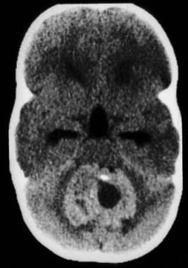
CT scan without contrast, axial view, demonstrates mixed but predominantly hyperdense tumor in the posterior fossa with severe obstructive hydrocephalus.
Click to see larger pictureClick to see detailView Full Size Image
Media type:  CT

Media file 2:  
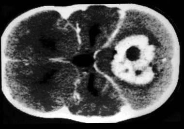
CT scan with contrast, axial view shows moderately intense contrast enhancement (compare with image 1).
Click to see larger pictureClick to see detailView Full Size Image
Media type:  CT

Media file 3:  MRI, T2-weighted image, axial view, showing mixed (isodensity and hyperdensity) heterogenous nature of the tumor with some peritumoral edema.
Click to see larger pictureClick to see detailView Full Size Image
Media type:  MRI

Media file 4:  MRI, T1-weighted image, without contrast, sagittal view, showing the posterior fossa location, mixed (hypodensity and isodensity) signal intensity and tending to grow out of the fourth ventricle.
Click to see larger pictureClick to see detailView Full Size Image
Media type:  MRI


  • Bhattacharjee MB, Armstrong DD, Vogel H, Cooley LD. Cytogenetic analysis of 120 primary pediatric brain tumors and literature review. Cancer Genet Cytogenet. Aug 1997;97(1):39-53. [Medline].
  • Bigner SH, McLendon RE, Fuchs H, et al. Chromosomal characteristics of childhood brain tumors. Cancer Genet Cytogenet. Sep 1997;97(2):125-34. [Medline].
  • Ernestus RI, Schroder R, Stutzer H, Klug N. The clinical and prognostic relevance of grading in intracranial ependymomas. Br J Neurosurg. Oct 1997;11(5):421-8. [Medline].
  • Graham DI, Lantos PL, eds. Greenfield's Neuropathology. 6th ed. Arnold Press;1997: 636-44.
  • Kaye AH, Laws E Jr, eds. Brain Tumors: An Encyclopedic Approach. 1st ed. Churchill Livingstone;1997: 493-504.
  • Kleihues P, Burger PC, Scheithauer BW. The new WHO classification of brain tumours. Brain Pathol. Jul 1993;3(3):255-68. [Medline].
  • Kleihues P et al. Pathology & Genetics. Tumors of the Nervous System. International Agency for Research on Cancer (IARC)/World Health Organization. 1997;96-109.
  • Kun LE. Brain tumors. Challenges and directions. Pediatr Clin North Am. Aug 1997;44(4):907-17. [Medline].
  • McLaughlin MP, Marcus RB, Buatti JM, et al. Ependymoma: results, prognostic factors and treatment recommendations. Int J Radiat Oncol Biol Phys. Mar 1 1998;40(4):845-50. [Medline].
  • Moynihan TJ. Ependymal tumors. Curr Treat Options Oncol. Dec 2003;4(6):517-23. [Medline].
  • Nazar GB, Hoffman HJ, Becker LE, et al. Infratentorial ependymomas in childhood: prognostic factors and treatment. J Neurosurg. Mar 1990;72(3):408-17. [Medline].
  • Osborn AG. Diagnostic Neuroradiology: A Text and Atlas. 1st ed. Mosby;1994: 566-70.
  • Russell DS, et al. Pathology of Tumors of the Nervous System. 4th ed. Arnold Press;1977: 203-26.

Ependymoma excerpt

Article Last Updated: Oct 3, 2006