Practice Essentials
Status epilepticus (SE) is a common, life-threatening neurologic disorder that is essentially an acute, prolonged epileptic crisis (see the image below). SE can represent an exacerbation of a preexisting seizure disorder or the initial manifestation of a seizure disorder (epilepsy), or it can represent an insult other than a seizure disorder. Among such insults, SE can occur in the context of an acute cerebral injury, or be due to a systemic process or illness, in a patient with or without a remote cerebral injury. In patients with known epilepsy, the most common cause of SE is a change in medication. Most seizures terminate spontaneously; SE represents a failure of seizure termination.
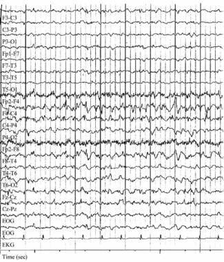 Focal status epilepticus. Electroencephalograph (EEG) in a patient with epilepsia partialis continua caused by Rasmussen encephalitis before hemispherectomy. The patient had long-standing, intractable partial epilepsy since the first decade of life. Seizures included complex partial with occasional secondary generalization and repetitive myoclonus involving the left side of the body. Note the frequent epileptiform discharges at 1-2 Hz involving the right frontocentral channels. These were evident on many of the patient's routine EEGs. Clinical myoclonus is often correlated with high-voltage bursts of such activity.
Signs and symptoms
By clinical history, nonmotor simple partial status epilepticus involves subjective sensory disturbances, including the following:
-
Focal or unilateral paresthesias or numbness
-
Focal visual changes, usually characterized by flashing lights
-
Focal visual obscuration or focal colorful hallucinations
-
Olfactory or gustatory hallucinations
-
Atypical rising abdominal sensations
Epilepsy partialis continua, or focal status epilepticus of the motor cortex, may occur in various contexts, with some authors subdividing it into type I (nonprogressive) and type II (progressive).
Type I epilepsy partialis continua features include the following:
-
Intermittent, semi-rhythmic, involuntary twitching involving a discrete subset of muscles
-
Most commonly affects the face and ipsilateral distal hand musculature
-
Myoclonus of this variety may evolve into partial or generalized convulsion
Type II epilepsy partialis continua features include the following:
-
Usually linked with Rasmussen encephalitis
-
Gradual loss of unilateral function, with parallel focal or unilateral hemispheric atrophy
-
Impaired intellectual skills to various degrees
-
Possible effect on language skills
Type I complex partial status epilepticus refers to recurrent, recognizable complex partial seizures without recovery between seizures. Type II represents continuous, ongoing complex partial seizure activity. The sequence of constellation of features in complex partial status epilepticus is as follows:
Serious medical, surgical, or neurologic illness
A brief convulsive seizure
Protracted stupor with fluctuating neurologic findings, subtle nystagmus, or focal twitching
In addition, complex partial status epilepticus may have the following characteristics:
-
History of recurrent or prolonged simple partial seizures or may follow or precede a generalized convulsive seizure
-
Confused and variable responsiveness; fluctuating or bizarre behavior
-
Impaired memory of the event
-
Clinical automatisms (eg, repetitive lip-smacking, fumbling, swallowing movements)
-
Subtle nystagmus
See Clinical Presentation for more detail.
Diagnosis
Examination for status epilepticus includes the following:
-
Generalized convulsive status epilepticus: Typical rhythmic tonic-clonic activity, impaired consciousness; rarely, may present as persistent tonic seizure
-
Status epilepticus due to the use of illicit, or street, drugs: needle-track marks
-
Status epilepticus due to possible mass lesion or brain infection: Papilledema, lateralized neurologic features
-
Subtle or transformed status epilepticus: Any patient without improving level of consciousness within 20–30 minutes of cessation of generalized seizure activity; ocular and fine motor findings such as nystagmus, pupillary hippus, or myoclonic or clonic movements of a hand, foot, digit or face.
-
Associated injuries in patients with seizures: May include tongue lacerations (typically lateral), shoulder dislocations, head trauma, facial trauma
Classification
The Luders and Rona semiologic classification consists of 3 axes, as follows [1] :
-
The type of brain function predominantly compromised
-
The body part involved
-
The evolution over time
The Treiman classification is as follows:
-
Generalized convulsive status epilepticus
-
Subtle status epilepticus
-
Nonconvulsive status epilepticus (eg, absence, complex partial)
-
Simple partial status epilepticus
The International League Against Epilepsy (ILAE) classification [2] consists of 4 axes, as follows:
-
Semiology - including those with or without prominent motor findings
-
Etiology - known and unknown causes
-
EEG correlates - description of the EEG
-
Age - neonatal, infancy, childhood, adolescent, adult, and elderly
Testing
The workup for potential status epilepticus is similar to that for any self-limited seizure but is done more expeditiously to confirm the diagnosis and to abort or limit the seizures.
Stat laboratory studies that should be obtained include the following:
-
Glucose and electrolyte levels (including calcium, magnesium)
-
Complete blood count
-
Renal and liver function tests
-
Toxicologic screening and anticonvulsant drug levels
-
Arterial blood gas results
Other tests that may be appropriate depending on the clinical setting include the following:
-
Blood cultures
-
Urinalysis and/or cerebrospinal fluid analysis
Imaging studies
Imaging modalities used to evaluate status epilepticus may include the following:
-
CT scanning and/or MRI of the brain
-
Chest radiography for etiology
Procedures
If a central nervous system infection is suspected, consider performing a lumbar puncture (after neuroimaging to rule out potential cerebral herniation).
See Workup for more detail.
Management
Aggressive treatment is necessary for status epileptics. Clinicians should not wait for blood level results before administering a loading dose of phenytoin, regardless of whether the patient is already taking phenytoin.
Pharmacotherapy
Most patients with status epilepticus who are treated aggressively with a benzodiazepine, fosphenytoin, and/or phenobarbital experience complete cessation of their seizures. If status epilepticus does not stop, general anesthesia is indicated.
Medications used in the treatment of status epilepticus include the following:
-
Benzodiazepines (eg, lorazepam, diazepam, midazolam): First-line agents
-
Anticonvulsant agents (eg, phenytoin, fosphenytoin)
-
Barbiturates (eg, phenobarbital, pentobarbital)
-
Anesthetics (eg, propofol)
Supportive therapy
Supportive care in patients with status epilepticus includes the following:
-
Maintenance of vital signs
-
Airway, breathing, circulation (eg, hemodynamic/cardiac monitoring)
-
Respiratory support, with intubation and/or mechanical ventilation if necessary
-
Periodic neurologic assessments
Surgery
Surgical intervention for status epilepticus is a last resort and rarely performed. [5, 6, 7, 8] Operative procedures depend on the etiology of this condition and may consist of ablating a structural abnormality, hemispherectomy, subpial resection, or placement of a vagus nerve stimulator.
See Treatment and Medication for more detail.
Background
Status epilepticus (SE) is a common, life-threatening neurologic disorder. It is essentially an acute, prolonged epileptic crisis. [2]
Etiologically, SE can be imperfectly divided into 3 groups. SE can represent an exacerbation of a pre-existing seizure disorder, the initial manifestation of a seizure disorder, or an insult other than a seizure disorder (see Etiology). In patients with known epilepsy, the most common cause is a change in medication.
Recognition of SE may be easy or difficult. SE in the patient with sequential, generalized major motor convulsions is obvious; the patient with nonconvulsive or subtle SE presents a diagnostic dilemma (see Differentials).
Aggressive treatment is necessary. Maintenance of vital signs, including respiratory function, is of major importance. Early treatment measures are performed in concert with diagnostic studies (see Treatment). [9, 10] Treatment guidelines for status epilepticus have been proposed [11] and have been confirmed in updated reviews. However, there is more to learn about the anticonvulsant of choice in cases of prolonged or refractory SE.
Go to Epilepsy and Seizures for an overview of this topic. Also see Pediatric Status Epilepticus.
Historical aspects
The first description of SE in the medical literature was in a Babylonian text from the first millennium BC. The author recognized the severity of the condition: "If an epilepsy demon falls many times upon him on a given day, he seven times punishes him and possesses him, his life will be spared. If he falls upon him eight times, his life may not be spared." [12]
Wolf et al described a case of probable 3-day absence stupor documented in Austria in 1501. [13] Several descendants of the affected person in this historical case have been shown to have a primary idiopathic epileptic syndrome.
Definition of status epilepticus
In early studies, SE was defined by its duration—that is, as continuous seizures occurring for longer than 1 hour. Clinical and animal experiences later showed that pathologic changes and prognostic implications occurred when SE persisted for 30 minutes. Therefore, the time for the definition was shortened.
The working group on SE of the Epilepsy Foundation (formerly the Epilepsy Foundation of America) formulated the current definition: "More than 30 minutes of continuous seizure activity or two or more sequential seizures without full recovery of consciousness between seizures." [14]
Many believe that a shorter period of seizure activity causes neuronal injury and that seizure self-termination is unlikely after 5 minutes. [15] Consequently, Lowenstein and others have suggested a duration of longer than 5 minutes as part of the a criterion for SE, if the seizure type is one in which typical generalized convulsive seizures resolve spontaneously after 3-5 minutes. [16] The Epilepsy Foundation working group recommended that emergency department physicians treat seizures as SE if seizure activity has continued for more than 10 minutes. [14]
Classification of status epilepticus
The term status epilepticus may be used to describe continuing seizure of any type.
The predominant type of seizure further refines the definition of SE, and several classification schemes have been proposed.
Categorization of SE cases is no simple matter because they often exhibit characteristics of both focal and generalized processes. Considerable literature has been devoted to this question over the last 30 years, beginning with Geier et al in 1976, [17] Ellis and Lee in 1978, [18] and Niedermeyer et al in 1979. [19]
Several investigators have suggested that the bulk of these indeterminate examples are instances of focal onset episodes of status that have secondarily generalized, in the same manner as many focal onset seizures. EEGs of patients with these conditions fail to capture the onset of status; therefore, this critical element is lost.
Some of these instances are characterized by diffuse, slow (< 3 Hz) spike-and-wave activity, albeit with focal predominance. In many instances, interictal recordings demonstrate focal discharges that further implicate a focal process. Whether such cases are best grouped with focal status remains controversial.
Luders and Rona [1] have suggested a detailed semiologic classification along 3 axes: (1) the type of brain function predominantly compromised, (2) the body part involved, and (3) The evolution over time. Celesia [20] and Treiman [21] proposed simpler schemes, which are more useful than other systems for emergency treatment decisions.
The Treiman classification is as follows:
-
Generalized convulsive SE
-
Subtle SE
-
Nonconvulsive SE (including absence SE and complex partial SE)
-
Simple partial SE
Generalized convulsive status epilepticus
Generalized convulsive SE is the most frequent and potentially dangerous type of SE. Generalized refers to the abnormal excessive cortical electrical activity, while convulsive refers to the motor activity of a seizure.
Subtle status epilepticus
Subtle SE consists of electrical seizure activity in the brain that endures when the associated motor responses are fragmentary or even absent.
The terminology is confusing, since subtle SE is sometimes designated a type of nonconvulsive SE (NCSE). Although subtle SE is, by definition, nonconvulsive, it should be distinguished from other NCSE. Subtle SE is considered the most severe clinical stage of generalized convulsive SE and patients with subtle SE, in contrast to that of those with NCSE, have a dismal prognosis.
Nonconvulsive status epilepticus
Nonconvulsive SE is divided into 2 categories: absence SE and complex partial SE. Differentiating these subtypes is important, since they indicate major differences in treatment, etiology, and prognosis.
In one review, [10] NCSE has been further subdivided according to the age of occurrence, as follows:
-
Neonatal and infantile
-
Only in childhood
-
In both childhood and adult life
-
In late adult life
Several epileptic syndromes, such as electrical status epilepticus in slow-wave sleep (ESES), would be classified under the "NCSE only in childhood" category.
Absence status epilepticus
On clinical presentation, a clear change in the level of consciousness is observed in patients with absence SE. Most patients are not comatose but are lethargic and confused, with decreased spontaneity and slow speech. Absence SE is also known as absence stupor because of the apparent state of low alertness.
The ictal electroencephalograph (EEG) during typical absence SE demonstrates generalized spike and wave discharges. The frequency may be slower than 3 Hz, and the waveforms (though bilaterally synchronous) are often irregular, poorly formed, and discontinuous, especially in the late stages. In adults and in some children, the apparently bisynchronous EEG discharges may represent complex partial SE as opposed to true absence SE.
About 2.6% of patients with absence seizures have had an episode of absence SE earlier in their lives. [22] Approximately 10% of adults with childhood-onset absence seizures experience absence SE. [23] About 75% of all cases of absence SE occur before the age of 20 years. When it occurs in adults, the patients are often elderly. The mean age of onset of absence SE in adults is 51 years.
Typical absence SE that occurs in children or adolescents who have primary or idiopathic generalized epilepsy (which includes absence seizures) readily responds to treatment. In contrast, absence SE in the symptomatic, primary generalized epilepsies (eg, Lennox-Gastaut syndrome) is often more difficult to control.
Four issues should be considered in the differential diagnoses of absence SE. First, complex partial SE usually manifests with recurring cycles of 2 separate phases: ictal and interictal. In contrast, absence SE usually occurs as 1 continuous episode of variable intensity.
Second, stereotyped automatisms can be seen in both complex partial and absence SE, though they tend to be richer in complex partial SE than in absence SE. Anxiety, aggression, fear, and irritability may be most common in complex partial SE, but they can be seen in both types.
Third, EEG is the best way to differentiate absence SE from complex partial SE.
Fourth, other possibilities include a postictal state and encephalopathies from toxic-metabolic causes, drugs, trauma, or infection. Psychiatric causes should be considered.
No deaths or long-term morbidity due to typical absence SE have been reported. Whether absence SE in children with developmental dementia and myoclonic/astatic epilepsy is injurious to the brain is controversial. Differentiating absence SE from other causes is important because many mimics of absence SE can lead to irreversible neuronal damage if they are not aggressively treated.
Complex partial status epilepticus
Complex partial SE is rare. Although many cases of prolonged complex partial SE without long-term neurologic sequelae have been described, negative outcomes can occur. No criteria for differentiating the cases associated with a poor outcome are known.
Complex partial SE that arises in the limbic cortex (eg, mesial temporal lobe) causes signs and symptoms such as staring, unresponsiveness, automatisms, atypical anxiety, rising abdominal symptoms, déjà vu, or more profound stupor. Complex partial SE of frontal-lobe origin may produce clinical symptoms indistinguishable from cases of temporal-lobe origin.
While isolated complex partial seizures usually originate in the temporal lobe, complex partial SE usually has an extratemporal focus. Shorvon believes that at least 15% of patients with complex partial epilepsy have a history of nonconvulsive SE. [24]
Simple partial status epilepticus
By definition, simple partial SE consists of seizures that are localized to a discrete area of cerebral cortex and produce no alteration in consciousness. Because this form of epilepsy is rare, no good studies have been done to determine its incidence.
Focal SE can arise in any region of the cortex. When motor cortex is affected, the condition is termed epilepsia partialis continua (EPC), which characteristically involves repetitive, often rhythmic, unilateral focal twitching of the limbs and/or face, usually with preservation of consciousness. This sparing of consciousness subcategorizes EPC as a form of simple partial SE.
Other regions of cortex similarly may generate focal SE. These cases are characterized by predictable phenotypes depending on the function of the particular region involved. For example, episodes of focal SE involving primary sensory cortex are expected to be associated with focal sensory symptoms, and occipital focal SE causes focal visual symptoms (eg, flashing spots of light, colorful visual hallucinations). Focal SE of language cortex typically causes aphasia, termed ictal aphasia.
Diagnosis is primarily based on clinical findings. Because of the relatively small area of cerebral cortical involvement, results of conventional scalp EEG are frequently uncharacteristic of the clinical ictal activity, or they may be normal.
In contrast to convulsive SE, simple partial SE is not associated with high rates of morbidity or mortality. Outcomes seem to be related to the underlying etiology, the duration of the SE, the age of the patient, and the medical complications, as in convulsive SE.
Treatment involves the same drugs and general pharmacologic principles as those used for convulsive SE. However, the relatively low morbidity and mortality rates suggest that aggressive treatment might not be needed. For example, if first-line drugs are ineffective, the clinician may elect not to use a general anesthetic agent to stop simple partial SE.
Pathophysiology
On a neurochemical level, seizures are sustained by excess excitation and reduced inhibition. Glutamate is the most common excitatory neurotransmitter and the NMDA (N-methyl-D-aspartate) receptor subtype is involved. Gamma-aminobutyric acid (GABA) is the most common inhibitory neurotransmitter. Failure of inhibitory processes is increasingly thought to be the major mechanism leading to status epilepticus.
Most seizures terminate spontaneously. Which processes are involved in seizure termination and why or how these processes fail in status epilepticus are active areas of inquiry.
Significant physiologic changes accompany generalized convulsive SE. Many of these systemic responses (eg, tachycardia, cardiac arrhythmias, hyperglycemia) are thought to result from the catecholamine surge that accompanies the seizures.
In the early stages of SE, prominent elevation in systemic arterial pressure is seen. In a study of 21 patients, White et al found a mean elevation of systolic pressure of 85 mm Hg and an elevation of diastolic pressure of 42 mm Hg. [25] As SE continues, blood pressures may decrease to levels below their former baseline.
Body temperature may increase in patients, as a result of the vigorous muscle activity and central sympathetic drive that accompany generalized convulsive SE (but, of course, infectious etiologies also must be considered in febrile patients). In a study by Aminoff and Simon, 75 of 90 patients with SE had hyperthermia, with temperatures reaching 42°C. [26] Hyperthermia has been correlated with poor neurologic outcomes and should be treated aggressively.
Marked acidosis usually occurs. In a study of 70 spontaneously ventilating patients with SE, 23 had a pH of less than 7.0. [26] The acidosis has both a respiratory and a metabolic component. The acidosis usually should not be treated; it does not correlate with the degree of neuronal injury, and acidosis is known to have an anticonvulsant effect. The acidosis resolves with termination of the seizure.
A mild leukocytosis (primarily due to demargination) is common in both blood and cerebrospinal fluid (CSF). In a study of 80 patients, 50 without evidence of infection had WBC count elevations from 12.7-28.8 X 10⁹/L (12,700-28,800 cells/µL). Bands should not be seen. CSF pleocytosis is common but the cell-count elevations are usually modest. In one study, only 4 of 65 patients had greater than 30 cells in the CSF. [26]
Convulsive SE affects not only the mechanical aspects of breathing but also causes pulmonary edema. Many of the medications used to treat SE (specifically, benzodiazepines and barbiturates) inhibit respiratory drive both individually and synergistically when given in combination. A patient with convulsive SE who has already received a full loading dose of benzodiazepines should be electively intubated before being given.
Cerebral metabolic demand increases greatly with generalize convulsive SE. However, cerebral blood flow and oxygenation are thought to be preserved or even elevated early in the course.
Research with paralyzed and artificially ventilated animals concluded that neuronal loss after focal or generalized SE is linked to the abnormal neuronal discharges and not simply to the systemic effects of the seizures. For example, Meldrum and Horton demonstrated that prolonged seizure activity results in pathologic changes after 30 minutes; after 60 minutes, neurons begin to die. [27] The hippocampus seems especially vulnerable to damage by this mechanism.
These observations parallel findings in human clinical studies, which have shown that the duration of SE correlates directly with morbidity and mortality rates. The longer the SE persists, the more likely that neurons will be damaged by excitatory neurotransmitters. Sustained seizure activity also progressively reduces GABA inhibition. On a receptor level, GABAergic mechanisms fail and seizures become pharmacoresistent. [28]
Neuronal death probably results from the inability to handle large increases in intracellular calcium brought about by prolonged exposure to excitatory neurotransmitters. However, changes in gene expression that are induced by SE result in alterations in the number or subunit composition of ion channels, receptors, cell metabolism, and neuronal connectivity. [10, 29]
The observation that prior history of epilepsy is associated with a better prognosis might be related to the fact that brief seizures might result in upregulation of neuroprotective mechanisms. This may serve as a form of adaptive tolerance. [10]
Alterations in the availability of existing receptors during SE might occur relatively quickly. This might contribute to responsiveness to benzodiazepines. [30]
SE in the developing brain seems to have lesser consequences despite a greater susceptibility to seizures. [31] This might be due to better adaptive mechanisms to cope with excitotoxicity.
Etiology
Etiologically, SE can be imperfectly divided into 3 groups. SE can represent an exacerbation of a pre-existing seizure disorder, the initial manifestation of a seizure disorder, or an insult other than a seizure disorder.
In patients with known epilepsy, the most common cause is a change in medication; the change may be directed by physician (eg, placing the patient on nothing-by-mouth [NPO] status before surgery) or may be due to abrupt cessation on the patient’s part, whether intentional or unintentional.
A myriad of other conditions may precipitate SE, including toxic or metabolic causes and anything that might produce cortical structural damage, as follows:
-
Stroke (remote or acute)
-
Hypoxic injury
-
Tumor
-
Head trauma
-
Drugs (eg, cocaine, theophylline); isoniazid (INH) may cause seizures and is unique in having a specific antidote, pyridoxine (vitamin B-6)
-
Alcohol withdrawal
-
Electrolyte abnormalities (eg, hyponatremia, hypernatremia, hypercalcemia, hepatic encephalopathy)
-
Neoplasms
-
CNS infections (eg, meningitis, brain abscess, encephalitis)
-
Toxins, notably sympathomimetics
In more recent series of SE, HIV infection and use of illicit drugs were reported with increased frequency.
Causes of SE vary significantly with age. DeLorenzo et al reported that in patients younger than 16 years, the most common cause was fever and/or infection (36%); in contrast, this accounted for only 5% of SE in adults. [32] In adults, the most common precipitant was cerebrovascular disease (25%), whereas this factor caused only 3% of pediatric cases.
In a more refined study that focused on children, Shinnar et al found that in children younger than 2 years with SE, more than 80% of cases were of febrile or acute symptomatic origin. [33] In contrast, cryptogenic and remote symptomatic causes were more common in older children than in younger children.
Epidemiology
Extrapolating from a population-based study in Richmond, VA, DeLorenzo et al estimated that 50,000-200,000 cases of SE occur annually in the United States. [34] In 1994, Shorvon estimated that cases of nonconvulsive SE occurred at an annual rate of 15-20 per 100,000 population, of which only 3-4 were clearly instances of complex partial SE. [35] This finding is in accordance with Celesia's early estimates in 1976 [20] and holds up in modern epidemiological studies. [36] Mortality rate from status epilepticus varies by etiology, clinical and electrographic features, and age, but ranges from 12–28%. [37]
True absence status (ie, generalized, ongoing, 3-Hz spike-and-wave activity) may account for fewer patients with nonconvulsive SE than previously believed. Nonconvulsive SE, and by extension focal SE, is believed to be frequently overlooked.
Epilepsy partialis continua is rare by comparison, even in pediatric epilepsy referral centers, though it is overwhelmingly a syndrome of children. In the author's series of 41 patients with focal SE who were referred from a tertiary referral center that treated adults over 15 years, only 3 had epilepsy partialis continua.
Sex and race in status epilepticus
SE affects males and females equally. SE is not believed to have a predilection for any particular racial or ethnic group.
Age-related differences in incidence
The age frequency of SE probably follows the same curve as that of the incidence of seizures generally. This J -shaped curve reflects the high frequency in the young and the increasing incidence with advancing age. Up to 70% of SE cases occur in children. However, the incidence of SE is highest in the population older than 60 years, at 83 cases per 100,000 population. [38]
Focal status epilepticus probably obeys a similar age relationship, though the data are understandably limited. In the author's study of adults with focal SE, the age range was 15-91 years with a mean age of 62 years. Most available studies are retrospective; prospective data on the age-related incidence of focal SE are still lacking.
Prognosis
Prognosis is related most strongly to the underlying process causing SE. For example, if meningitis is the etiology, the course of that disease dictates outcome. Patients with SE from anticonvulsant irregularity or those with alcohol-related seizures generally have a favorable prognosis if treatment is commenced rapidly and complications are prevented.
A multivariate analysis by Drislane et al identified presentation in coma and SE caused by anoxia/hypoxia as indicators of a poor prognosis. [39] However, in a small case series of cardiac arrest patients who developed postanoxic SE, predictors of a favorable outcome included preserved brainstem reactions, cortical somatosensory evoked potentials, and EEG reactivity. [40] These patients were treated with therapeutic hypothermia.
The more advanced the stage of SE, the less favorable the response to treatment. In the Veterans Affairs Status Epilepticus Cooperative study, 56% of patients who were first seen with overt, generalized convulsive SE responded to initial treatment. Only 15% of the individuals with subtle, generalized convulsive SE responded to initial treatment. [41] Treating nonconvulsive SE is urgent because longer duration of this condition correlates with a worse prognosis. [42]
Mortality from status epilepticus
Mortality rates related to SE have decreased over the last 60 years, probably in relation to faster diagnosis and more aggressive treatment. The probability of death is closely correlated with age. In prospective population-based studies, DeLorenzo et al found mortality rates of 13% for young adults, 38% for the elderly, and >50% for those older than 80 years. [34]
In 1998, the Veterans Affairs Status Epilepticus Cooperative Study Group reported mortality rates of 27% for overt generalized convulsive SE and 65% for subtle generalized convulsive SE. [41] DeLorenzo et al reported a mortality rate of 21% in patients with generalized SE, defining mortality as death occurring within 30 days. [32]
Aicardi and Chevrie examined 239 children with generalized convulsive SE that lasted longer than an hour; 26 died, and 88 had permanent neurologic damage (47 of whom had been neurologically intact before the episode). [43]
Death most often is related to an underlying cause of brain injury. [44] According to Hauser, no more than 2% of patients die directly from SE. [45]
In a prospective study of 24 SE patients who died, 10 had a gradual decrease in mean arterial pressure and/or heart rate. The remaining 14 had no cardiac changes until the time of death. About 90% of patients with cardiac decompensation had a history of many risk factors for atherosclerotic cardiovascular disease, whereas only 30% of those without acute cardiac decompensation had clinically significant risk factors. [46]
Prognosis in nonconvulsive status epilepticus
Models of partial epilepsy have demonstrated profound and long-lasting neurologic changes after experimental SE. In human studies, occasional patients have reportedly had profound memory and behavioral changes after episodes of complex partial SE. In some reports, the duration of the status was linked with these lasting memory deficits. However, most cohorts of patients with nonconvulsive did not undergo prestatus and poststatus neuropsychologic testing to permit direct comparison.
Krumholz and colleagues described 7 cases of serious morbidity and 3 deaths in patients with complex partial SE. [47] The study has been criticized because many of the patients had severe neurologic or medical insults in addition to status, which may have been pivotal in the genesis of their residual neurologic deficits. Nonetheless, 3 patients had prolonged memory and/or other cognitive deficits, possibly provoked by their SE.
Data from available studies suggest that nonconvulsive SE alone usually does not cause irreversible neurologic injury, though rare instances may occur. However, nonconvulsive SE appears so often in the company of serious neurologic or medical injury that clinically significant morbidity and mortality are common.
Patients with focal motor SE (ie, epilepsy partialis continua) have a particularly poor prognosis if they are untreated in the setting of Rasmussen encephalitis.
In the author's series of patients with focal SE, patients with new neurologic insults (eg, acute stroke) or those whose SE occurred postoperatively had a mortality rate of 67%. Those with a history of epilepsy did well overall. In this group, SE was usually precipitated by a new toxic and/or metabolic or other medical aggravator and had little to no lasting neurologic aftereffects.
The author compared patients with recurrent seizures with those who had ongoing, continuous seizure activity. No difference in outcome was observed between the subgroups of focal SE.
Patient Education
Reinforcement of compliance with prescribed medications at routine clinical encounters may be helpful in preventing SE. For patient education information, see the Brain and Nervous System Center, as well as Seizures Emergencies and Epilepsy.
-
Treatment algorithms for convulsive status epilepticus.
-
Focal status epilepticus. Electroencephalograph (EEG) in a patient with epilepsia partialis continua caused by Rasmussen encephalitis before hemispherectomy. The patient had long-standing, intractable partial epilepsy since the first decade of life. Seizures included complex partial with occasional secondary generalization and repetitive myoclonus involving the left side of the body. Note the frequent epileptiform discharges at 1-2 Hz involving the right frontocentral channels. These were evident on many of the patient's routine EEGs. Clinical myoclonus is often correlated with high-voltage bursts of such activity.
-
Focal status epilepticus. Electroencephalograph (EEG) in a 35-year-old patient with a history of intractable partial epilepsy, in complex partial status epilepticus. The patient underwent a rapid antiepileptic drug taper as an inpatient for long-term video/EEG monitoring as a presurgical candidate. On clinical observation, the patient abruptly stopped and stared, exhibiting automatisms. This first of 2 EEG fragments covers approximately 30 seconds and illustrates the start and evolution of a seizure in the right temporal lobe. The onset appears to be at Sp2 and T4. Note the time of the event, 18:35 on May 9.
-
Focal status epilepticus. This electroencephalographic (EEG) fragment was obtained at approximately 12:39 on May 10, 18 hours after the onset of complex partial status epilepticus originating in the right temporal lobe, in a 35-year-old patient with a history of intractable partial epilepsy. Other EEG acquisitions over the interval were identical. On clinical observation, the patient was lethargic, sluggish, and vague, with variable responsivity to examiners. Note the persistent epileptiform discharges at 1.5-2.5 Hz with phase reversal mainly at Sp2 though infrequently shifting to Sp1 and F7. The bulk of the discharges are maximal at Sp2, reflecting their mesial temporal origin, with rare, subtle, and low-amplitude reflection from lateral neocortical channels (F8). Background activities are slow with admixed beta frequencies. This finding corresponds to complex partial status epilepticus.


