Practice Essentials
Foix-Alajouanine syndrome is an arteriovenous (AV) malformation of the spinal cord predominantly affecting the lower thoracic and/or lumbosacral levels; cervical cord involvement is rare. Findings include necrosis of the affected cord regions. Grey matter (as compared with white matter) structures are more severely involved.
Masses of enlarged, tortuous, thick-walled subarachnoid veins are observed overlying the surface of the cord (primarily on the posterior aspect). Smaller blood vessels with thickened fibrotic walls also are present within the affected spinal cord segments (see the image below).
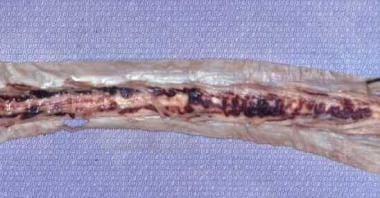 Gross photograph of the dorsal surface of the spinal cord showing dilated and tortuous vessels.
Signs and symptoms
Patients present with increasing unilateral and/or bilateral weakness, dysesthesias, and numbness or tingling in the lower extremities, which may be symmetrical or asymmetrical. [6] Early problems with bowel, bladder, and sexual function are common. [7]
After brief exertion, symptoms begin as a heavy feeling in the legs that generally improves with rest. The symptoms gradually worsen over months, and the patient may have difficulty standing for long periods. Frequent falls can be a problem. Urinary and fecal incontinence eventually occur.
Diagnosis
Catheter spinal angiography remains the criterion standard for the diagnosis of Foix-Alajouanine syndrome and spinal dural AV fistulas. It may demonstrate specific arterial feeders and draining dorsal veins.
Management
The choice of treatment for Foix-Alajouanine syndrome is either endovascular embolization or surgical ligation of the fistula; [5] in some cases, both modalities are used. [7]
Background
Foix-Alajouanine syndrome is an arteriovenous (AV) malformation of the spinal cord predominantly affecting the lower thoracic and/or lumbosacral levels; cervical cord involvement is rare. Findings include necrosis of the affected cord regions. Grey matter (as compared with white matter) structures are more severely involved. (See Etiology.)
Masses of enlarged, tortuous, thick-walled subarachnoid veins are observed overlying the surface of the cord (primarily on the posterior aspect). Smaller blood vessels with thickened fibrotic walls also are present within the affected spinal cord segments (see the image below). (See Workup.)
Gross photograph of the dorsal surface of the spinal cord showing dilated and tortuous vessels.
Foix and Alajouanine first described the syndrome in 2 young men (aged 29 y and 27 y), in 1926. [3] Historically eponymic, the syndrome now has many other names in the literature, including angiodysgenetic necrotizing myelopathy, subacute necrotizing myelopathy, and venous congestive myelopathy. [4]
Etiology
The etiology of Foix-Alajouanine syndrome is not well understood. Most patients have an AV fistula in the lower thoracic dura. One etiologic hypothesis is that the higher arterial pressure within the dura is transmitted to the spinal venous plexus via the intradural venous system, compromising perfusion and leading to infarction of the spinal cord parenchyma. Arterial blood originating from the dural fistula enters the venous system, increasing pressure and impairing normal drainage from the cord parenchyma. [4]
Thrombosis may occur, but not until late in the course of the disease. Venous stasis, not thrombosis, may be the primary cause of infarction. The preferential involvement of the distal cord is presumably due to orthostasis.
The onset in middle age suggests that the syndrome is acquired, in contrast to other vascular malformations, which are presumed to be congenital abnormalities, although the specificity for the spinal cord is not easily explained.
Pathophysiology
The enlarged, abnormal veins are associated with dural AV shunts or fistulas, usually intradurally, but rarely extradurally. [1] These AV shunts are associated with reflux of arterial blood into the venous drainage of the cord. This leads to congestion of venous outflow and results in increased venous pressure in the affected regions of the spinal cord, often leading to ischemic injury. (See Etiology and Treatment.) [2, 5]
Epidemiology
Frequency
Foix-Alajouanine syndrome is a rare entity. No specific statistics are available with regard to its frequency in the United States, but the condition is likely underdiagnosed.A retrospective study from 2001 in Germany estimated a prevalence of 5-10 cases per one million people in the general population, though this disease is probably underdiagnosed. [12]
Mortality/morbidity
Outcomes after diagnosis and treatment depend on factors such as the duration of symptoms, the pre-treatment disability, and the success of the procedure to close the fistula. Symptoms that generally improve after treatment are gait difficulties and muscle strength. Micturition, pain, and muscle spasms are symptoms that often do not respond as well. [5]
Demographics
No data is currently available indicating if Foix-Alajouanine syndrome is more prevalent in specific ethnic/racial groups.
The male to female ratio for Foix-Alajouanine syndrome is approximately 5:1. [5]
Foix-Alajouanine syndrome usually occurs in older patients (>50 y). Patients younger than 30 years are rarely reported. [2, 5]
Patient Education
Patient education
Patients should be taught proper bladder and bowel care. Education regarding the identification of early symptoms and signs of disease recurrence also is required. Educate the patient’s family and/or caregiver about proper skin care and nutrition and about patient transfer requirements. (See Presentation.)
Prognosis
Foix-Alajouanine syndrome is a subacute disorder that gradually evolves over 1-5 years. [2] Affected patients initially are spastic but eventually develop flaccid paralysis of the limbs and may become wheelchair bound. Death can occur with terminal sepsis or other sequelae.
The prognosis is poor if treatment is not administered before neurologic deterioration occurs. Successful embolization and/or surgical interruption of the draining veins can halt the progression of symptoms and provide clinical improvement in many patients. Operations, if successful, are permanent modalities of treatment. After embolization, recanalization may occur, but this is rarely seen if the draining vein is filled with glue. [5]
-
Gross photograph of the dorsal surface of the spinal cord showing dilated and tortuous vessels.
-
Photomicrograph of the cervical spinal cord region showing a thickened subarachnoid vein with a thrombotic occlusion (hematoxylin and eosin stain).
-
Photograph of the cervical spinal cord illustrating dilated, abundant subarachnoid veins (hematoxylin and eosin stain).
-
Photomicrograph of the cervical spinal cord region demonstrating several dilated, hyalinized intraparenchymal vessels (hematoxylin and eosin stain).
-
Photomicrograph of the cervical spinal cord depicting ischemic necrosis of the parenchyma (hematoxylin and eosin stain).


