You are in: eMedicine Specialties >
Neurology > Neurological Infections
Viral Meningitis
Article Last Updated: Aug 23, 2007
AUTHOR AND EDITOR INFORMATION
Section 1 of 11
Author: Amir Vokshoor, MD, Staff Neurosurgeon, Department of Neurosurgery, Spine Surgeon, Diagnostic and Interventional Spinal Care, Saint John's Health Center
Amir Vokshoor is a member of the following medical societies: Alpha Omega Alpha, American Association of Neurological Surgeons, and American Medical Association
Coauthor(s):
Cordia Wan, MD, Resident Physician, Department of Neurology, Kaiser Permanente, Los Angeles Medical Center
Editors: J Stephen Huff, MD, Associate Professor of Emergency Medicine and Neurology, Department of Emergency Medicine, University of Virginia Health Sciences Center; Francisco Talavera, PharmD, PhD, Senior Pharmacy Editor, eMedicine; Florian P Thomas, MD, MA, PhD, Drmed, Director, Spinal Cord Injury Unit, St Louis Veterans Affairs Medical Center; Associate Program Director, Associate Professor, Departments of Neurology, Molecular Virology, and Molecular Microbiology and Immunology, St Louis University School of Medicine; Matthew J Baker, MD, Consulting Staff, Collier Neurologic Specialists, Naples Community Hospital; Nicholas Y Lorenzo, MD, Chief Editor, eMedicine Neurology; Consulting Staff, Neurology Specialists and Consultants
Author and Editor Disclosure
Synonyms and related keywords:
aseptic meningitis, serous meningitis, nonpyogenic leptomeningitis, abacterial meningitis, enterovirus, coxsackievirus, echovirus, viral meningitis, viral infection, herpes viruses, HSV-1, HSV-2, varicella zoster virus, VZV, B virus
Background
Viral meningitis is the inflammation of leptomeninges as a manifestation of CNS infection. Viral names the causative agent, and the term meningitis implies lack of parenchymal and spinal cord involvement (otherwise called encephalitis and myelitis, respectively). Certainly, viral pathogens can cause a combination of meningoencephalitis or meningomyelitis, and a partially treated bacterial meningitis can present with an aseptic (or nonbacterial) picture suggestive of viral meningitis.
In true viral meningitis, the clinical course is usually self-limited, with complete recovery in 7-10 days.
More than 85% of cases today are caused by nonpolio enteroviruses; thus, disease characteristics, clinical manifestations, and epidemiology mimic those of enteroviral infections.
Mumps, polio, and lymphocytic choriomeningitis viruses (LCMV) are now rare offenders in developed countries.
Polio remains a major cause of debilitating myelitis in some regions of the world. For the clinician, consideration of other meningitis causes, such as bacteria, mycoplasma, or fungi, is crucial, since these can have devastating outcomes if left untreated. The physician should also realize that the picture of aseptic meningitis is created not only by infectious agents, but also by chemical irritation (chemical meningitis), neoplasm (meningitis carcinomatous), granulomatous disorders, and any other inflammatory conditions. This discussion, however, will focus on meningitis by viral agents.
Pathophysiology
Viral pathogen may gain access to the CNS via 2 major routes: hematogenous or neural. Hematogenous is the most common route for penetration of most known viral pathogens. Neural penetration refers to spread along nerve roots and usually is limited to herpes viruses (HSV-1, HSV-2, and varicella zoster virus [VZV] B virus), and possibly some enteroviruses. Multiple host defenses prevent viral inoculum from causing clinically significant infection. These include local and systemic immune responses, skin and mucosal barriers, and the blood-brain barrier (BBB). The virus replicates in the initial organ system (ie, respiratory or gastrointestinal mucosa) and gains access to the bloodstream. Primary viremia introduces the virus to the reticuloendothelial organs (liver, spleen, and lymph nodes.) If the replication persists despite immunologic defenses, secondary viremia occurs, which is thought to be responsible for seeding of the CNS. Rapid viral replication likely plays a major role in overcoming the host defenses. The actual mechanism of viral penetration into the CNS is not well understood. The virus may cross the BBB directly at the capillary endothelial level or through natural defects (ie, area postrema and other sites that lack a BBB). The inflammatory response is seen in the form of pleocytosis; polymorphonuclear leukocytes (PMNs) lead the differential cell count in the first 24-48 hours, followed later by increasing numbers of monocytes and lymphocytes. The cerebrospinal fluid (CSF) lymphocytes have been recognized as T cells, although B cell immunity is also important in defending against some viruses. Evidence exists that some viruses may gain access to the CNS by retrograde transport along nerve roots. For example, the likely pathway for HSV-1 encephalitis is via the olfactory or trigeminal nerve roots, with the virus being transported by the olfactory fibers to the basal frontal and anterior temporal lobes.
Frequency
United States
More than 10,000 cases are reported annually, but the actual incidence may be as high as 75,000. Lack of reporting is due to the uneventful clinical outcome of most cases and the inability of some viral agents to grow in culture. According to CDC reports, inpatient hospitalizations resulting from viral meningitis range from 25,000-50,000 each year. An incidence of 11 per 100,000 population per year has been estimated in some reports.
International
Obtaining accurate international prevalence and incidence of this clinically heterogenous and often benign disease is difficult. Worldwide causes of viral meningitis include enteroviruses, mumps virus, measles virus, VZV, and HIV. Meningitis symptoms may develop in as few as 1 in 3,000 cases of infection by these agents. Studies from Finland have estimated the incidence to be 19 per 100,000 population in children aged 1-4 years. This is in significant contrast to 219 cases per 100,000 population estimated for children younger than 1 year. Japanese B encephalitis virus, the most common pathogen in epidemic viral meningitis worldwide, accounts for more than 35,000 infections annually throughout Asia but is estimated to cause 200-300 times that number of subclinical infections. The distribution and attack characteristics of some agents, such as arboviruses, which are transmitted by arthropod vectors, show strong geographic variability. Lack of effective vaccination policies in some Third World countries plays a role in the geographic discrepancy of other infectious agents.
Mortality/Morbidity
- Excluding the neonatal period, the mortality rate associated with viral meningitis is less than 1%; the morbidity rate is also low.
- Some controversy exists as to the long-term effects on children, with some studies attributing learning disabilities, neuromuscular impairments, and deafness to viral meningitis. Investigators believe that most of these cases must involve the CNS parenchyma, causing encephalitis or encephalomyelitis. Children may complain of irritability, incoordination, and inability to concentrate for several weeks or longer. Infants with enteroviral meningitis during the first few months of life may have an increased risk of altered language development.
- Physicians must realize that viruses capable of causing meningitis also can cause more serious infections of the CNS as well as other organs. The World Health Organization (WHO) statistical reports from 1997 reported enteroviral meningitis with sepsis as the fifth most frequent cause of neonatal mortality. Complications such as brain edema, hydrocephalus, and seizures can occur in the acute period and are discussed later in this article.
Race
No specific racial predilection has been identified.
Sex
Depending on the type of viral pathogen, the ratio of affected males to females can vary. Enteroviruses are thought to affect males 1.3-1.5 times more often than females. Mumps virus is known to affect males 3 times more frequently than females. Most arboviruses have diverse attack characteristics, affecting both sexes but at different ages.
Age
- The incidence of viral meningitis drops with age.
- Neonates are at greatest risk and have the most significant risk of morbidity and mortality.
- In neonates older than 7 days, enteroviruses are the most common cause of aseptic meningitis. Vaccination has greatly reduced the incidence of meningitis from mumps, polio, and measles viruses.
- The incidence during the first year of life is 20 times higher than in older children and adults.
- Some of the arboviruses strike at the extremes of age, with the elderly at greater risk of infection, while mumps and measles peak in the later teenage years.
History
- Upon presentation, most patients report fever, headache, irritability, nausea, vomiting, stiff neck, rash, or fatigue within the past 18-36 hours.
- Headache is almost always present and often reported as severe. However, the classic description of abrupt onset "worst headache of my life," attributable to aneurysmal subarachnoid hemorrhage, is uncommon.
- Constitutional symptoms of vomiting, diarrhea, cough, and myalgias appear in more than 50% of patients.
- History of temperature elevation occurs in 76-100% of patients who come to medical attention. A common pattern is low-grade fever in the prodromal stage and higher temperature elevations at the onset of neurological signs.
- Younger children may not report headache and may simply be irritable.
- Newborns may present with poor feeding and lethargy.
- Some viruses cause rapid onset of the above symptoms, while others manifest as nonspecific viral prodromes, such as malaise, myalgia, and upper respiratory symptoms. In many cases, symptoms have a biphasic pattern; the nonspecific flu-like symptoms and low-grade fever precede neurologic symptoms by approximately 48 hours. With the onset of neck stiffness and headache, the fever usually returns.
- Meticulous history taking is essential and must include evaluation of exposure to ill contacts, mosquitoes, ticks, outdoor activity in areas of endemic Lyme disease, travel history with possible exposure to tuberculosis, as well as history of medication use, intravenous drug use, and sexually transmitted disease risk.
- An important part of history is prior antibiotic use, which may alter the clinical picture of bacterial meningitis.
Physical
General physical findings in viral meningitis are common to all causative agents, but some viruses have unique clinical manifestations that help in focusing diagnostic approach. The classically taught triad of meningitis include fever, nuchal rigidity, and altered mental status, though not all patients have all three symptoms, and headache is almost always present. The examination reveals no focal neurological deficits in the majority of cases.
- Fever is common (80-100% of cases) and usually ranges between 38ºC and 40ºC.
- Nuchal rigidity or other signs of meningeal irritation (Brudzinski or Kernig sign) may be seen in more than half of patients but is generally less severe than in bacterial meningitis. Pediatric patients, especially neonates, tend not to exhibit nuchal rigidity on examination.
- Irritability, disorientation, and altered mentation may be seen. Severe lethargy or bulging fontanelle in neonates are signs of increased intracranial pressure but may be absent in more than half of cases. The neonate may exhibit hypotonia, irritability, and poor feeding. The clinical picture can mimic neonatal bacterial septicemia accompanied by multiple organ system involvement.
- Headache is common and severe.
- Photophobia is relatively common but may be mild. Phonophobia may also be present.
- Seizures occur occasionally and are usually from the fever, although the involvement of brain parenchyma (encephalitis) should be considered. Global encephalopathy and focal neurological deficits are rare but can be present. Deep tendon reflexes are usually normal but may be brisk.
- Other signs of specific viral infection can aid in diagnosis. These include pharyngitis and pleurodynia in enteroviral infections, skin manifestations such as zoster eruption in VZV, maculopapular rash from measles and enteroviruses, vesicular eruption by herpes simplex, and herpangina in coxsackievirus A infections. Epstein-Barr virus (EBV) infections are suggested by pharyngitis, lymphadenopathy, and splenomegaly. Immunodeficiency and pneumonia should suggest adenovirus, cytomegalovirus (CMV), or HIV as the causative agent. Parotitis and orchitis can occur with mumps, while most enteroviral infections have associated gastroenteritis and rash.
Causes
Multiple viruses are capable of causing meningitis. This discussion attempts to simplify the microbiology of each viral family with emphasis on disease manifestations and risk factors. Note that in as many as one third of cases, no causative agents are identified. This number is improving with new testing methodologies. - Enteroviruses account for more than 85% of all cases of viral meningitis. They are part of the viral family Picornaviridae ("pico" for small, "rna" for ribonucleic acid), and include echoviruses, coxsackieviruses A and B, polioviruses, and the numbered enteroviruses. Nonpolio enteroviruses are common viruses, nearly as prevalent as rhinoviruses (the common cold).
- The overwhelming majority of meningitis cases are caused by serotypes of coxsackie and echoviruses. Coxsackievirus B subgroups alone account for more than 60% of meningitis cases in children younger than 3 months.
- Enteroviruses enter the human host usually via the oral-fecal route, but can also spread through the respiratory route.
- Enteroviruses are ubiquitous in the summer and early fall; their propensity to cause infection during the warmer months is the major factor in the higher incidence of aseptic meningitis during that time.
- The associated clinical findings in enteroviral infections may include pharyngitis, pleurodynia, rash, and pericarditis.
- Expectant mothers infected with coxsackievirus B may remain minimally symptomatic, but their infants can acquire the infection perinatally and develop a potential fatal illness, with the infection targeted mainly toward the heart.
- Enteroviruses numbers 70 and 71, which exhibit strong neurotropism, are associated with meningoencephalitis, poliolike paralytic syndromes, and Guillain-Barre syndrome, as well as aseptic meningitis.
- Arboviruses account for about 5% percent of cases in North America
- Arboviruses consist of more than 500 viruses from different viral families, all given the common name of "ar-bo" for arthropod-borne disease. Blood-sucking arthropods, usually mosquitoes, serve as vectors for transmission.
- Since exposure to mosquito or ticks is the risk factor for transmission, the number of infections is highest in summer and early fall in concordance with high mosquito populations.
- Some of the important arboviruses include the eastern and western equine encephalitis viruses in the Togavirus family; St. Louis encephalitis, West Nile, Japanese B, and Murray Valley viruses of the Flavivirus family; and California group and Jamestown Canyon viruses from the Bunyaviridae family. Colorado tick fever is caused by a coltivirus in the western regions of the United States.
- The most common clinical manifestation is meningoencephalitis rather than pure meningitis
- Seizures are more common with arboviral meningitis than any other group of viruses.
- Some agents preferentially infect certain age groups, such as St. Louis encephalitis, which affects the extremes of age, and California virus, which infects young children.
- Children with St. Louis or California group encephalitis viruses may not exhibit any neurological signs or altered mental status.
- St. Louis encephalitis (SLE) virus is the most common cause of arboviral meningitis, and is also the most common overall mosquito-transmitted disease in the United States. Japanese B virus is the biggest offender of this group internationally.
- Of the arboviruses, West Nile virus caused much recent attention as it was first recognized in the United States only in 1999 and quickly became an epidemic in 2002 with more than 4,000 reported cases.
- Infection with the West Nile virus is usually asymptomatic or manifests as mild symptoms of nonspecific fever, myalgia, fatigue. However, 1 in 150 cases develop into severe disease involving the nervous system, with encephalitis reported more than meningitis.
- These cases of neuroinvasive West Nile disease occur more often in elderly persons.
- Cases of West Nile neuroinvasive disease were highest in the states of Idaho, Illinois, and Texas during 2006. States with higher incidences of West Nile infection overall also included California, Colorado, Louisiana, and Mississippi.
- Mumps: A member of the Paramyxovirus family, mumps virus was one of the first known causative agents of meningitis and meningoencephalitis.
- The incidence of mumps in the vaccination era has decreased significantly to 1 per 100,000 population in the United States. Nonetheless, mumps continues to be the cause in 10-20% of cases in parts of the world where vaccines are not readily accessible.
- Males aged 16-21 years are at highest risk of developing this infection, with a 3:1 male/female ratio.
- Clusters of cases occur in schools and colleges in the winter months.
- Concomitant parotitis is a helpful clinical tool but may be absent in as many as half of cases with CNS involvement.
- A cohort study of 12,000 unvaccinated children from northern Finland revealed mumps meningoencephalitis accounting for 40.9% of all viral CNS infections. It also remains an important cause of aseptic meningitis in England and Japan.
- in 2003, epidemics of aseptic meningitis following MMR vaccination campaigns in various nations (including Brazil and the UK) prompted the Global Advisory Committee on Vaccine Safety to conduct a review of vaccine-derived mumps meningitis. At the time, the committee stated that certain strains of the mumps vaccine (Urabe, Leningrad-Zagreb, and Leningrad-3 strains) were associated with higher incidences of postvaccination aseptic meningitis. Later in 2006, the committee determined that the international literature reviewed were actually inconclusive and further studies were needed. Even so, replacement mumps components were developed and vaccines reformulated worldwide.
- Herpes family viruses: HSV-1, HSV-2, VZV, EBV, CMV, and human herpesvirus 6 collectively cause approximately 4% of cases of viral meningitis, with HSV-2 being the most common offender.
- They may attack any time of the year.
- The disease is often self-limited. When associated with encephalitis, however, mortality rate can be high, and early treatment with acyclovir can significantly reduce morbidity.
- HSV-1 remains the most common cause of sporadic encephalitis, while HSV-2 infections of CNS mostly are restricted to aseptic meningitis.
- HSV-2 genital infection often precedes meningitis; sexual contact with actively infected individuals is one of the known risk factors. Maternal-fetal transmission can occur, leading to significant systemic sequelae including infantile septicemia and death.
- EBV, HSV-1, and especially HSV-2 have been associated with Mollaret meningitis, a rare, benign, recurrent meningitis that resolves spontaneously. Mollaret cells (activated monocytes with atypical appearance of enlarged, bilobed nuclei and amorphous cytoplasm) are found in the CSF usually on the first day of symptoms. Herpesvirus 6, EBV, and HIV have also been implicated. These viruses are all known to remain latent within the nervous system.
- CMV infections occur mostly in immunocompromised hosts. CMV may cause a subacute encephalitis in patients with AIDS. Congenital CMV, which is a much more serious form of infection, has significant associated morbidity and mortality.
- Childhood or adult chickenpox infections by VZV rarely are complicated by meningitis. Adult zoster involving any dermatome may lead to meningitis or meningoencephalitis.
- Lymphocytic choriomeningitis virus: LCMV belongs to the family of arenaviruses. Now a rare cause of meningitis, the virus is transmitted to humans by contact with rodents (eg, hamster, rats, mice) or their excreta. Those at highest risk are laboratory workers, pet owners, or persons living in nonhygienic areas.
- Adenovirus: Adenovirus is a rare cause of meningitis in immunocompetent individuals but a major cause in AIDS patients. The infection may occur simultaneously with an upper respiratory infection.
- Measles: This Morbillivirus is another now rare cause. The characteristic maculopapular rash aids in the diagnosis. Most cases occur in younger people in schools and colleges. Measles remains a worldwide health threat with the highest attack rate of any infection; eradication of measles is an important public health goal of the WHO.
- HIV: HIV may be a cause of atypical meningitis characterized by chronicity and recurrence. About the time of seroconversion, patients may present with CSF pleocytosis, elevated protein level and, occasionally, high intracranial pressure. Reports have suggested that as many as 5-10% of HIV infections can be heralded by meningitis. Aside from the usual meningeal signs, HIV infections may also cause global encephalopathy, seizures, and focal neurologic deficits. Some patients develop chronically abnormal CSF findings with mild or no symptoms. HIV often can be isolated from the CSF.
- Tuberculous, fungal, mycoplasmal, and other causes of nonbacterial meningitis are not included in this discussion since they are not viruses. However, they are important causes of aseptic meningitis and should be suspected in the appropriate clinical setting. For example, Lyme borreliosis causes a significant number of cases of aseptic meningitis in the Northeast and Mid-Atlantic states. The diagnosis is suggested by the history of tick bite or outdoor activity in these areas of endemic disease, and presence of erythema chronicum migrans at the site of tick bite is pathognomonic. Lyme meningitis has a predilection to cause focal cranial nerve palsies, with the seventh nerve most commonly affected.
- Clinicians must consider partially treated bacterial meningitis as possible etiology for the aseptic nature of their patient's disease; for example, patients with bacterial otitis and sinusitis who have been taking antibiotics may present with meningitis and CSF findings identical to viral meningitis.
Abnormal Neonatal EEG
Acute Disseminated Encephalomyelitis
Aseptic Meningitis
Brucellosis
Cytomegalovirus Encephalitis
EEG in Status Epilepticus
Haemophilus Meningitis
Herpes Simplex Encephalitis
HIV-1 Associated CNS Conditions: Meningitis
HIV-1 Associated Opportunistic Infections: CNS
HIV-1 Associated Opportunistic Infections: CNS Cryptococcosis
HIV-1 Associated Opportunistic Infections: CNS Toxoplasmosis
Hydrocephalus
Leptomeningeal Carcinomatosis
Low-Grade Astrocytoma
Lumbar Puncture (CSF Examination)
Lyme Disease
Migraine Variants
Neurocysticercosis
Neurosarcoidosis
Neurosyphilis
Staphylococcal Meningitis
Subdural Empyema
Systemic Lupus Erythematosus
Tuberculous Meningitis
Varicella Zoster
Other Problems to be Considered
Partially treated bacterial meningitis
Parameningeal infection
Coccidioides immitis infection
Cryptococcus neoformans infection
Histoplasma capsulatum infection
Candida species infection
Blastomyces dermatitidis infection
Mycoplasma infection
Listeria infection
Leptospira infection
Drugs
Heavy metals
Surgically implanted materials
Sjögren syndrome
Behçet disease
EEG in neurological infections
Lab Studies
- Routine chemistry and hematology tests should be performed.
- In neonatal and severe cases, arterial blood gases analysis, coagulation studies, and liver function tests should also be considered.
- Serum WBC count is not a sensitive indicator of the severity of infection, especially in the immunocompromised, neonatal, or elderly patient.
- Serum sodium level may be abnormal because of dehydration or the rare occurrence of syndrome of inappropriate antidiuretic hormone secretion (SIADH).
- Serum amylase level may be elevated in cases caused by mumps even in the absence of parotitis.
- Recent reports have shown high C-reactive protein (CRP) levels in the serum of children with bacterial meningitis whose CSF Gram stain findings were negative for bacteria; a comparable group of children with viral meningitis did not have similar elevations in serum CRP (ie, 50-150 in bacterial meningitis group vs <20 in viral meningitis group).
- CSF examination is the most important test in differentiating the cause of meningitis. CT scan should be performed in cases associated with any abnormal neurological sign to exclude an intracranial lesion or obstructive hydrocephalus prior to lumbar puncture (LP). CSF culture remains the criterion standard in discerning bacterial or pyogenic from aseptic meningitis. Again, a partially treated bacterial meningitis may present with a negative Gram stain result and thus appear aseptic. The following are some CSF characteristics used to support the diagnosis of viral meningitis:
- Cells: Pleocytosis with WBC counts in the range of 50 to >1000 x 109/L of blood has been reported in viral meningitis. Mononuclear cell predominance is the rule, but PMNs may comprise the majority of cells in the first 12-24 hours; the cell count usually is then dominated by lymphocytes in the classic CSF pattern of viral meningitis. This helps to distinguish viral from bacterial meningitis, which has a much higher cell count and a predominance of PMNs in the cell differential; this is by no means an absolute rule, however.
- Protein: CSF protein level usually is only slightly elevated, but can range from being normal to as high as 200 mg/dL.
- Glucose: Normal in most cases, but severe hypoglycorrhachia has been reported, especially with LCMV or mumps virus. Very low glucose levels with a lymphocytic pleocytosis may be seen in tuberculous meningitis.
- Culture, Gram stain, and acid-fast stain: These tests should be performed, as well as polymerase chain reaction (PCR) testing for the likely viral pathogens in selected cases.
- Antigens: CSF latex antigen testing helps to rule out bacterial causes such as Haemophilus influenzae and Neisseria meningitidis. Addition of a drop of CSF sediment to an India ink preparation may aid in the diagnosis of cryptococcal meningitis, although antigen assay testing for cryptococci is the preferred test.
- Consider saving CSF for less common tests (ie, PCR for HIV and CMV) if the cause of meningitis is not certain after initial tests.
- The exact sequence of testing for these agents depends on the clinical condition and suggestive facts in history and examination. For example, most cases of viral meningitis do not require PCR testing for HIV.
- High WBC count in the CSF (especially neutrophils), high protein level, and low glucose level should suggest the diagnosis of a bacterial meningitis, although some viral pathogens may present with similar CSF profiles.
- The Xpert EV test, approved for use in March 2007, can rapidly test for enteroviral meningitis. The test uses a reverse-transcription PCR disposable cartridge on which CSF is applied, and enteroviral genetic material is identified if present. Results are ready in 2.5 hours, as opposed to days to weeks in traditional PCR studies. No published trials are available at the time of this article, but per FDA's statement, 255 patients were tested across 6 medical institutes; 96% of patients who tested positive did have viral meningitis, and 97% of patients who tested negative did not have viral meningitis. This quick test may become useful in directing treatment.
- Also in 2007, JAMA published a Bacterial Meningitis Score that clinically predicts whether patients were at high or low risk for having bacterial meningitis versus aseptic meningitis. Patients had a very low risk for bacterial meningitis if all of the following were absent: positive CSF Gram stain, CSF absolute neutrophil count (ANC) of ≥1000, CSF protein of ≥80 mg/dL, a peripheral ANC of ≥10,000 cells/mcL, or a seizure before or at time of presentation. The higher number of criteria present, the higher the chance the patient has bacterial meningitis. This bacterial meningitis score may be used as an additional tool to aid in further management.
Imaging Studies
- Imaging for suspected viral meningitis and encephalitis may include CT scan of the head with and without contrast, or MRI of the brain with gadolinium.
- CT scan with contrast helps in ruling out intracranial pathology. Contrasted scans should be obtained to evaluate for any enhancement along the meninges and to exclude cerebritis, intracranial abscess, subdural empyema, or other lesions. Alternatively, and if readily available, an MRI of the brain with gadolinium may be performed.
- MRI with contrast is the criterion standard in visualizing intracranial pathology in viral encephalitis (see Media file 1). HSV-1 commonly affects basal frontal and temporal lobes with a typical picture of diffusely enhancing bilateral lesions.
Other Tests
- All patients whose condition is not improving clinically within 24-48 hours should have more extensive work-up to discern the cause of meningitis.
- Blood, feces, and throat swabs may be sent for viral serology and cultures.
- Acid-fast staining of CSF should be performed and the remaining fluid should be sent for PCR testing for HIV and CMV.
- Serum titers of antibodies against HIV and toxoplasma should be obtained.
- Additional serum collection 10-21 days later may aid in discerning rising titers in the antibodies against specific viral pathogens; a 4-fold increase in viral antibodies confirms the diagnosis. This is particularly useful for arboviral and LCMV cases, but also is helpful in ruling out toxoplasmosis, leptospirosis, borreliosis, and rickettsial infections. Although some of these studies do not yield an immediate result for clinical decision making, they may be useful for prognostication.
- In case of suspected encephalitis, MRI with contrast enhancement and adequate visualization of the basal frontal and temporal areas is necessary.
- EEG may be performed if encephalitis or subclinical seizures are suspected in the altered patient. Periodic lateralized epileptiform discharges (PLEDs) are often seen in herpetic encephalitis.
Procedures
- Lumbar puncture is the most important procedure used in diagnosis of viral meningitis. Other potential procedures, depending on individual indications and disease severity, include intracranial pressure monitoring, brain biopsy, and ventricular drainage or shunting.
- CT scan usually is performed prior to LP to rule out intracranial hematoma, mass effect, or obstructive hydrocephalus. The LP itself may provide significant symptomatic relief, presumably due to the decrease in intracranial pressure. LP should be performed in the standard sterile fashion, and the CSF opening pressure should be measured. Coagulopathy due to intrinsic or extrinsic factors (eg, warfarin) is a relative contraindication to LP. The clinician should exercise caution and, as for all medical procedures, weigh the risks and benefits associated with each individual case.
- Intracranial pressure monitoring rarely is needed in patients with meningoencephalitis complicated by cerebral edema. The risks of intracranial hemorrhage in cases with coagulopathy often outweigh the diagnostic benefit of the monitor. The monitor should be placed under strictly sterile conditions by a neurosurgeon or neuro-intensivist.
- Operative brain biopsy for confirmation of herpetic encephalitis largely has been replaced by PCR testing for viral DNA. In some cases, encephalomalacia due to an unknown viral infection may be confused with vascular infarction or rarely a tumor; a biopsy may be helpful. With the use of stereotactic localization and a needle biopsy, morbidity is minimal.
Histologic Findings
Because of the low mortality rate associated with acute viral meningitis, pathologic features other than lymphocytic response within the CSF are generally not in evidence. The leptomeninges undergo inflammation with PMNs and later mononuclear cells in the acute phase of the disease. Perivascular cuffing, neuronophagia, and increased number of microglial cells have been noted in specimens from patients who died of viral encephalitis.
Medical Care
Treatment for viral meningitis is mostly supportive. Rest, hydration, antipyretics, and pain or anti-inflammatory medications may be given as needed. The most important decision is whether to initiate antimicrobial therapy empirically for bacterial meningitis while waiting for the cause to be identified. Intravenous (IV) antibiotics should be administered promptly if bacterial meningitis is suspected. Patients with signs and symptoms of meningoencephalitis should receive acyclovir early to possibly curtail HSV encephalitis. Therapy can be modified as the results of Gram stain, cultures, and PCR testing become available. Patients in unstable condition need critical care unit admission for airway protection, neurologic checks, and prevention of secondary complications. Enteroviruses and HSV are both capable of causing viral septic shock in newborns and infants. In these young patients, broad-spectrum antibacterial coverage and acyclovir should be instituted as soon as the diagnosis is suspected. Special attention should be paid to fluid and electrolyte balance (especially sodium), since SIADH has been reported. Fluid restriction, diuretics, and rarely hypertonic saline infusion may be used to correct the hyponatremia. Prevention of secondary infections of urinary tract and pulmonary systems is of paramount importance.
- Waiting for lumbar puncture results should not delay administration of antibiotics when warranted on clinical grounds. Broad-spectrum coverage is attained with ampicillin and a third-generation cephalosporin (ceftriaxone or cefotaxime; ceftazidime can also be used). Aminoglycosides are used in severe infections in neonates or children. Antituberculous, antifungal, and antiretroviral medications are reserved for clinically-suggested or laboratory-confirmed cases. Please see the article on bacterial meningitis for specific recommendations.
- Seizures should be treated immediately with IV anticonvulsants such as lorazepam, phenytoin, midazolam, or a barbiturate. Unconscious patients with viral encephalitis may be in nonconvulsive status epilepticus, and EEG is used to reveal and monitor subclinical seizures. Cerebral edema does occur in cases of severe encephalitis and may require intracranial pressure control by infusion of mannitol (1 g/kg initial dose followed by 0.25-0.5 g/kg q6h), IV dexamethasone, or intubation and mild hyperventilation, with arterial PCO2 around 28-30 mm Hg. Placement of an intracranial pressure monitor with transduced intraparenchymal pressure is recommended in these cases.
- Multiple antiviral medications are currently being tested in the general population; their impact on preventing the potential rare sequelae of viral meningitis have not yet been established.
- In herpetic viral infections, use of acyclovir is significantly beneficial only if given very early in the course of the infection. Suspected cases should be treated as soon as possible; in cases complicated by seizures, encephalitis is assumed and acyclovir should be initiated.
- Anti-HIV therapy is initiated when the history or associated risk factors suggest the early phases of HIV meningoencephalitis.
- Ganciclovir for CMV-related infections is reserved for severe cases with positive CMV culture or when a congenital infection or an AIDS-related infection is strongly suspected.
- Administration of IVIg to neonates with overwhelming enteroviral meningitis has met with occasional success and is reserved for severe cases lacking other therapeutic options.
Surgical Care
No surgical therapy is usually indicated. In rare patients in whom viral meningitis is complicated by hydrocephalus, a CSF diversion procedure, such as ventriculoperitoneal (VP) or LP shunting, may be required. Ventriculostomy with an external collection system is indicated in the rare cases of acute hydrocephalus. Occasionally meningeal or parenchymal biopsy for definitive diagnosis of rare viral infections is required. Intracranial pressure monitoring, required for some cases of encephalitis, usually can be performed at bedside.
Consultations
- Neurology - Seizure control, EEG, management of brain edema in refractory cases, neuro-intensive care
- Neurosurgery - Placement of intracranial pressure monitor, CSF shunting or temporary drainage in cases with hydrocephalus, neuro-intensive care
- Infectious disease - Control of epidemics, isolation of patient and contacts, choice of antibiotics in refractory or atypical cases
- Neonatology - Any newborn or infant with severe viral meningitis requiring intensive care
Diet
No special diet is required.
Activity
The activity limitations should be individualized based on each patient's clinical picture. Bed rest is recommended for the acute phase of infection.
Symptomatic control with antipyretics, analgesics, and antiemetics is usually all that is needed in the management of uncomplicated viral meningitis. The decision to start antibacterial therapy for treatment of possible bacterial meningitis is the most crucial; empiric antibacterial therapy for likely pathogens should be considered in the context of the clinical setting (see articles on bacterial meningitis for details). Acyclovir should be used in cases suspicious for HSV (patients with herpetic lesions), and is usually used empirically in more severe cases complicated by encephalitis or sepsis.
Anti-HIV therapy is initiated when the history is strongly suggestive and/or confirmatory tests have proven an infection. These medications are covered in other articles. Ganciclovir for CMV-related infections is reserved for severe cases with positive CMV culture, congenital infection, or immunocompromised patients.
Pleconaril is an antipicornavirus drug that held potential for treatment of enteroviral meningitis. However, to date, no study has demonstrated clear efficacy, and pleconaril trials have now shifted focus to treatment of rhinovirus upper respiratory infections.
Administration of IVIg to neonates with overwhelming enteroviral meningitis has met with only occasional success and is not covered in this section. For anticonvulsant therapy, refer to articles covering status epilepticus and pediatric seizure disorders.
Drug Category: Antiemetic agents
These agents are used mostly to prevent chemotherapy-induced nausea and vomiting.
| Drug Name | Ondansetron (Zofran) |
| Description | Selective 5-HT3-receptor antagonist that blocks serotonin both peripherally and centrally. Has efficacy in patients who do not respond well to other antiemetics. |
| Adult Dose | 4-8 mg IV q8h/q12h |
| Pediatric Dose | 0.1 mg/kg slow IV maximum of 4 mg/dose; may repeat q12h |
| Contraindications | Documented hypersensitivity |
| Interactions | Stimulators of cytochrome P-450 system, including barbiturates, rifampin, phenytoin, and phenylbutazone, change clearance; inhibitors of hepatic P-450 system, such as cimetidine, allopurinol, and disulfiram, increase toxicity |
| Pregnancy | B - Fetal risk not confirmed in studies in humans but has been shown in some studies in animals
|
| Precautions | Should be scheduled rather than given prn; data support prevention of chemotherapy-induced nausea and vomiting rather than delayed treatment; administer for prevention of nausea and vomiting, not for rescue of nausea and vomiting |
| Drug Name | Droperidol (Inapsine) |
| Description | Neuroleptic agent that may reduce emesis by blocking dopamine stimulation of chemoreceptor trigger zone. Also has antipsychotic and sedative properties. |
| Adult Dose | 2.5-5 mg IV/IM q4-6 prn |
| Pediatric Dose | <6 months: Not established > 6 months: 0.05-0.06 mg/kg/dose IV/IM q4-6 prn |
| Contraindications | Documented hypersensitivity |
| Interactions | Atropine and lithium increase toxicity; fentanyl and other analgesics may cause increased BP (administration with epinephrine may decrease BP) |
| Pregnancy | C - Fetal risk revealed in studies in animals but not established or not studied in humans; may use if benefits outweigh risk to fetus
|
| Precautions | Safety in children <6 mo not established; caution in patients with seizures, bone marrow suppression, or severe liver disease; significant hypotension can occur; tardive dyskinesia, extrapyramidal reactions, Parkinson-like state, and akathisia have been reported, especially in elderly; orthostatic hypotension and altered state of mind can occur, especially in elderly May cause QT prolongation (delayed recharging of heart between beats) within minutes following injection at doses at or below recommended levels; prolonged QT can cause potentially fatal heart arrhythmia known as torsades de pointes (TdP); all patients should undergo a 12-lead ECG prior to administration of drug to determine if QT interval is prolonged (ie, QTc >440 msec for males or 450 msec for females); if QT interval is prolonged, droperidol should not be administered; for patients in whom potential benefit of droperidol treatment is felt to outweigh risks of potentially serious arrhythmias, ECG monitoring should be performed prior to treatment and continued for 2-3 h after completing treatment to monitor for arrhythmias |
Drug Category: Antiviral agents
Anti-enteroviral therapy is under investigation for viral meningitis and may soon become available. Anti-HIV and anti-tuberculosis regimens are not covered here, but should be instituted if these infections are strongly suggested clinically or confirmed by testing. Empiric therapy can be discontinued once the cause of viral meningitis has been established and bacterial meningitis excluded.
| Drug Name | Acyclovir (Zovirax) |
| Description | To be started as soon as diagnosis of herpetic meningoencephalitis suspected. Inhibits activity of both HSV-1 and HSV-2. |
| Adult Dose | 30 mg/kg/d IV divided q8h for 10-14 d |
| Pediatric Dose | 30 mg/kg/d IV divided q8h for 10 d |
| Contraindications | Documented hypersensitivity |
| Interactions | Probenecid or zidovudine prolongs half-life and increases CNS toxicity |
| Pregnancy | C - Fetal risk revealed in studies in animals but not established or not studied in humans; may use if benefits outweigh risk to fetus
|
| Precautions | Caution in renal failure or when using nephrotoxic drugs |
Further Inpatient Care
- If the CSF Gram stain result is negative but moderate to severe pleocytosis is noted (WBC >1000 x 109/L), a repeat LP should be considered in 12-18 hours if patient is not clinically improved. All patients with suspected bacterial meningitis should be treated empirically with appropriate antibiotics. After the bacterial Gram stain, latex antigen tests, and cultures return negative, antibacterial therapy can be discontinued. If results of PCR testing of CSF and viral culture for herpes simplex are negative, acyclovir can be discontinued; otherwise a 10-day course is recommended. If no clinical improvement is noted and all the common bacterial and viral pathogens have been ruled out, the following tests should be performed and the therapy modified depending on their results:
- CSF - Venereal Disease Research Laboratories test (VDRL), PCR for CMV, acid-fast stain
- Skin - Purified protein derivative (PPD) to help exclude tuberculosis
- Blood - HIV antibody and PCR, rapid plasma reagent (RPR), Lyme antibody (in areas of endemic disease or if history suggestive), toxoplasmosis antibody (especially in infants and newborns)
- Prevention of secondary infections, control of seizures, management of electrolyte abnormalities such as SIADH, and adequate nutritional support are paramount for successful management of these patients.
Further Outpatient Care
- Although most patients with signs of meningitis are hospitalized, a subgroup with aseptic meningitis are treated appropriately in an ambulatory setting. Absolute criteria for discharge of these patients from the emergency department (ED) have not been established, but recent investigations in children suggest that age > 1 year, nontoxic clinical appearance, normal serum WBC count, mild CSF pleocytosis, negative CSF Gram stain, adequate control of symptoms, and a reliable family setting may serve as some useful factors in the decision to discharge. Prospective studies would aid in further delineating guidelines for patient discharge and follow-up. Most admissions are for IV hydration, empiric antibiotics, and observation, or if a diagnosis other than viral meningitis is being considered.
- Arrange follow-up with the primary care physician in 1-3 days with explicit instructions to return to the ED in case of any clinical worsening. A follow-up call in a day to report on the status of the patient seems like a common-sense recommendation.
- In selected patients, additional serum specimens 10-21 days later may reveal a specific viral antibody titer rise, which is useful in arboviral, LCMV, and some nonviral causes of aseptic meningitis.
- In cases complicated by seizures, outpatient anticonvulsants should be continued and close follow-up should be considered in the first week after discharge.
In/Out Patient Meds
- Outpatient supplies of antipyretics such as acetaminophen and antiemetics such as promethazine may be given to ambulatory patients who do not appear clinically toxic. No strict criteria exist for discharging patients with viral meningitis. Outpatient medications also may include anticonvulsants in cases complicated by seizures. Inpatient medications include empiric antibiotics in selected cases as already discussed.
Transfer
- Patients with focal signs, severe lethargy, or headache should be transferred to the closest institution with CT capability. Children younger than 1 year and neonates should be transferred to a hospital equipped with pediatric intensive care capability.
- Medications should be instituted prior to transfer in select cases, particularly empiric therapy for bacterial meningitis, if indicated.
Deterrence/Prevention
- Pregnant women should avoid exposure to rodents, rats, and house mice, which carry LCMV. Some investigators even suggest avoidance of young children and public pools by pregnant women in the third trimester to decrease the risk of enteroviral colonization and transmission to the fetus. Neonates should be kept away from exposure to mosquitos for prevention of arboviral infection.
Complications
- Communicating hydrocephalus is a rare complication of viral meningitis, and is due to obstruction of arachnoid granulations by inflammatory debris. The usual time of onset is within weeks of the original symptoms. VP shunting is usually successful in relieving the hydrocephalus. Less common is acute hydrocephalus with onset within hours to days of original symptoms, which may require ventriculostomy with an external collection system.
- Long-term neurological sequelae from uncomplicated viral meningitis are rare. Sequelae including seizure disorders, hydrocephalus, sensorineural hearing loss, weakness, paralysis, cranial nerve palsy, learning disabilities, blindness, behavior disorders, and speech delay in children have been reported in the literature, especially for infants and young children.
Prognosis
- The prognosis for viral meningitis is usually excellent, with most cases resolving in 7-10 days. Implicit in the diagnosis is the self-limited nature of this disease. The exception falls with the neonatal patients, in whom viral meningitis can be fatal or associated with significant morbidity. Children with viral meningitis may suffer from neuromuscular impairment (ie, mild paresis or loss of coordination) as well as learning disabilities as reported in the literature. Concomitant encephalitis adds significant potential for adverse outcomes. Concurrent systemic manifestations such as pericarditis and hepatitis are other indicators of poor prognosis.
Patient Education
- Vaccination remains the most potent means of combating infections by polio, measles, mumps, and varicella viruses.
- Strict handwashing is effective in controlling the spread of enterovirus-related infections, but maintaining public hygiene remains a problem in some developing countries. Although enteroviruses are ubiquitous, some reports suggest pregnant women in the third trimester should avoid public swimming pools to decrease the risk of enteroviral colonization.
- The education of sex partners about the use of barrier devices can significantly decrease the incidence of HSV-2 infections.
- Protection against mosquito exposure (using insect sprays, netting, eradication of breeding sites) should be exercised to prevent arbovirus infection, and is especially important in vulnerable patients such as the young.
- Avoidance of exposure to rodents can decrease the incidence of LCMV meningoencephalitis. Infected pets, house mice, and rats pose a risk to pregnant women.
- For excellent patient education resources, visit eMedicine's Brain and Nervous System Center, Children's Health Center, and Bites and Stings Center. Also, see eMedicine's patient education articles Meningitis in Adults, Meningitis in Children, Encephalitis, and Ticks.
Medical/Legal Pitfalls
- Failure to recognize partially treated bacterial meningitis, fungal meningitis, tuberculous meningitis, and other causes associated with significant morbidity and mortality
- Failure to start antibiotics early to prevent potentially treatable causes of meningitis
- Failure to examine the patient, arrange follow-up, keep adequate records, or act with or convey the necessary degree of urgency
- Complications following LP in a patient with an intracranial lesion; in many cases obtaining neuroimaging prior to LP is prudent
Special Concerns
- The signs and symptoms in the very young are not "textbook," and a high index of suspicion is required for accurate diagnosis and management. The elderly also may present with atypical signs and symptoms.
| Media file 1:
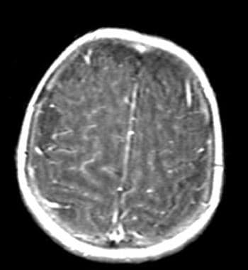
T1-weighted MRI of brain demonstrates diffuse enhancement of the meninges in viral meningoencephalitis. |
 | View Full Size Image | |
Media type: MRI
|
| Media file 2:
This rash consists of an enlarging annular plaque. Image courtesy of Lyme Disease Network (http://www.lymenet.org/). |
 | View Full Size Image | |
Media type: Photo
|
- Bobek MP, Sagher O. Aseptic meningitis caused by Teflon implantation for microvascular decompression. Case report. J Neurosurg. Jan 1999;90(1):145-7. [Medline].
- Cherry JD. Aseptic meningitis and viral meningitis. In: Feigin RD, Cherry JD, eds. Textbook of Pediatric Infectious Diseases. 2. 4th ed. Philadelphia: Saunders; 1998:450-57.
- DeNoon DJ. Wonder Drug Coming in for the Cold. WebMD Medical News. 2000;[Full Text].
- Doherty RL, Jordan MC. Viral meningoencephalitis. In: Infectious Diseases. 5th ed. 1994:1092-100.
- Eppes SC, Nelson DK, Lewis LL, Klein JD. Characterization of Lyme meningitis and comparison with viral meningitis in children. Pediatrics. May 1999;103(5 Pt 1):957-60. [Medline].
- Hamilton MS, Jackson MA, Abel D. Clinical utility of polymerase chain reaction testing for enteroviral meningitis. Pediatr Infect Dis J. Jun 1999;18(6):533-7. [Medline].
- Huang CC, Liu CC, Chang YC, et al. Neurologic complications in children with enterovirus 71 infection. N Engl J Med. Sep 23 1999;341(13):936-42. [Medline].
- Ishii K, Kamei S, Takasu T. [Epidemiology of virus-related nervous infections in Japan]. Nippon Rinsho. Apr 1997;55(4):839-48. [Medline].
- Karande S, Patil S, Kulkarni M. Acute aseptic meningitis as the only presenting feature of leptospirosis. Pediatr Infect Dis J. Apr 2005;24(4):390-1. [Medline].
- Mohle-Boetani JC, Matkin C, Pallansch M, et al. Viral meningitis in child care center staff and parents: an outbreak of echovirus 30 infections. Public Health Rep. May-Jun 1999;114(3):249-56. [Medline].
- Moris G, Garcia-Monco JC. The challenge of drug-induced aseptic meningitis. Arch Intern Med. Jun 14 1999;159(11):1185-94. [Medline].
- Muir P, Kammerer U, Korn K. Molecular typing of enteroviruses: current status and future requirements. The European Union Concerted Action on Virus Meningitis and Encephalitis. Clin Microbiol Rev. Jan 1998;11(1):202-27. [Medline].
- Norris CM, Danis PG, Gardner TD. Aseptic meningitis in the newborn and young infant. Am Fam Physician. May 15 1999;59(10):2761-70. [Medline].
- Oostenbrink R, Moll HA, Moons KG. Predictive model for childhood meningitis. Pediatr Infect Dis J. Nov 2004;23(11):1070-1. [Medline].
- Powell S, Schochet SS. Selected pediatric viral infections. Semin Pediatr Neurol. Sep 1995;2(3):211-9. [Medline].
- Rotbart HA. Viral meningitis and the aseptic meningitis syndrome. In: Lambert HP, ed. Infections of the Central Nervous System. Philadelphia: Decker; 1991:19-39.
- Sawyer MH. Enterovirus infections: diagnosis and treatment. Pediatr Infect Dis J. Dec 1999;18(12):1033-9; quiz 1040. [Medline].
- Sormunen P, Kallio MJ, Kilpi T, Peltola H. C-reactive protein is useful in distinguishing Gram stain-negative bacterial meningitis from viral meningitis in children. J Pediatr. Jun 1999;134(6):725-9. [Medline].
- Takeoka T. [Cerebrospinal fluid examination in the infectious meningitis and encephalitis]. Nippon Rinsho. Apr 1997;55(4):809-14. [Medline].
- Waisman Y, Lotem Y, Hemmo M, et al. Management of children with aseptic meningitis in the emergency department. Pediatr Emerg Care. Oct 1999;15(5):314-7. [Medline].
- 2006 West Nile Activity in the United States [database online]. Center for Disease Control; Updated May 30, 2007.
- Nigrovic, Kuppermann, Macias et al. Clinical Prediction Rule for Identifying Children With Cerebrospinal Fluid Pleocytosis at Very Low Risk of Bacterial Meningitis. JAMA. Jan 3, 2007;297:52-60. [Full Text].
- FDA Clears Rapid Test for Meningitis. FDA; March 16, 2007. [Full Text].
- INTERNATIONAL NOTEGLOBAL ADVISORY COMMITTEE ON VACCINE SAFETY, 11-12 JUNE, 2003. Canada Communicable Disease Report; April 1, 2004. [Full Text].
- Weekly Epidemiological Record. No. 3, 2007, 82. World Health Organization; January 19, 2007. 18-22. [Full Text].
Viral Meningitis excerpt Article Last Updated: Aug 23, 2007
|