Continually Updated Clinical Reference
 
 
  All Sources     eMedicine     Medscape     Drug Reference     MEDLINE
 
eMedicine - Heavy Chain Disease, Mu : Article by

Quick Find
Authors & Editors
Introduction
Clinical
Differentials
Workup
Treatment
Medication
Follow-up
Miscellaneous
Further Reading
References

Related Articles
Chronic Lymphocytic Leukemia

Lymphoma, B-Cell

Lymphoma, Non-Hodgkin

Monoclonal Gammopathies of Uncertain Origin

Multiple Myeloma

Waldenstrom Hypergammaglobulinemia


Patient Education
Click here for patient education.


Author: Ajeet Gajra, MD, Assistant Professor of Medicine, Director of Hematology/Oncology Fellowship Program, State University of New York Upstate Medical University; Consulting Staff, Department of Internal Medicine, Division of Hematology and Oncology, Veterans Affairs Medical Center

Ajeet Gajra is a member of the following medical societies: American Association for Cancer Research, American College of Physicians-American Society of Internal Medicine, American Medical Association, American Society of Clinical Oncology, and American Society of Hematology

Coauthor(s): Sara Grethlein, MD, Associate Dean for Graduate Medical Education, Associate Professor, Department of Internal Medicine, Division of Hematology and Oncology, State University of New York at Upstate

Editors: Paul Schick, MD, Emeritus Professor, Department of Internal Medicine, Thomas Jefferson University Medical College; Research Professor, Department of Internal Medicine, Drexel University College of Medicine; Francisco Talavera, PharmD, PhD, Senior Pharmacy Editor, eMedicine; Troy H Guthrie, Jr, MD, Director of Cancer Institute, Baptist Medical Center; Rajalaxmi McKenna, MD, FACP, Consulting Staff, Department of Medicine, Southwest Medical Consultants, SC, Good Samaritan Hospital, Advocate Health Systems; Emmanuel C Besa, MD, Professor, Department of Medicine, Division of Hematologic Malignancies, Kimmel Cancer Center, Thomas Jefferson University

Author and Editor Disclosure

Synonyms and related keywords: mu-HCD, heavy chain disease, HCD, B-cell proliferative disorder, immunoglobulin heavy chain, Ig heavy chain, plasma cell disorder, monoclonal gammopathy of unknown significance, gammopathies, chronic lymphocytic leukemia, CLL, multiple myeloma, plasmacytoma, splenomegaly, peripheral lymphadenopathy, hepatomegaly, osteolytic lesion, pathologic fractures

Background

Heavy chain diseases (HCDs) are rare B-cell proliferative disorders characterized by the synthesis and secretion of incomplete immunoglobulin heavy chains. These disorders initially were recognized as gammopathies due to the presence of monoclonal proteins in the patient's serum or urine. The disorders were defined in terms of the production of structurally aberrant immunoglobulin molecules.

Normal immunoglobulin molecules are symmetrical and are composed of 2 pairs of polypeptide chains designated the light and heavy chains, which are interconnected by disulfide bonds. The heavy chains are the larger polypeptide subunits; they are specific and distinctive structures that distinguish the major classes of immunoglobulins.
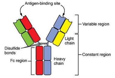
 Reductive cleavage of the immunoglobulin molecule by papain yields 2 Fab fragments (consisting of a light chain and an Fd fragment) and one Fc fragment (consisting of portions of the 2 heavy chains). Plasma cell disorders characterized by an anomalous serum and urinary protein that is immunochemically related to the Fc fragment of the immunoglobulin molecule are known as HCDs. When the anomalous protein structurally resembles the heavy chain fragment of immunoglobulin M (IgM) molecule, it is designated as mu-HCD. Ballard and colleagues first described this entity in 1970.1 This article focuses on mu-HCD; however, other heavy chain diseases are described (eg, see Heavy Chain Disease, Gamma).

Pathophysiology

Heavy chain disease mu was first described in 1969. The characteristic feature of HCD is the production of a monoclonal immunoglobulin molecule in which the heavy chain is truncated and the covalent attachment of light chains is absent. This may be due to lack of light chain production or the failure of heavy-light disulfide bond formation.

The mu heavy chains analyzed to date have an absent variable region and a shortened constant domain. The reasons a complete immunoglobulin fails to assemble are poorly understood. A defect at the level of immunoglobulin gene structure and assembly has been shown to be responsible for the synthesis of the truncated mu-HCD protein, caused by deletion of coding information and formation of an aberrant RNA molecule.2 The available data do not allow for an accurate description of the molecular defects involved in mu-HCD proteins. Despite certain similarities, every case seems to have an individual pattern.3

Frequency

United States

It is a rare disease, with 34 cases reported in the literature. However, many cases likely have not been reported, especially in the past decade. Given the difficulty in diagnosing this disorder, most reports are from the United States, western Europe, and Scandinavia.

International

True international incidence is difficult to ascertain for reasons stated above. A single case has been reported from Japan.

Mortality/Morbidity

Given its infrequent presentation, mu-HCD is not a major cause of morbidity or mortality. The disease course can be extremely variable, and the survival of patients in the literature reports varies from 1 month to 11 years. Reports of cures are also described.

Race

Most patients with mu-HCD reported in the literature were white, but at least 3 black patients and 1 Asian patient have also been reported.

Sex

No obvious sex predilection has been described; half the patients with mu-HCD are male.

Age

The age of reported patients varies from 15-80 years; mu-HCD does not have an obvious age predilection.


History

  • Initial reported cases had a clinical picture consistent with chronic lymphocytic leukemia (CLL); however, with better diagnostic procedures and a high index of suspicion, the defining protein abnormality has been noted in a broader range of clinical settings. Currently, only one third of patients with mu-HCD appear to have CLL. In 150 consecutive patients with CLL, thorough investigation of serum proteins failed to identify a single instance of mu-HCD, which suggests an incidence of less than 1% in this population.4
  • Other clinical presentations vary, one of which may be essential monoclonal gammopathy with no clinical symptoms of B-cell lymphoma (20%). In 10% of cases, an intact IgM protein was simultaneously detected in the serum. Another 10% of cases were associated with clinical multiple myeloma or plasmacytoma. Mu-HCD is also reported to be associated with systemic amyloidosis in rare instances.5
  • Patients usually present with evidence of systemic disease, such as weight loss, fever, anemia, and recurrent infection. Splenomegaly is detected in almost all cases, hepatomegaly is present in approximately 75% of cases, and peripheral lymphadenopathy is present in approximately 40% of patients.
  • Bone involvement with osteolytic lesions and pathologic fractures has been noted in approximately 40% of patients. These patients usually present with bone pain, especially in the back.
  • Renal complications seem infrequent, with cast nephropathy and renal failure reported in a single case. Two patients, including the first reported case, had amyloidosis with renal impairment.
  • Some patients present with other pathologic conditions such as systemic lupus erythematosus, hepatic cirrhosis, and myelodysplastic syndrome.

Physical

  • Physical examination findings are variable and depend on patient presentation.
  • Pallor may be noted if significant anemia is present, which is usually observed only in advanced disease.
  • Lymphadenopathy is noted in 40% of patients.
  • Splenomegaly is almost universal, and hepatomegaly is noted in most cases.

Causes

The cause of mu-HCD is not known. No viruses, chemical, physical, or genetic factors have been identified.6


Chronic Lymphocytic Leukemia
Lymphoma, B-Cell
Lymphoma, Non-Hodgkin
Monoclonal Gammopathies of Uncertain Origin
Multiple Myeloma
Waldenstrom Hypergammaglobulinemia


Lab Studies

Laboratory studies should include complete blood cell count with differential, screening chemistries to ascertain renal function (eg, BUN and creatinine) and hepatic function (eg, total protein, albumin, aspartate aminotransferase, alanine aminotransferase, lactate dehydrogenase, alkaline phosphatase), calcium levels, and beta-2 microglobulin values.

Imaging Studies

  • No definitive guidelines are available regarding the extent of imaging studies that should be performed when diagnosing patients suggested to have mu-HCD.
  • Performing a chest radiograph is reasonable, and a skeletal survey is considered essential given the fact that 40% of patients present with osteolytic lesions.
  • Obtaining CT scans of the thorax and abdomen is appropriate because hepatosplenomegaly and lymphadenopathy are common. CT scan findings help to objectively quantify disease and are useful for assessing response to therapy.

Other Tests

  • Serum protein electrophoresis (SPEP) or urine protein electrophoresis (UPEP) and immunofixation are essential tests.
    • As a rule, the neoplastic cells do not produce large amounts of immunoglobulin, which may make detecting the abnormal immunoglobulin produced in a patient with HCD difficult. Performing a combination of electrophoretic, immunoelectrophoretic, and immunofixation techniques can help establish the diagnosis.
    • In a minority of cases, the proteins can initially be identified as a discrete homogenous band of mobility on SPEP or UPEP findings.
    • When developed with specific antiheavy and antilight sera, the immunoelectrophoretic pattern reveals a heavy chain–specific arc that does not react with either antikappa or antilambda antisera.
    • IgM M-proteins sometimes do not react with certain antilight sera. This is more common with immunoelectrophoresis than with immunofixation techniques. In these cases, separating the monoclonal proteins from the serum, treating them with reducing agents to cleave disulfide bonds, and subjecting them to gel electrophoresis to determine the size of the immunoglobulin heavy chain polypeptide may be necessary.
    • More commonly, the proteins are present in smaller amounts and give a heterogenous pattern on electrophoresis findings. A monoclonal spike was detected in 8 of 19 patients, and 3 of 28 patients had a biclonal gammopathy.7 Again, immunoelectrophoresis or immunofixation findings showing the development of patterns with a panel of antiheavy/antilight antibodies can strongly suggest the diagnosis. In specialized laboratories, more detailed structural analysis can be performed on the isolated, reduced, and alkylated heavy chain monomer to confirm the presence or absence of immunoglobulin light chains.
    • Urinary excretion of the mu fragment has been noted in only 2 patients; this presumably is because the polymers of the carboxy-terminal mu fragment are too large to be filtered by intact renal glomeruli. Monoclonal light chains have been found in the urine in two thirds of cases. Thus, Bence Jones proteinuria is a common occurrence in patients with this disorder. Nonetheless, renal complications are infrequent. Immunoglobulin light chains capable of producing amyloid are found in approximately 12% of cases, an incidence that is similar to that observed in patients with multiple myeloma.
    • Immunoglobulin gene rearrangement may be used to differentiate a B-cell lymphoproliferative process from a monoclonal or reactive proliferation of lymphocytes. This technique not only provides a specific marker for B cells, but it also is a true marker for monoclonality.

Procedures

  • Almost all patients should undergo bone marrow aspiration and biopsy. Certain histologic features, as outlined below, may aid in making the diagnosis of mu-HCD.
  • When mu-HCD is not considered based on the patient's presentation, as is commonly the case, biopsy of the appropriate involved area (eg, lymph node mass) is required to establish the diagnosis of a lymphoproliferative disorder.

Histologic Findings

Marrow involvement is characterized by infiltration with lymphocytes and plasma cells. Cells that often are described as lymphocytic plasmacytes or plasmacytoid lymphocytes are prominent. Although the marrow of almost all patients contains the multivacuolated plasma cells described in the index case, the vacuoles are not universally apparent. The identification of these vacuoles sometimes offers a clue to the diagnosis, which requires confirmation by appropriate electrophoretic and immunoelectrophoretic studies.

Staging

Given the rarity of mu-HCD, a clinical staging system has not been developed. Accompanying lymphoproliferative disorders such as CLL, non-Hodgkin lymphoma, and multiple myeloma should be appropriately staged.


Medical Care

  • The clinical course is variable, with survival ranging from 1 month to more than 10 years after the appearance of symptoms. Because of its low incidence, no large series of patients treated in a systematic way at a single center has been reported. Common drugs used include alkylating agents such as chlorambucil, melphalan, and cyclophosphamide, but patient responses have been inconsistent. More recently, fludarabine phosphate (Fludara) has been used.
  • No specific treatment exists. Currently, the finding of a mu-HCD protein in the serum of an apparently healthy patient should be considered a monoclonal gammopathy of unknown significance, and the patient should be monitored closely for the development of a symptomatic lymphoproliferative disorder. If this develops, chemotherapy targeted at the specific disorder is used. The same holds true if a patient presents with a treatable clinical disease such as CLL and the presence of mu-HCD.

Surgical Care

Surgical care usually is not required, although special circumstances may require surgery (eg, surgery needed to fix a pathologic fracture).

Consultations

  • Diagnosis of mu-HCD often can be difficult to establish. Consulting a hematopathologist is appropriate to facilitate an appropriate and adequate workup.
  • Occasionally, consultation with a radiation oncologist may be required to treat a pathologic fracture site after surgical correction or to prophylactically treat a site of bony involvement to prevent a pathologic fracture.

Diet

No dietary restrictions are necessary.

Activity

No activity restrictions are necessary.


No specific treatment exists. In the absence of a demonstrated lymphoproliferative disorder, the condition is monitored as a monoclonal gammopathy of unknown significance. Once a lymphoproliferative disorder develops, chemotherapeutic agents are used as appropriate for the patient's disorder, stage, and clinical situation (eg, melphalan and prednisone or vincristine, doxorubicin, and dexamethasone for multiple myeloma; chlorambucil or fludarabine for CLL). Successful treatment with fludarabine phosphate has recently been reported  in two cases.8 The details of some of these therapies can be found in Chronic Lymphocytic Leukemia and Multiple Myeloma.


Further Inpatient Care

Inpatient care is usually not required.

Further Outpatient Care

  • Once the diagnosis of mu-HCD is established, any accompanying lymphoproliferative disorder should be treated. Typically, this involves chemotherapy administered in an outpatient setting.
  • If no overt lymphoproliferative disorder is found, the patient should be evaluated every few months to assess the abnormal protein. Reasonable follow-up intervals are every 3 months in the first year following diagnosis, every 4 months for the next year, and every 6 months thereafter. The patient should be evaluated earlier if symptoms occur. If the patient is symptomatic in any way, then radiologic assessment and other diagnostic procedures for lymphoproliferative disorders are essential.

Complications

  • Possible complications include pathologic fracture secondary to osteolytic lesions.
  • Renal insufficiency occasionally is associated with mu-HCD. Etiologies include cast glomerulopathy, nodular glomerulosclerosis, and amyloidosis.
  • Positive results from a direct antiglobulin test have also been reported.

Prognosis

The clinical course is variable, with survival ranging from 1 month to 11 years after the appearance of symptoms with a median survival of 24 months in the well studied cases. Reports of cures are described, primarily in the setting of a curable lymphoproliferative disorder. Because of its low incidence, no large series of patients treated in a systematic way at a single center has been reported.


Medical/Legal Pitfalls

This is a rare disorder that is relatively difficult to diagnose. If a diagnosis is made, evaluating the patient at regular intervals is essential, even in the absence of an associated lymphoproliferative disorder, because such a disorder may develop.

Special Concerns

The diagnosis of mu-HCD requires clinician awareness. The finding of Bence Jones proteinuria in a patient with a lymphoproliferative disorder and vacuolated plasma cells in bone marrow warrants further investigation to exclude mu-HCD. Once mu-HCD is considered, immunoelectrophoretic and immunofixation testing of both urine and serum should be performed.


O'Connell, Horita TJ, Kasravi B. Understanding and Interpreting Serum Electrophoresis. American Family Physician. January 2005;71 (1):105-12


  1. Ballard HS, Hamilton LM, Marcus AJ, Illes CH. A new variant of heavy-chain disease (mu-chain disease). N Engl J Med. May 7 1970;282(19):1060-2. [Medline].
  2. Bakhshi A, Guglielmi P, Coligan JE, et al. A pre-translational defect in a case of human mu heavy chain disease. Mol Immunol. Jul 1986;23(7):725-32. [Medline].
  3. Mihaesco C, Ferrara P, Guillemot JC, et al. A new extra sequence at the amino terminal of a mu heavy chain disease protein (DAG). Mol Immunol. Aug 1990;27(8):771-6. [Medline].
  4. Bonhomme J, Seligmann M, Mihaesco C, et al. MU-chain disease in an African patient. Blood. Apr 1974;43(4):485-92. [Medline].
  5. Kinoshita K, Yamagata T, Nozaki Y, et al. Mu-heavy chain disease associated with systemic amyloidosis. Hematology. Apr 2004;9(2):135-7. [Medline].
  6. Witzig TE, Wahner-Roedler DL. Heavy chain disease. Curr Treat Options Oncol. Jun 2002;3(3):247-54. [Medline].
  7. Wahler-Roedler, DLKyle RA. Heavy hain Diseases. Best Practice & Research Clinical Hematology. 2005;18:729-46.
  8. Yanai M, Maeda A, Watanabe N, et al. Successful treatment of mu-heavy chain disease with fludarabine monophosphate: a case report. Int J Hematol. Feb 2004;79(2):174-7. [Medline].
  9. Buxbaum JN, Alexander A. Heavy Chain Diseases. In: Beutler B, Lichtman MA, Coller BS, Kipps TJ, and Seligsohn U, eds. Williams Hematology. 6th ed. New York, NY: McGraw-Hill; 2001:1327-36.
  10. Fermand JP, Brouet JC. Heavy-chain diseases. Hematol Oncol Clin North Am. Dec 1999;13(6):1281-94. [Medline].
  11. Iwasaki T, Hamano T, Kobayashi K, Kakishita E. A case of mu-heavy chain disease: combined features of mu-chain disease and macroglobulinemia. Int J Hematol. Oct 1997;66(3):359-65. [Medline].
  12. Liapis H, Papadakis I, Nakopoulou L. Nodular glomerulosclerosis secondary to mu heavy chain deposits. Hum Pathol. Jan 2000;31(1):122-5. [Medline].
  13. O'Connell, Horita TJ, Kasravi B. Understanding and Interpreting Serum Electrophoresis. American Family Physician. January 2005;71 (1):105-12.
  14. Preud'homme JL, Bauwens M, Dumont G, et al. Cast nephropathy in mu heavy chain disease. Clin Nephrol. Aug 1997;48(2):118-21. [Medline].
  15. Wahner-Roedler DL, Kyle RA. Mu-heavy chain disease: presentation as a benign monoclonal gammopathy. Am J Hematol. May 1992;40(1):56-60. [Medline].
  16. Witzens M, Egerer G, Stahl D, et al. A case of mu heavy-chain disease associated with hyperglobulinemia, anemia, and a positive Coombs test. Ann Hematol. Nov 1998;77(5):231-4. [Medline].

Heavy Chain Disease, Mu excerpt

Article Last Updated: Sep 21, 2007