Background
Oral hairy leukoplakia (OHL) is a disease of the mucosa first described in 1984. This pathology is associated with Epstein-Barr virus (EBV) and occurs mostly in people with HIV infection, including those who do not have a diagnosis of AIDS. HIV-negative people can have oral hairy leukoplakia, especially individuals with organ transplants and other immunocompromised disease. [1, 2] The condition has also been reported in individuals not immunocompromised. [3] The first case in an HIV-negative patient was reported in 1999 in a 56-year-old patient with acute lymphocytic leukemia. Later, many cases were reported in heart, kidney, and bone marrow transplant recipients and in patients with hematological malignancies. [4, 5]
Pathophysiology
The Epstein-Barr virus (EBV), a ubiquitous herpesvirus estimated to infect 90% of the world's population, is linked to a growing number of diseases, especially in immunocompromised hosts. Like all herpesviruses, EBV establishes a life-long, persistent infection of its host. The pathogenesis of hairy leukoplakia is clearly complex, potentially requiring a convergence of factors including EBV co-infection, productive EBV replication, EBV genetic evolution, expression of specific EBV "latent" genes, and immune escape. All of these factors are likely facilitated by local and systemic host immunodeficiency. [6]
EBV initially infects basal epithelial cells in the pharynx, where it enters a replicative state leading to the release of infectious virus into the saliva throughout the life of the infected person. In the pharynx, the virus also enters B cells, where it persists indefinitely in a latent state. Cytotoxic T lymphocytes cannot eliminate EBV from the body, but they are essential in maintaining the latent state of the infection. In states of immune dysfunction in which the number of EBV-specific cytotoxic T lymphocytes is decreased, there is an increase in the number of circulating EBV-infected B cells.
In addition, a marked decrease or an absence of Langerhans cells occurs in hairy leukoplakia biopsy tissues. [7, 8] Langerhans cells are the antigen-presenting immune cells that are required for an immune system response to the viral infection and their deficiency may permit EBV to persistently replicate and escape immune recognition.
Etiology
Oral hairy leukoplakia (OHL) is associated with HIV infection and/or immunosuppression. [9] The risk of developing oral hairy leukoplakia doubles with each 300-unit decrease in CD4 count. A high viral load was strongly associated to the oral lesions occurrence independently of CD4+ cell count. [10] More recently, it was described in patients with other forms of severe immunodeficiency including those associated with chemotherapy, organ transplant, and leukemia. Rarely, it may occur in patients who are immunocompetent. [3]
Oral hairy leukoplakia also is associated with Behçet syndrome and ulcerative colitis.
Smoking more than a pack of cigarettes a day is positively correlated with the development of oral hairy leukoplakia in HIV-positive men.
No increase in oral hairy leukoplakia was observed when controlled for number of oral sex partners. [11]
Oral hairy leukoplakia may not be the traditional lesion reported in only HIV-positive individuals and other immunosuppressed individuals. Increased reports of oral hairy leukoplakia are now published in other populations. Four cases were reported in Bristol Dental Hospital over a 6-month period in immunocompetent individuals. All lesions were on the tongue and tested positive for EBV. The authors concluded that oral hairy leukoplakia diagnosis should not be limited to HIV-positive individuals or those with immunosuppression. [12]
An oral and maxillary pathology archive spanning 1994-2020 was investigated looking for cases of oral hairy leukoplakia in immunocompetent individuals. Eleven cases were found, all positive for EBV. Of these, 63.6% were males with a mean age of 62 years, and all patients were White. All lesions were located on the lateral borders of the tongue. The etiology of oral hairy leukoplakia in this group of patients was not clearly understood. These authors concluded that oral hairy leukoplakia should not be considered pathognomonic for HIV infection and should be included in the differential diagnosis of other keratotic lesions, especially in elderly individuals. [13]
Recipients of solid-organ transplants on immunosuppressives and those with hematologic malignancies, autoimmune diseases, and other systemic inflammatory conditions, in addition to immunocompetent individuals, are now identified with oral hairy leukoplakia. Long-term inhaled topical and systemic steroids were identified as a risk factor in the development of oral hairy leukoplakia. Hypogammaglobulinemia secondary to long-term anticonvulsant treatment using lamotrigine was also associated with oral hairy leukoplakia. [14]
In another study investigating non-HIV–associated oral hairy leukoplakia, 28 patients had respiratory problems requiring long-term steroid inhaler use, four had autoimmune diseases requiring immunosuppressant therapy, and four had diabetes. Most of the lesions were located on the tongue, and 24 showed Candida co-infection. In their results with 35 patients 25 were identified as males and 10 females. The average age at the time of diagnosis was 61 years (range 33-86 years). Thirty-four of 35 had lesions located on the tongue, where the lateral border was the most common sight. Twenty-four lesions had Candida co-infection. A wide variety of clinical diagnoses were given on the pathology request forms, but none of the 35 cases where oral hairy leukoplakia was suspected clinically. The authors concluded the presence of oral hairy leukoplakia should not be regarded as pathognomic for HIV infection or significant systemic immunosuppression. Local and systemic immunosuppression caused by steroid inhaler use is a risk factor for the development of oral hairy leukoplakia. [14] See the image below.
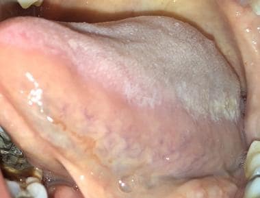 Oral hairy leukoplakia (OHL) on left lateral tongue in patient who used topical steroid inhaler.
Epidemiology
Frequency
United States
Hairy leukoplakia is one of the most common virally induced, oral diseases of HIV-infected individuals, with a point prevalence as high as 25-53%. [10] The 6-year incidence of oral hairy leukoplakia (OHL) in this patient population was reported to be around 32%. A significant trend to a lower prevalence of oral hairy leukoplakia was observed in the group of patients who were already taking combined antiretroviral therapy (cART), non–highly active antiretroviral therapy (HAART) and HAART (P< .001 and P = .004, respectively). [15, 16]
Fewer cases of oral hairy leukoplakia were reported in non-HIV–infected patients. This is probably due to underdiagnosis and underreporting of this disease in patients with hematological malignancies or solid organ transplantation. Some studies showed the prevalence of oral hairy leukoplakia in renal transplant recipients to be more than 11%. [17]
International
The incidence of oral hairy leukoplakia is similar to that in the United States and thereby reflects the prevalence of HIV. In populations where the prevalence of HIV is low, oral mucosal lesions alone are poor prognostic predictors of HIV infection. [18]
A cross-sectional study from Brazil reported on data collected from clinical examinations, interviews, and medical records for adult patients treated an HIV/AIDS clinic at the University Hospital of the Federal University in Rio Grande. Three hundred persons were observed (April 2006 to January 2007). Of these patients, 51% were male and the mean age was 40 years. Thirty-nine percent presented with oral lesions. The most common was candidiasis (59.1%), followed by hairy leukoplakia (19.5%). [19]
A study from Saudi Arabia reported that compared with age and sex-matched healthy control subjects (N = 52), 8.6% of stable renal transplantation patients (N = 58) had oral leukoplakia. Other oral lesions reported were gingival hyperplasia (74.1%) and erythematous candidiasis (15.5%). [20] However, a study from Spain reported only one case of hairy leukoplakia in 500 renal transplant recipients studied. [21]
When associated with HIV infection, hairy leukoplakia is more common in Europe and the United States compared with Africa and Asia. [22]
Race
No racial predilection is established for oral hairy leukoplakia.
Sex
Oral hairy leukoplakia is most commonly observed in homosexual men who are HIV positive, especially in those who smoke.
Age
No age predilection is established for oral hairy leukoplakia.
Prognosis
The majority of patients with oral hairy leukoplakia (OHL) tend to have significant immunosuppression at the time of diagnosis. Oral hairy leukoplakia occurs relatively soon after HIV seroconversion, typically before AIDS. Median CD4 count when oral hairy leukoplakia is first detected is 235-468/µL. Another study showed oral hairy leukoplakia occurred mostly in patients with CD4 counts of 200-500/µL. [23]
In patients with HIV, the median CD4 count when oral hairy leukoplakia is first detected is 468/µL. If these patients do not have AIDS-defining disease at the time oral hairy leukoplakia is diagnosed, the probability of developing AIDS if not receiving highly active antiretroviral therapy (HAART) is 48% by 16 months and 83% at 31 months. In addition, studies have shown that patients with AIDS with oral hairy leukoplakia have a shorter lifespan than those who do not present with this lesion. Furthermore, if these patients are concomitantly co-infected with hepatitis B virus, the risk of early progression to AIDS increases 4-fold. Further studies in HIV-seropositive patients show that the median survival after the diagnosis of oral hairy leukoplakia is around 20 months. In patients with CD4 count greater than or equal to 300/μL, oral hairy leukoplakia is associated with median survival time of 25 months, compared with 52 months in patients with normal counts. [24]
-
Oral leukoplakia.
-
Lateral tongue in oral hairy leukoplakia.
-
Morsicatio linguarum, or tongue biting.
-
Ventral tongue in oral leukoplakia.
-
Lateral tongue in lichen planus.
-
Proliferative verrucous leukoplakia of the lateral tongue; biopsy showed squamous cell carcinoma.
-
Proliferative verrucous leukoplakia of the gingiva; biopsy showed squamous cell carcinoma.
-
White sponge nevus.
-
Hyperplastic candidiasis.
-
Oral hairy leukoplakia (OHL) on left lateral tongue in patient who used topical steroid inhaler.


