Goals and Options
The goal of surgical treatment of basal cell carcinoma (BCC) is to destroy or remove the tumor so that no malignant tissue is allowed to proliferate further. Factors to consider when choosing therapy include the histologic subtype of BCC, the location and size of tumors, the age of the patient, the patient's ability to tolerate surgery, likely outcome to the patient's appearance, and the expense. [1]
Recurrent tumors are generally more aggressive than primary lesions, and subclinical extension also tends to be increased. Tumors that are aggressive and those occurring near vital or cosmetically sensitive structures are best treated with methods that allow for an examination of the tissue margins.
Knowledge of the behavior of the different clinical and pathologic types of BCC is essential in choosing the appropriate therapy. The most common surgical methods of treating BCC are curettage, excision with margin examination, and Mohs micrographic surgery (see the image below). Radiotherapy is another common BCC treatment, and cryotherapy is sometimes used. BCC recurrence after irradiation makes surgery mandatory.
 Postoperative wound after Mohs micrographic surgery demonstrates the extensive subclinical involvement that is typical of many infiltrating and morpheaform basal cell carcinomas.
In patients with negative margins on excision of high-risk BCCs but histologic evidence of extensive perineural or large-nerve involvement, National Comprehensive Cancer Network (NCCN) guidelines suggest considering adjuvant radiation therapy. [2] However, a systematic review found that in patients with histologic perineural invasion, surgery alone and surgery plus radiation therapy demonstrated statistically equivalent outcomes at median 5-year follow-up. These authors do not recommend adjuvant radiation therapy for BCC with solely histologic perineural invovlement, provided that surgical excision achieves clear margins. [3]
For more detailed information regarding the treatment of BCC, refer to guidelines and appropriate use criteria from the American Academy of Dermatology and other recognized sources, such as the NCCN. [2, 4, 5, 6, 7]
Go to Basal Cell Carcinoma for more complete information on this topic.
Electrodesiccation and Curettage
Electrodesiccation and curettage is the most widely used method for removing primary BCCs. Although this technique is quick to destroy tumor cells, its adequacy cannot be assessed immediately, as the surgeon cannot visually detect the depth of microscopic tumor invasion and surgical margin.
Technique
A looped blade (curette) is used to vigorously scrape tumor away from adjacent normal skin. [8] One may start with a larger curette to debulk the tumor and then follow with a smaller curette to better remove smaller fragments of tumor from surrounding stroma. This technique works best on nodular or superficial BCC, because these tumors tend to be friable and do not tend to be embedded in fibrous stroma. The instrument is applied firmly and used in multiple directions over the tumor and immediately adjacent skin.
Curetting is often followed by electrodesiccation, and the entire process may be repeated 1-2 more times. If electrodesiccation is not performed, vigorous and firm scraping in several directions is especially important. Many recurrences after curettage are believed to be due to insufficient aggressiveness on the part of the surgeon. The overall cure rate exceeds 90% for low-risk BCCs. The method is quick, simple, and less expensive than most other methods.
Limitations
Curettage is a relatively blind technique in which the specimen cannot be examined for margin control. This lack of microscopic margin control limits the usefulness of curettage in high-risk areas, such as the face and ears.
The National Comprehensive Cancer Network (NCCN) advises that the use of curettage and electrodesiccation or shave excision should be limited to BCCs that pose low risk of recurrence (ie, primary BCCs that are located on the trunk or extremities and are well defined and < 2 cm in size). In addition, margin control with curettage relies on the operator’s ability to distinguish between firm normal dermis and soft tumor tissue; however, subcutaneous adipose is softer than tumor tissue, so that ability is lost with BCCs that extend into adipose. In such cases, the NCCN recommends that surgical excision should generally be performed. [2]
Furthermore, the aggressive subtypes of BCC, such as morpheaform (sclerosing), infiltrating, micronodular, and recurrent tumors, are usually not friable and therefore unlikely to be removed by using the curette. The success of this treatment, even in nonaggressive tumors at low-risk sites, is highly dependent on the operator's experience and technique. [6]
Finally, healing by secondary intention (granulation) often leads to atrophic white scars that may not be satisfactory in aesthetically important areas.
Advantages
Electrodesiccation and curettage is a brief procedure (< 5 min) and is effective in treating primary nodular and superficial BCC. Cure rates are as high as 95%. Tumors ranging from 2-5 mm in diameter have a 15% recurrence rate, whereas tumors with a diameter of 3 cm or greater have a 50% recurrence rate within 5 years when treated by this procedure.
Disadvantages
The success of electrodesiccation and curettage is operator dependent and often leaves a white atrophic scar. The procedure is less effective on the nose, and the tumor often tracks down pilosebaceous units.
Electrodesiccation and curettage is less effective than Mohs micrographic surgery in treating the infiltrating, micronodular, morpheaform (sclerosing), and recurrent types of BCC; Mohs micrographic surgery is considered the treatment of choice in most of those cases.
The curettage and cauterization procedure is not suitable for patients with cardiac pacemakers or for the treatment of morpheaform lesions, deeply invasive and ulcerated lesions, or recurrent tumors with ill-defined borders.
Curettage Without Desiccation
After adequate anesthesia is administered to the patient, the tumor is scraped using a curette. [9] This is often repeated twice more.
Advantages
This procedure is brief (< 5 min) and is effective in treating primary nodular and superficial BCC. Cure rates may be as high as 95%, although this method has been studied less than has electrodesiccation and curettage. Curettage alone is believed by some authors to have a better cosmetic outcome than that of electrodesiccation and curettage.
Disadvantages
This procedure is neither widely accepted nor commonly performed. The success of curettage is operator dependent and often leaves a white, atrophic scar. It is less effective on the nose, and the tumor often tracks down pilosebaceous units.
Curettage is less effective than Mohs micrographic surgery for treatment of the infiltrating, micronodular, morpheaform, and recurrent types of BCC. Mohs micrographic surgery is considered the treatment of choice in most instances.
Curettage With Er:YAG Laser Ablation
After adequate anesthesia has been administered to the patient, the tumor is scraped using a curette. The newly formed ulcer is then ablated, along with a narrow (< 1 mm) margin of adjacent epidermis. This is often repeated 2 more times.
Advantages
Curettage with Er:YAG laser ablation is brief (< 5 min), is effective in treating primary nodular and superficial BCC, and is believed by some authors to have a better cosmetic outcome than does electrodesiccation and curettage. Cure rates may be as high as 95%, although this procedure has been studied less than has electrodesiccation and curettage.
Disadvantages
Curettage with Er:YAG laser ablation is less commonly performed than electrodesiccation and curettage. The success of the procedure is operator dependent and may leave a white, atrophic scar. It is less effective on the nose, and the tumor often tracks down pilosebaceous units.
Curettage with Er:YAG laser ablation is less effective in treating the infiltrating, micronodular, morpheaform, and recurrent types of BCC than is Mohs micrographic surgery, which is considered the treatment of choice in most instances.
Laser Ablation Without Curettage
A carbon dioxide laser is applied most frequently to the superficial type of BCC. This procedure may be considered when a bleeding diathesis is present, as bleeding is unusual when this laser is used. This procedure provides a bloodless field, minimal postoperative pain, and good postoperative appearance without scar formation. In cases of superficial T1-T2 BCC, laser microsurgery appears to be a safe and effective treatment modality without conjunctival extension.
The carbon dioxide laser is best known for its cosmetic use in laser resurfacing, [10, 11] as well as for its use in ablating superficial and nodular BCCs, with reported recurrence rates varying from 50%, with older devices, to 3%. [12]
Limitations
After treatment, changes in skin color may develop, which may be seen within a few weeks or may take at least 1 year or longer to be apparent. Additionally, scars may be seen and hypertrophic (thick) scars have been reported.
Advantages
Laser ablation without curettage may have less scarring than traditional electrodesiccation and curettage, but comparative studies are needed.
Disadvantages
This procedure has been studied less than other methods, and a great variance in recurrence rates has been reported. Hypopigmentation may develop, which may not be apparent for a year or more.
Surgical Excision
This method can usually be performed in an ambulatory setting and provides the pathologist with a specimen to examine the tissue margins. Healing time is generally shorter with sutured closure than with granulation, and cosmesis compares favorably with that of curettage.
Technique
After adequate anesthesia has been administered to the patient, a No. 15 or No. 10 blade is used to incise down to the subcutis. [13]
Limitations
Surgical excision is operator dependent, as those who are more experienced may be better at detecting the clinical margins. To increase the likelihood of complete tumor removal, one must include a margin of normal-appearing skin in order to remove all clinically invisible tumor extension. The larger the amount of clinically normal-appearing skin removed, the higher the cure rate, although the more extensive removal leaves a larger surgical defect and a poorer cosmetic result in most patients. The American Academy of Dermatology (AAD) recommends surgical excision with 4-mm clinical margins and histologic margin assessment for low-risk primary BCC. Standard excision may be considered for select high-risk tumors. However, AAD guidelines advise strong caution when selecting treatment for high-risk tumors without complete margin assessment. [6]
(See the images below.)
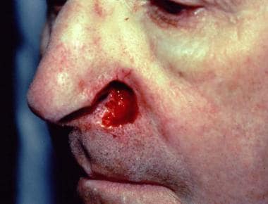
 Basal cell carcinoma of the skin. Intraoperative view showing the extent of resection (inferior view).
Pathologic evaluation of the excised tissue can be performed via routine paraffin processing, which may take 1 day or longer, or by using frozen sections, in which the results are available in as little as 15 minutes.
Advantages
Surgical excision usually produces good to excellent cosmetic results and cure rates as high as 95%.
Disadvantages
Surgical excision is operator-dependent, as those who are more experienced may be better at clinically detecting tumor margins and thus require smaller margins (removal of normal-appearing skin). Excision is less effective in treating tumors without clearly defined clinical margins (eg, infiltrating BCC, micronodular BCC, morpheaform BCC), and is far less effective in treating recurrent BCC than it is in treating primary BCC. If a positive margin occurs, additional surgery is needed.
When standard excision is performed for high-risk tumors, a linear repair, skin graft, or healing by second intention is recommended. If a repair requiring significant tissue rearrangement is indicated, closure should be delayed until negative histologic margins are confirmed. [6, 2]
Surgical excision is more time-consuming and costly than curettage. In addition, this method requires the sacrifice of normal tissue to obtain acceptable cure rates. Margins of at least 4 mm are needed, even with the least aggressive BCCs, to achieve 95% cure rates and as much at least 8 mm for more aggressive tumors. [14]
If standard "bread-loaf" tissue sectioning is used, areas of margin involvement may be missed under microscopy, because only a small percentage of the peripheral and deep margins are evaluated.
The 5-year recurrence rate for primary tumors greater than 1.5 cm in diameter is approximately 12%. Primary tumors that measure greater than 3 cm in diameter have a 5-year recurrence rate of 23%. [13]
Mohs Micrographically Controlled Surgery
In 2015, the American Society for Dermatologic Surgery (ASDS) released consensus guidelines for the treatment of basal cell carcinomas that recommended MMS as the treatment of choice for high-risk BCCs and for those in cosmetically sensitive locations. The guidelines also noted that MMS offers complete tumor margin analysis while other surgical options do not. [5]
The 2018 American Academy of Dermatology (AAD) guidelines for management of BCC found evidence to strongly support the use of MMS for both primary and recurrent BCC at increased risk for recurrence on the basis of factors such as anatomic location and histologic growth pattern. [6]
The American Academy of Dermatology, in collaboration with the American College of Mohs Surgery, the American Society for Dermatologic Surgery Association, and the American Society for Mohs Surgery, has developed criteria for rating the appropriateness of Mohs micrographic surgery in 69 BCC scenarios. [4]
Mohs micrographic surgery is indicated for the following tumors:
-
Primary BCCs occurring at sites known to have a high initial treatment failure rate with traditional methods
-
Tumors with poorly defined clinical borders
-
Tumors anywhere on the face with diameters greater than 1 cm
-
Tumors with diameters greater than 2 cm
-
Tumors with infiltrative or morpheaform/sclerotic histopathologic patterns
-
Tumors arising in regions where maximum preservation of uninvolved tissue is desirable, as is a good cosmetic outcome (including, but not limited to, the eyelids, nose, ears, lips)
Note the image below.
Postoperative wound after Mohs micrographic surgery demonstrates the extensive subclinical involvement that is typical of many infiltrating and morpheaform basal cell carcinomas.
Technique
Mohs surgery involves removal of the clinically apparent tumor and a thin rim of normal-appearing skin around the defect. [15, 16] This saucer-shaped tissue specimen represents tissue adjacent to the tumor or the margin surrounding the tumor and is sectioned and marked so that the entirety of the undersurface and outer edges of the tumor are examined microscopically to minimize sampling error. Use of the frozen-section technique allows for an examination of tissue while the patient is in the office. The tissue is mapped microscopically so that if any foci of tumor persist, further excision can be directed to only those areas, to spare the normal tissue.
After adequate anesthesia is administered to the patient, the clinically apparent tumor is often removed by curettage or excision. [17] The surgeon then removes a thin layer of tissue (called a stage), usually less than 1 mm in thickness, from the surrounding epidermis and either the dermis or subcutis, which is then examined under the microscope.
The tumor is removed and processed to allow for localization of any tumor that might persist. This process allows the surgeon to take additional stages (pieces or sections) from the location where the tumor persists. According to many authors, Mohs micrographic surgery is the criterion standard of care for BCC treatment, because it is associated with the lowest recurrence rate. [18]
Sometimes, massive, invasive BCC tumors may be treated with Mohs surgery to clear peripheral margins with deeper aspects that have been treated surgically by other disciplines. [19]
Limitations
A limitation of MMS is the unavailability of tissue blocks for molecular testing or further evaluation of high-risk or unusual features by use of paraffin sections. [6]
Advantages
Mohs micrographically controlled surgery has the highest cure rate of any treatment modality for BCC (99% for primary BCC, 90-95% for recurrent BCC) and it spares as much uninvolved skin as possible. It is the treatment of choice for infiltrating, micronodular, morpheaform, and recurrent types of BCC.
With the Mohs technique, almost 100% of the tissue margins are examined, compared with standard vertical (bread-loaf) sectioning, in which less than 1% of the outer margins are examined. Because relatively thin layers are taken only in areas of proven tumor, this technique is tissue sparing. Excision and repair can usually be performed on the same day. Of most importance, Mohs micrographic surgical excision has the best long-term cure rates of any treatment modality for BCC.
In an analysis, the 5-year recurrence rate for BCC treated with Mohs micrographic surgery was 1%, compared with 7.5% after cryosurgery, 7.7% after desiccation and curettage, 8.7% after radiotherapy, and 10.1% after surgical excision. [20] In another study, Malhotra et al reported a 5-year recurrence rate of 0% for primary tumors and of 7.8% for recurring tumors after Mohs microsurgery. [21]
Disadvantages
The chief disadvantages of Mohs surgery are its expense and time requirement compared with curettage. However, Mohs excision compares favorably with standard surgical excision when the savings of treating fewer recurrences with this technique are factored in. [20, 5] In addition, with standard surgical excision, 32-39% of cases of nonmelanoma skin cancer require a second excision to obtain clear margins. [20]
A review of 20,821 Mohs procedures reported 149 adverse events (0.72%), four of which were serious (0.02%), but none fatal. Infections accounted for 61.1% of adverse events; dehiscence and partial or full necrosis, for 20.1%; and bleeding and hematoma for 15.4%. Bleeding and wound-healing complications occurred mostly in patients receiving anticoagulation therapy. Risk for adverse adverse was modestly lower with the use of some antiseptics and antibiotics and sterile gloves, but the clinical significance and cost-effectiveness of those measures remains uncertain. [22, 23]
Cryosurgery
Cryosurgery may be considered for small, clinically well-defined primary tumors. This procedure is especially useful for patients who are debilitated with medical conditions that preclude other types of surgery. Cryosurgery is a viable alternative for treating BCC involving the inner canthus, thereby minimizing damage to the lacrimal system. Generally, this procedure is not recommended for BCC, especially for morpheaform lesions, deeply invasive and ulcerated lesions, or recurring tumors with ill-defined borders. Although tissue sparing, this technique temporary leaves the wound open, and scarring or hypopigmentation may result.
Technique
At least 3 freeze-thaw cycles with liquid nitrogen (−196°C, freezing duration of 40-60 seconds) with tissue temperature of −50°C are required to destroy basal cell carcinoma. A margin of skin that clinically appears normal also must be destroyed to fully eradicate subclinical extension. Most patients experience pain and swelling after the area thaws. [24]
Contraindications
Absolute contraindications for cryosurgery include patients with cold intolerance, cryoglobulinemia, cryofibrinogenemia, Raynaud disease (only for treatment of lesions on the hands and feet), platelet deficiency disorders, and morphea- or sclerosing-type of BCC. Relative contraindications to cryosurgery include tumors of the free eyelid margin.
Limitations
Caution should be used before treating nodular, ulcerative BCC that is greater than 3 cm or recurrent carcinomas following surgical excision. Edema is common following treatment, especially around the periorbital region. Eschar may form and may persist for about 4 weeks. Following treatment, permanent pigment loss at the treatment site is sometimes unavoidable. Atrophy and hypertrophic scarring, as well as instances of motor and sensory neuropathies, have been reported.
Advantages
Cryosurgery has good cosmetic results and cure rates when used to treat tumors with well-defined clinical margins (eg, nodular BCC). The procedure is a good option for patients who are not surgical candidates and who are unable or unwilling to undergo radiation.
Overall, BCC of the eyelid treated with cryosurgery has a high cure rate; in a study, 92% of BCCs treated did not recur during a mean follow-up period of 5 years (range, 1.6-8.4 y). Cryosurgery is also cost-effective and well tolerated.
Disadvantages
Cryosurgery success is operator-dependent, as accurate clinical detection of tumor margins increases the effectiveness of treatment. In addition, patients must be willing to endure the immediate posttreatment swelling, resultant necrosis of treated areas, and unpredictable scarring that can occur with this approach. Other concerns include the lack of histologic confirmation of malignancy removal, and recurrent carcinoma can become extensive because it can be obscured by fibrous scar tissue. [24]
This method is not commonly used for the treatment of BCC, except by a few experienced cryosurgeons.
Immunocryosurgery
This newer therapy combines the use of the topically applied imiquimod with cryosurgery and may be considered for small, clinically well-defined primary tumors, although larger studies are needed to fully evaluate the efficacy of this treatment. Like cryosurgery, this procedure may be useful for patients who are debilitated with medical conditions that preclude other types of surgery. Use in high-risk lesions should be with extreme caution until further studies are available. Such high-risk lesions may include morpheaform or sclerotic lesions, deeply invasive and ulcerated lesions, or recurring tumors with ill-defined borders. Although tissue sparing, this technique temporarily leaves the wound open, and scarring or hypopigmentation may result. [25]
Technique
Patients apply imiquimod 5% gel topically to the tumor daily for 5 weeks. At the conclusion of the second week, liquid nitrogen is applied to the clinically apparent tumor. If available, a temperature probe may be inserted into the skin at a lateral margin. Treatment stops when the temperature at the lateral margins reaches -60°C. Most patients experience pain and swelling after the area thaws. One month after the completion of imiquimod, a second treatment with cryosurgery may be performed if the tumor appears to have persisted.
Contraindications
Absolute contraindications for cryosurgery include the following:
-
Cold intolerance
-
Cryofibrinogenemia
-
Raynaud phenomenon (only for treatment of lesions on the hands and feet)
-
Platelet deficiency disorders
-
Morphea- or sclerosing-type of BCC
-
Allergy to imiquimod or any of the ingredients contained in the preparation
Relative contraindications to cryosurgery include tumors of the free eyelid margin.
Limitations
As few studies exist, treatment should be used with caution, especially if one does not use a temperature probe. Other limitations are identical to those seen with cryosurgery.
Advantages
Both imiquimod and cryosurgery have good cosmetic results and cure rates when used independently for appropriate tumors. The procedure may be an option for patients who are not surgical candidates and are unable or unwilling to undergo radiation.
Reported cure rates are somewhat promising, although larger studies are needed.
Disadvantages
Cryosurgery success is operator dependent, as accurate clinical detection of tumor margins increases the effectiveness of treatment. Therefore, cure rates may vary among providers. In addition, patients must be willing to endure the irritation that is common during imiquimod treatment and the immediate post-procedure swelling, resultant necrosis of treated areas, and unpredictable scarring that can occur with cryosurgery.
Photodynamic Therapy
Although not a surgical procedure, photodynamic therapy (PDT) is approved by the US Food and Drug Administration (FDA) for the treatment of superficial or nodular BCC. Cure rates for those cases range from 70% to 90%. [26, 1]
Technique
PDT is a 2-part treatment consisting of topical application of a photosensitizer, either 5-ALA or MAL, followed by one to several hours of incubation by light irradiation, typically with a blue, red, or broadband light source. Application of the photosensitizer is often preceded by light curettage of BCCs. Usually a single treatment cycle is performed, but treatments may be repeated. [6]
Advantages
For patients who are not surgical candidates or cannot apply medication, photodynamic therapy is a viable option.
Disadvantages
Two treatment sessions are recommended. Moderate to severe pain and burning sensation is common, as is local redness. Patients must strictly avoid sunlight for at least 48 hours, or ultraviolet exposure may further activate the medication, causing severe sunburn. [1] Cure rates are typically less than other commonly used modalities.
-
Postoperative wound after Mohs micrographic surgery demonstrates the extensive subclinical involvement that is typical of many infiltrating and morpheaform basal cell carcinomas.
-
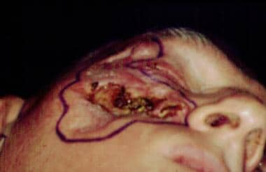
Basal cell carcinoma of the skin. Intraoperative view outlining the extent of resection (lateral view). The surgical resection involved zygomatic bone and the lateral orbit. The eye was spared.
-
Basal cell carcinoma of the skin. Intraoperative view showing the extent of resection (inferior view).