Ise S, Ofuji S. Subcorneal pustular dermatosis. A follicular variant?. Arch Dermatol. 1965 Aug. 92(2):169-71. [QxMD MEDLINE Link].
Ofuji S, Ogino A, Horio T, et al. Eosinophilic pustular folliculitis. Acta Derm Venereol. 1970. 50(3):195-203. [QxMD MEDLINE Link].
Nervi SJ, Schwartz RA, Dmochowski M. Eosinophilic pustular folliculitis: a 40 year retrospect. J Am Acad Dermatol. 2006 Aug. 55(2):285-9. [QxMD MEDLINE Link].
Yamamoto Y, Nomura T, Kabashima K, Miyachi Y. Clinical epidemiology of eosinophilic pustular folliculitis: results from a nationwide survey in Japan. Dermatology. 2015. 230(1):87-92. [QxMD MEDLINE Link].
Lo CS, Yang CY, Ko JH, Lee WY, Shih IH, Lin YC. Hyperimmunoglobulin E syndrome presenting as eosinophilic pustular folliculitis: a case report. Int J Dermatol. 2015 Feb. 54(2):211-4. [QxMD MEDLINE Link].
Yoshida S, Yatsuzuka K, Chigyo K, Kuroo Y, Takemoto K, Sayama K. A Case of Eosinophilic Pustular Folliculitis since Birth. Children (Basel). 2021 Jan 7. 8 (1):[QxMD MEDLINE Link].
Suresh MS, Arora S, Nair RR. Doctor I am on fire: eosinophilic folliculitis in HIV negative. Indian J Dermatol Venereol Leprol. 2009 Mar-Apr. 75(2):194-6. [QxMD MEDLINE Link].
Fraser SJ, Benton EC, Roddie PH, Krajewski AS, Goodlad JR. Eosinophilic folliculitis: an important differential diagnosis after allogeneic bone-marrow transplant. Clin Exp Dermatol. 2009 Apr. 34(3):369-71. [QxMD MEDLINE Link].
Uchiyama M, Mitsuhashi Y, Okubo Y, Tsuboi R. Eosinophilic pustular folliculitis (Ofuji's disease) without macroscopic pustules. Int J Dermatol. 2012 Jan. 51(1):50-2. [QxMD MEDLINE Link].
Scavo S, Magro G, Caltabiano R. Erythematous and edematous eruption of the face. Eosinophilic pustular folliculitis. Int J Dermatol. 2010 Sep. 49(9):975-7. [QxMD MEDLINE Link].
Tsuboi H, Niiyama S, Katsuoka K. Eosinophilic pustular folliculitis (Ofuji disease) manifested as pustules on the palms and soles. Cutis. 2004 Aug. 74(2):107-10. [QxMD MEDLINE Link].
Sufyan W, Tan KB, Wong ST, et al. Eosinophilic pustular folliculitis. Arch Pathol Lab Med. 2007 Oct. 131(10):1598-601. [QxMD MEDLINE Link].
Mizoguchi S, Setoyama M, Higashi Y, Hozumi H, Kanzaki T. Eosinophilic pustular folliculitis induced by carbamazepine. J Am Acad Dermatol. 1988. 38:641-3.
Maejima H, Mukai H, Hikaru E. Eosinophilic pustular folliculitis induced by allopurinol and timedium bromide. Acta DermVenereol. 2002. 82:316-7.
Ooi CG, Walker P, Sidhu SK, Gordon LA, Marshman G. Allopurinol induced generalized eosinophilic pustular folliculitis. Australas J Dermatol. 2006 Nov. 47(4):270-3.
Roos TC, Albrecht H. Foscarnet-associated eosinophilic folliculitis in a patient with AIDS. J Am Acad Dermatol. 2001 Mar. 44(3):546-7.
Laing ME, Laing TA, Mulligan NJ, Keane FM. Eosinophilic pustular folliculitis induced by chemotherapy. J Am Acad Dermatol. 2006 Apr. 54(4):729-30.
Fujii M, Takahashi I, Kishiyama K, Honma M, Ishida-Yamamoto A. Case of generalized eosinophilic pustular folliculitis induced by allopurinol. J Dermatol. 2016 Apr. 43 (4):458-9. [QxMD MEDLINE Link].
Brenner S, Wolf R, Ophir J. Eosinophilic pustular folliculitis: a sterile folliculitis of unknown cause?. J Am Acad Dermatol. 1994 Aug. 31(2 Pt 1):210-2. [QxMD MEDLINE Link].
Veraldi S, Ferrante P, Mancuso R, et al. Evidence of retroviral involvement in an Italian patient with Ofuji's disease. Dermatology. 1999. 198(1):86-9. [QxMD MEDLINE Link].
Gul U, Kilic A, Demiriz M. Eosinophilic pustular folliculitis: the first case associated with hepatitis C virus. J Dermatol. 2007 Jun. 34(6):397-9. [QxMD MEDLINE Link].
Matsudate Y, Miyaoka Y, Urano Y. Two cases of eosinophilic pustular folliculitis associated with pregnancy. J Dermatol. 2016 Feb. 43 (2):218-9. [QxMD MEDLINE Link].
Sugaya M, Suga H, Miyagaki T, Fujita H, Sato S. Eosinophilic pustular folliculitis associated with Sézary syndrome. Clin Exp Dermatol. 2014 Jun. 39(4):536-8. [QxMD MEDLINE Link].
Patrizi A, Chieregato C, Visani G, et al. Leukaemia-associated eosinophilic folliculitis (Ofuji's disease). J Eur Acad Dermatol Venereol. 2004 Sep. 18(5):596-8. [QxMD MEDLINE Link].
Kimoto M, Ishihara S, Konohana A. Eosinophilic pustular folliculitis with polycythemia vera. Dermatology. 2005. 210(3):239-40. [QxMD MEDLINE Link].
Takamura S, Teraki Y. Eosinophilic pustular folliculitis associated with hematological disorders: A report of two cases and review of Japanese literature. J Dermatol. 2016 Apr. 43 (4):432-5. [QxMD MEDLINE Link].
Cunningham F, Gupta P, Becher G, Magro CM. Necrotizing eosinophilic folliculitis: a new manifestation of the atopic diathesis?. Clin Exp Dermatol. 2017 Jan. 42 (1):100-101. [QxMD MEDLINE Link].
Jiang YY, Zeng YP, Jin HZ. Eosinophilic Pustular Folliculitis Associated with Cutaneous Angiosarcoma. Chin Med J (Engl). 2018 Jan 5. 131 (1):115-116. [QxMD MEDLINE Link].
Owen RL, Shah KK, Sosis AC, Purcell SM. Eosinophilic pustular folliculitis with underlying mantle cell lymphoma. Cutis. 2018 Jun. 101 (6):454-457. [QxMD MEDLINE Link].
Goiriz R, Guhl-Millan G, Penas PF, et al. Eosinophilic folliculitis following allogeneic peripheral blood stem cell transplantation: case report and review. J Cutan Pathol. 2007 Dec. 34 Suppl 1:33-6. [QxMD MEDLINE Link].
Kim BW, Lee JH, Won CH, Chang SE, Lee MW, Choi JH, et al. Eosinophilic pustular folliculitis: association with long-term immunosuppressant use in a solid organ transplant recipient. Ann Dermatol. 2014 Aug. 26(4):520-1. [QxMD MEDLINE Link]. [Full Text].
Chuh A. Eosinophilic folliculitis due to wearing protective gear in citizens volunteering for sanitation services during the COVID-19 pandemic - an original epidemiological, clinical, dermoscopic, and laboratory-based study. Int J Dermatol. 2020 Dec. 59 (12):1468-1474. [QxMD MEDLINE Link].
Fujiyama T, Tokura Y. Clinical and histopathological differential diagnosis of eosinophilic pustular folliculitis. J Dermatol. 2013 Jun. 40(6):419-23. [QxMD MEDLINE Link].
Takashima S, Nishie W, Morita Y, Osawa R, Iwata H, Fujita Y, et al. Eosinophilic pustular folliculitis in a patient with mycosis fungoides. J Eur Acad Dermatol Venereol. 2015 Feb 11. [QxMD MEDLINE Link].
Chopra D, Kumari S, Kundal RK, Bahl RK, Aggarwal S, Singh H, et al. A study of clinicopathological correlation of pruritic papular eruptions in HIV patients. Indian J Sex Transm Dis AIDS. 2018 Jan-Jun. 39 (1):44-49. [QxMD MEDLINE Link].
Boone SL, Guitart J, Gerami P. Follicular mycosis fungoides: a histopathologic, immunohistochemical, and genotypic review. G Ital Dermatol Venereol. 2008 Dec. 143(6):409-14. [QxMD MEDLINE Link].
Schwartz RA, Janniger CK. Generalized pustular figurate erythema: A newly delineated severe cutaneous drug reaction linked with hydroxychloroquine. Dermatol Ther. 2020 May. 33 (3):e13380. [QxMD MEDLINE Link].
Fertitta, Laura; Bodemer, Christine; Molina, Thierry; Frassati-Biaggi, Annonciade; Fraitag, Sylvie; Leclerc-Mercier, et al. Eosinophilic Pustular Folliculitis of Infancy: A Histologic Assessment of 43 Cases. A J Derm. June 2022. 44(6):395-403(9). [Full Text].
Piantanida EW, Turiansky GW, Kenner JR, Mather MK, Sperling LC. HIV-associated eosinophilic folliculitis: Diagnosis by transverse histologic sections. J Am Acad Dermatol. 1998 Jan. 38(1):124-6.
Junqueira Magalhães Afonso JP, Tomimori J, Michalany NS, Nonogaki S, Porro AM. Pruritic papular eruption and eosinophilic folliculitis associated with human immunodeficiency virus (HIV) infection: A histopathological and immunohistochemical comparative study. J Am Acad Dermatol. 2012 Apr 19. [QxMD MEDLINE Link].
Satoh T, Ito Y, Miyagishi C, Yokozeki H. Basophils Infiltrate Skin Lesions of Eosinophilic Pustular Folliculitis (Ofuji's Disease). Acta Derm Venereol. 2011 Jan 31. [QxMD MEDLINE Link].
Annam V, Yelikar BR, Inamadar AC, Palit A, Arathi P. Clinicopathological study of itchy folliculitis in HIV-infected patients. Indian J Dermatol Venereol Leprol. 2010 May-Jun. 76(3):259-62. [QxMD MEDLINE Link].
Dwivedi M, Sharma V, Pathak K. Pilosebaceous targeting by isotretenoin-loaded invasomal gel for the treatment of eosinophilic pustular folliculitis: optimization, efficacy and cellular analysis. Drug Dev Ind Pharm. 2017 Feb. 43 (2):293-304. [QxMD MEDLINE Link].
Satoh T, Shimura C, Miyagishi C, Yokozeki H. Indomethacin-induced reduction in CRTH2 in eosinophilic pustular folliculitis (Ofuji's disease): a proposed mechanism of action. Acta Derm Venereol. 2010. 90(1):18-22. [QxMD MEDLINE Link].
Nomura T, Katoh M, Yamamoto Y, Miyachi Y, Kabashima K. Eosinophilic pustular folliculitis: Trends in therapeutic options. J Dermatol. 2016 Feb 15. [QxMD MEDLINE Link].
Nomura T, Katoh M, Yamamoto Y, Miyachi Y, Kabashima K. Eosinophilic pustular folliculitis: A proposal of diagnostic and therapeutic algorithms. J Dermatol. 2016 Mar 30. [QxMD MEDLINE Link].
Fukamachi S, Kabashima K, Sugita K, Kobayashi M, Tokura Y. Therapeutic effectiveness of various treatments for eosinophilic pustular folliculitis. Acta Derm Venereol. 2009. 89(2):155-9. [QxMD MEDLINE Link].
Otsuka A, Doi H, Miyachi Y, Kabashima K. Treatment of eosinophilic pustular folliculitis with ciclosporin: suppression of mRNA expression of IL-4 and IL-13. J Eur Acad Dermatol Venereol. 2010 Dec. 24(12):1489-91. [QxMD MEDLINE Link].
Patel NP, Laguda B, Roberts NM, Francis ND, Agnew K. Treatment of Eosinophilic Pustulosis of Infancy with Topical Tacrolimus. Br J Dermatol. 2012 May 7. [QxMD MEDLINE Link].
Ng SS, Tay YK. Successful treatment of eosinophilic pustular folliculitis with topical tacrolimus 0.1 percent ointment. Dermatol Online J. 2012 Feb 15. 18(2):10. [QxMD MEDLINE Link].
Toyonaga E, Abe R, Moriuchi R, Ito K, Abe Y, Shimizu H. Indomethacin for refractory infantile eosinophilic pustular folliculitis. JAMA Dermatol. 2013 Mar. 149(3):367-8. [QxMD MEDLINE Link].
Ishiguro N, Shishido E, Okamoto R, Igarashi Y, Yamada M, Kawashima M. Ofuji's disease: A report on 20 patients with clinical and histopathologic analysis. J Am Acad Dermatol. 2002 Jun. 46(6):827-33.
Inaoka M, Hayakawa J, Shiohara T. HIV seronegative eosinophilic pustular folliculitis successfully treated with metronidazole. J Am Acad Dermatol. 2002 May. 46(5 Suppl):S153-5.
Lim HW, Vallurupalli S, Meola T, et al. UVB phototherapy is an effective treatment for pruritus in patients infected with HIV. J Am Acad Dermatol. 1997 Sep. 37(3 Pt 1):414-7. [QxMD MEDLINE Link].
Misago N, Narisawa Y, Matsubara S, et al. HIV-associated eosinophilic pustular folliculitis: successful treatment of a Japanese patient with UVB phototherapy. J Dermatol. 1998 Mar. 25(3):178-84. [QxMD MEDLINE Link].
Hara D, Kuroda K, Mieno H, et al. Treatment of eosinophilic pustular folliculitis with tacrolimus ointment. J Am Acad Dermatol. 2004 Nov. 51(5 Suppl):S143-5. [QxMD MEDLINE Link].
Kabashima K, Sakurai T, Miyachi Y. Treatment of eosinophilic pustular folliculitis (Ofuji's disease) with tacrolimus ointment. Br J Dermatol. 2004 Oct. 151(4):949-50. [QxMD MEDLINE Link].
Kawaguchi M, Mitsuhashi Y, Kondo S. Successful treatment of eosinophilic pustular folliculitis with topical tacrolimus. Int J Dermatol. 2004 Aug. 43(8):608-10. [QxMD MEDLINE Link].
Brazzelli V, Barbagallo T, Prestinari F, et al. HIV seronegative eosinophilic pustular folliculitis successfully treated with doxicycline. J Eur Acad Dermatol Venereol. 2004 Jul. 18(4):467-70. [QxMD MEDLINE Link].
Wilson BD, Kucera JC, Shin PJ. The role of radiation treatment in the management of eosinophilic pustular folliculitis. J Med. 2002. 33(1-4):111-3. [QxMD MEDLINE Link].
Odyakmaz Demirsoy E, Demirsoy U, Ozod U, Kiran R. Eosinophilic pustular folliculitis of infancy suppressed with cetirizine. Pediatr Dermatol. 2019 Feb 21. [QxMD MEDLINE Link].
Dong RJ, Huang SZ, Upadhyay P, Shrestha S, Zhai YJ, Li YY. Thalidomide in the Treatment of Sweet's Syndrome and Eosinophilic Folliculitis Associated With Immune Reconstitution Inflammatory Syndrome. Front Med (Lausanne). 2019. 6:343. [QxMD MEDLINE Link].
Ramondetta A, Giuffrida G, Di Vincenzo S, De Pasquale R. A case of eosinophilic pustular folliculitis successfully treated with adalimumab. Dermatol Ther. 2022 May 4. e15550. [QxMD MEDLINE Link].
Bürgler C, Guillet C, Kolm I, Theiler M, Schmid-Grendelmeier P, Kroiss S, et al. Treatment of eosinophilic pustular folliculitis with benralizumab in a 13-year-old girl. J Eur Acad Dermatol Venereol. 2021 Feb 17. [QxMD MEDLINE Link].
Hernández-Martín Á, Nuño-González A, Colmenero I, Torrelo A. Eosinophilic pustular folliculitis of infancy: a series of 15 cases and review of the literature. J Am Acad Dermatol. 2013 Jan. 68 (1):150-5. [QxMD MEDLINE Link].
Hernández-Martín Á, Nuño-González A, Colmenero I, Torrelo A. Eosinophilic pustular folliculitis of infancy: a series of 15 cases and review of the literature. J Am Acad Dermatol. 2013 Jan. 68 (1):150-5. [QxMD MEDLINE Link].
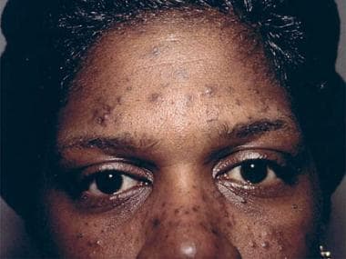 Eosinophilic pustular folliculitis in a patient infected with HIV. Note acneiform hyperpigmented papules. Photograph courtesy of Sarah A. Myers, MD.


