Continually Updated Clinical Reference
 
 
  All Sources     eMedicine     Medscape     Drug Reference     MEDLINE
 
eMedicine - Empyema, Pleuropulmonary : Article by

Quick Find
Authors & Editors
Introduction
Clinical
Differentials
Workup
Treatment
Medication
Follow-up
Miscellaneous
Multimedia
References

Related Articles
Boerhaave Syndrome

Hemothorax

Intra-abdominal Sepsis

Lung Abscess

Lung Cancer, Non-Small Cell

Lung Cancer, Oat Cell (Small Cell)

Pleural Effusion

Pleurodynia

Pneumococcal Infections

Pneumonia, Aspiration

Pneumonia, Bacterial

Pneumonia, Community-Acquired

Pneumonia, Fungal

Secondary Lung Tumors

Tuberculosis


Patient Education
Click here for patient education.


Author: Sat Sharma, MD, FRCPC, Professor and Head, Division of Pulmonary Medicine, Department of Internal Medicine, University of Manitoba; Site Director, Respiratory Medicine, St. Boniface General Hospital

Sat Sharma is a member of the following medical societies: American Academy of Sleep Medicine, American College of Chest Physicians, American College of Physicians-American Society of Internal Medicine, American Thoracic Society, Canadian Medical Association, Royal College of Physicians and Surgeons of Canada, Royal Society of Medicine, Society of Critical Care Medicine, and World Medical Association

Editors: Michael Peterson, MD, Chief of Medicine, Vice-Chair of Medicine, University of California at San Francisco; Endowed Professor of Medicine, University of California at San Francisco-Fresno; Francisco Talavera, PharmD, PhD, Senior Pharmacy Editor, eMedicine; Om Prakash Sharma, MD, FRCP, FCCP, DTM&H, Professor, Department of Medicine, Division of Pulmonary and Critical Care Medicine, University of Southern California Keck School of Medicine; Timothy D Rice, MD, Associate Professor, Departments of Internal Medicine and Pediatrics and Adolescent Medicine, Saint Louis University School of Medicine; Zab Mosenifar, MD, Director, Division of Pulmonary and Critical Care Medicine, Director, Women's Guild Pulmonary Disease Institute, Executive Vice Chair, Department of Medicine, Cedars Sinai Medical Center; Professor of Medicine, David Geffen School of Medicine at UCLA

Author and Editor Disclosure

Synonyms and related keywords: pleuropulmonary empyema, complicated parapneumonic pleural effusion, Staphylococcus aureus, S aureus, Streptococcus pneumoniae, S pneumoniae, Klebsiella, Pseudomonas, Haemophilus, Bacteroides, Peptostreptococcus, uncomplicated parapneumonic effusion, thoracic empyema, thoracoplasty, decortication, pneumonia, parapneumonic effusion, esophageal perforation, trauma, surgical procedure on pleural space, septicemia

Background

For centuries, empyema has been recognized as a serious problem. Around 500 BC, Hippocrates recommended treating empyema with open drainage. Since then, the treatment of empyema remained essentially unchanged until the middle of the 19th century. In 1876, Hewitt described a method of closed drainage of the chest in which a rubber tube was placed into the empyema cavity and drained via the water seal drainage. In the early 20th century, surgical therapies for empyema (eg, thoracoplasty, decortication) were introduced.

Parapneumonic pleural effusions are divided into 3 groups or stages based on pathogenesis: uncomplicated parapneumonic effusion, complicated parapneumonic effusion, and thoracic empyema.

Uncomplicated parapneumonic effusion is an exudative predominantly neutrophilic effusion that occurs as the lung interstitial fluid increases during pneumonia. These effusions are resolved with appropriate antibiotic treatment of pneumonia.

Complicated parapneumonic effusion is a bacterial invasion of the pleural space that leads to an increased number of neutrophils, pleural fluid acidosis, and elevated lactic dehydrogenase (LDH) concentration. These effusions often are sterile because bacteria are usually cleared rapidly from the pleural space.

Thoracic empyema is characterized by either aspiration of pus on thoracentesis or the presence of bacterial organisms on Gram stain. A positive culture is not required for diagnosis.

Pathophysiology

The evolution of a parapneumonic pleural effusion (see Image 1) can be divided into 3 stages, including exudative, fibropurulent, and organization stages.

During the exudative stage, sterile pleural fluid rapidly accumulates in the pleural space. The pleural fluid originates in the interstitial spaces of the lung and in the capillaries of the visceral pleura because of increased permeability. The pleural fluid has a low WBC and LDH level, and the glucose and pH levels are within the reference range. These effusions resolve with antibiotic therapy, and chest tube insertion is not required.

During the fibropurulent stage, bacterial invasion of the pleural space occurs, with accumulation of polymorphonuclear leucocytes, bacteria, and cellular debris. A tendency toward loculation exists, pleural fluid pH and glucose levels are lower, and the LDH level increases.

During the organization stage, fibroblasts grow into the exudates from both the visceral and parietal pleural surfaces, and they produce an inelastic membrane called pleural peel. Pleural fluid is thick. In an untreated patient, pleural fluid may drain spontaneously through the chest wall (ie, empyema necessitatis). Empyema may arise without an associated pneumonic process. The most common causes are esophageal perforation, trauma, surgical procedure on pleural space, and septicemia.

Bacteriologic features of culture-positive parapneumonic effusions have changed over time. Prior to the antibiotic era, Streptococcus pneumoniae and hemolytic streptococci were common. Presently, aerobic organisms are isolated slightly more frequently than anaerobic organisms. Staphylococcus aureus and S pneumoniae account for approximately 70% of aerobic gram-positive cultures.

Bacteriology of parapneumonic effusions is closely related to the bacteriology of a pneumonic process. Gram-positive aerobic organisms are isolated twice as frequently as gram-negative aerobic organisms. Klebsiella, Pseudomonas, and Haemophilus species are the 3 most commonly isolated aerobic gram-negative organisms. Bacteroides and Peptostreptococcus species are the 2 most commonly isolated anaerobic organisms. A mixed bacterial flora containing aerobes and anaerobes is more likely to produce an empyema than a single-organism infection. Anaerobic bacteria have been cultured in 36-76% of empyemas.

Frequency

United States

Incidence of pleural effusion with various pneumonias depends on the infecting organism. The pleural space is commonly infected in patients with anaerobic pneumonia. In one series of patients with anaerobic infections of the lung, 35% had pleural effusions, and 94% of these were positive for organisms. Aerobic organisms were cultured from the pleural fluid in 40% of patients. Generally speaking, pleural effusions occur in 40% of bacterial pneumonias.

Mortality/Morbidity

Mortality rates associated with empyema depend upon severity of the underlying disease and prompt therapy. A mortality rate of 11-50% has been reported. In patients who are older and debilitated, mortality is high.


History

Clinical manifestations of parapneumonic effusion and empyema largely depend on whether the patient has an aerobic or anaerobic infection. If fever persists for more than 48 hours after initiation of antibiotic treatment, a complicating parapneumonic effusion or empyema likely exists.

  • Aerobic bacterial pneumonia
    • Clinical presentation in patients with aerobic bacterial pneumonia is similar to that of bacterial pneumonia.
    • Patients present with an acute febrile illness with chest pain, sputum production, and leukocytosis.
    • A complicated parapneumonic effusion is suggested by the presence of a fever lasting more than 48 hours after initiation of antibiotic therapy.
  • Anaerobic bacterial infection
    • Patients with anaerobic bacterial infections involving the pleural space usually present with a subacute illness.
    • Most of these patients have symptoms persisting for more than 7 days.
    • Approximately 60% of patients have weight loss.
    • Most of these patients have poor oral hygiene, many suffer from alcoholism, and others have factors that predispose them to recurrent aspiration.

Physical

  • Patients may have a fever and appear toxic.
  • The signs of pleural effusion, including dullness on percussion and absence of breath sounds, are evident.

Causes

In the preantibiotic era, as many as 11% of incidents of pneumococcal pneumonia were associated with empyema, and 64% of empyemas were caused by S pneumoniae. Beta-hemolytic streptococci caused 15% of empyemas, and staphylococci caused 8% of empyemas.

  • Anaerobic infections currently comprise as many as 40% of empyemas.
  • Empyema is most often associated with pneumonia, particularly aspirational events with anaerobic microbiology.
  • Increasingly, empyema is a complication of previous surgery, which accounts for 30% of cases. The usual organisms are Staphylococcus species and gram-negative bacteria.
  • Trauma may also be associated with superinfection of pleural space.
  • In the absence of trauma or surgery, the infecting organism may spread from blood or other organs into the pleural space. These are subdiaphragmatic abscesses (eg, ruptured esophagus, mediastinitis, osteomyelitis, pericarditis, cholangitis, diverticulitis, pericarditis).


Boerhaave Syndrome
Hemothorax
Intra-abdominal Sepsis
Lung Abscess
Lung Cancer, Non-Small Cell
Lung Cancer, Oat Cell (Small Cell)
Pleural Effusion
Pleurodynia
Pneumococcal Infections
Pneumonia, Aspiration
Pneumonia, Bacterial
Pneumonia, Community-Acquired
Pneumonia, Fungal
Secondary Lung Tumors
Tuberculosis

Other Problems to be Considered

Malignant pleural effusion
Tuberculous pleural effusion
Esophageal perforation (Boerhaave syndrome)
Drug-induced pleural effusion
Pleuritis secondary to systemic collagen vascular diseases
Subdiaphragmatic abscess


Lab Studies

  • Consider the possibility of a parapneumonic effusion during the initial evaluation of every patient with bacterial pneumonia because a delay in instituting proper pleural drainage substantially increases morbidity.
  • The sputum is purulent and may help with the identification of underlying bacteriology.
  • Peripheral leukocytosis (15,000/µL) usually exists.

Imaging Studies

  • Chest radiography
    • The lateral chest radiograph usually suggests the presence of a significant amount of pleural fluid (see Image 2).
    • If either of the diaphragms is not visible throughout their entire length or the posterior costophrenic angles are blunted, obtain bilateral decubitus chest radiographs.
    • Free pleural fluid is indicated by the presence of fluid between the chest wall and the inferior part of the lung. If this distance measures more than 10 mm, perform a diagnostic thoracentesis.
  • Ultrasonography
    • Loculated pleural effusions appear as pleural-based masses without air bronchogram on standard chest radiographs.
    • Ultrasonography can effectively distinguish loculated pleural fluid from an infiltrate. Therefore, if loculated pleural effusion is suspected, perform an ultrasonographic examination.
  • CT scanning of the thorax
    • CT scanning of the chest with contrast enhances the pleural surface and assists in delineating the pleural fluid loculations (see Image 4).
    • CT scanning of the chest may also detect airway or parenchymal abnormalities such as endobronchial obstruction or the presence of lung abscesses.

Procedures

  • A thoracentesis is recommended in the workup of any undiagnosed pleural effusion (see Image 3).
    • Pleural fluid may vary from a clear yellow liquid to thick foul-smelling pus. Foul-smelling fluid indicates an anaerobic infection.
    • Determine pleural fluid pH. A complicated parapneumonic effusion has a pH of less than 7.2.
    • Send pleural fluid for WBC count and protein, LDH, and glucose levels. Results in patients with parapneumonic effusions reveal WBC count of greater than 500 cells/µL, elevated protein and LDH levels, and decreased glucose level.


Medical Care

Individualize treatment of each patient, depending on the type or stage of parapneumonic effusion.

  • Initial treatment
    • The initial treatment of a patient with pneumonia and pleural effusion involves 2 major decisions. First, select an appropriate antibiotic. Second, decide whether to initiate tube drainage of the pleural space. The initial antibiotic selection is usually based on whether the pneumonia is community-acquired pneumonia or hospital-acquired pneumonia and the severity of the patient's illness. For a patient with community-acquired pneumonia, the recommended agents are second-generation or third-generation cephalosporins in addition to a macrolide. For patients hospitalized with severe community-acquired pneumonia, initiate treatment with a macrolide plus a third-generation cephalosporin with antipseudomonal activity. Enteric gram-negative bacilli frequently cause pneumonia acquired in institutions (eg, hospitals, nursing homes). Therefore, initial antibiotic coverage should include an antibiotic effective against pseudomonads.
    • Base the decision to institute tube drainage on examination of the pleural fluid. Identify patients who need tube drainage as soon as possible because the pleural effusions become loculated if tube thoracostomy is delayed.
    • Patients with pleural effusions that have a pleural fluid thickness greater than 10 mm on lateral decubitus radiographs must have a diagnostic thoracentesis. Effusions with pleural fluid thickness less than 10 mm on decubitus chest radiographs almost always resolve with appropriate systemic antibiotics.
    • If the diagnostic thoracentesis yields thick pus, the patient has an empyema. Institute tube thoracostomy immediately. If the pleural fluid is not thick pus, then the results of the pleural fluid Gram stain and the pleural fluid glucose, pH, and LDH levels should guide the course of action.
  • Uncomplicated parapneumonic effusions
    • If the pleural fluid pH is greater than 7.20, the pleural fluid glucose is greater than 40 mg/dL, and the pleural fluid LDH is less than 1000 IU/L, the parapneumonic effusion is in the exudative stage, and no further therapeutic intervention is required. However, if the pleural effusion increases in size or the patient remains or becomes febrile, repeat the thoracentesis.
    • Uncomplicated effusions resolve with antibiotics alone.
    • Patients with uncomplicated effusions can be monitored with serial radiography or meticulous physical examination to document resolution of effusion.
  • Complicated parapneumonic effusions
    • If the initial thoracentesis reveals pleural fluid with a pH less than 7.20 or a glucose level greater than 40 mg/dL, immediately perform tube thoracostomy. If the Gram stain of the pleural fluid is positive, tube thoracostomy is recommended.
    • Complicated effusions have a variable response to appropriate antibiotic therapy, although some patients can be treated with antibiotics alone. Patients with complicated parapneumonic effusions are treated as though they have thoracic empyema.
  • Chest tubes (tube thoracostomy)
    • Insert chest tubes immediately after a complicated parapneumonic effusion or empyema is diagnosed because delay leads to formation of loculated pleural effusion. Position the chest tube in a dependent part of the pleural effusion. Although traditionally large-bore (38-32F) tubes are recommended, smaller catheters (8.5-16F) can also be used successfully in draining the pleural space.
    • If the patient responds clinically and radiologically to closed tube drainage of the pleural space, leave the chest tubes in place until the volume of the pleural drainage is less than 50 mL/24 h and the drainage fluid becomes clear yellow.
    • If the patient has not demonstrated clinical or radiologic improvement, perform ultrasonic examination or CT scanning of the pleural space to detect remaining loculi of pleural fluid and to ensure that the tube is in the proper place. If multiple loculi are identified, administer thrombolytic therapy intrapleurally. Closed chest tube drainage yields satisfactory results in approximately 60% of patients with aerobic infections and 25% of patients with anaerobic infections.
  • Intrapleural thrombolytic agents
    • Since the 1970s, several studies have reported success of thrombolytic therapy for loculated complicated parapneumonic effusions. The thrombolytic agents appeared to be more effective if they were administered early in the course of parapneumonic effusions. In a randomized trial of patients with multiloculated pleural effusions, those in the urokinase group drained significantly more pleural fluid, required less surgical intervention, and required fewer days in the hospital.
    • With fibrinolytic therapy, success rates of 70-90% have been quoted. Streptokinase is used in a dose of 250,000 IU in 100 mL of isotonic sodium chloride solution once or twice a day. Following instillation, the chest tube is clamped for 2-4 hours before draining spontaneously. These agents may be administered daily for as many as 14 days. Both streptokinase and urokinase appear to be equally effective, although streptokinase may lead to sensitization with production of an antibody response and subsequent allergic reaction if employed for systemic thrombolysis. Multiple tubes may be inserted under CT scan guidance for multiloculated pleural space.
    • A recent prospective randomized trial of intrapleural fibrinolytic agent streptokinase was conducted in the drainage of infected pleural-fluid collections. In this double-blind trial, 454 patients with pleural infection (either purulent pleural fluid or pleural fluid with a pH <7.2 with signs of infection) received either intrapleural streptokinase (250,000 IU bid for 3 days) or placebo. Among the 427 patients who received streptokinase or placebo, no benefit was reported for streptokinase in terms of mortality, rate of surgery, radiographic outcomes, or length of hospital stay; serious adverse events (chest pain, fever, or allergy) were more common with streptokinase (Maskell, 2005).
    • Tokuda et al performed a meta-analysis of all properly randomized trials comparing intrapleural fibrinolytic agents with placebo in adult patients with empyema and complicated parapneumonic effusions. The outcome of primary interest was the reduction of death and surgical intervention. Five trials totaling 575 patients were included. The meta-analysis did not support the routine use of fibrinolytic therapy for all patients who required chest tube drainage for empyema or complicated parapneumonic effusions. However, because of significant heterogeneity of the treatment effects, selected patients might benefit from the treatment (Tokuda, 2006).

Surgical Care

Several surgical procedures are available to treat a patient with empyema. The definite indication for surgical therapy is persistent pleural sepsis despite antibiotics and attempts at pleural drainage with thoracoscopy.

  • Thoracoscopy
    • Thoracoscopy is an alternate therapy for multiloculated empyema.
    • In patients with multiloculated parapneumonic effusion, the loculations in the pleural space can be disrupted with a thoracoscope, and the pleural space can be drained completely.
    • If extensive adhesions are present or thick pleural peel entraps the lung, the procedure may be converted to open thoracostomy and decortication.
    • Luh et al published their experience in treatment of complicated parapneumonic effusion and pleural empyema by video-assisted thoracoscopic surgery (VATS) in 234 patients (108 women, 126 men). More than 85% (200 patients) received preoperative diagnostic or therapeutic thoracentesis, tube thoracostomy, or fibrinolytics. Out of 234 patients, 202 patients (86.3%) achieved satisfactory results with VATS treatment. Only 40 patients required open decortication or repeat procedures. VATS is safe and effective for treatment; earlier intervention with VATS can produce better clinical results (Luh, 2005).
    • Hope et al reviewed outcomes of surgical treatment for parapneumonic empyema. The use of VATS was compared with thoracotomy. Morbidity and mortality rates were similar among all groups. Conversion rate to open thoracotomy was 21%. Based on a shorter postoperative length of stay with similar morbidity and mortality in patients operated on within 11 days of admission, early aggressive surgery treatment for parapneumonic empyema is recommended (Hope, 2005).
  • Rib resection and drainage of pleural space
    • Open drainage of the pleural space may be employed when closed tube drainage of the pleural infection is inadequate and the patient does not respond to intrapleural thrombolytic agents. This procedure is recommended only when the patient is too ill to tolerate a decortication. The resection of 1-3 ribs overlying the lower part of the empyema cavity is performed, a large-bore chest tube is inserted into the empyema cavity, and the tube is drained into a colostomy bag.
    • When a patient is treated by open drainage, expect an open chest wound for a prolonged period. In one series, the median time for healing the drainage site was 142 days. With decortication, the period of convalescence is much shorter, although patients who are markedly debilitated do not tolerate decortication.
  • Decortication
    • In decortication, all the fibrous tissue is removed from the visceral pleural peel, and all pus is evacuated from the pleural space. Decortication is a major thoracic operation requiring full thoracotomy; therefore, do not perform decortication on patients who are markedly ill.
    • Decortication is the procedure of choice for patients in whom pleural sepsis is not controlled by closed tube thoracostomy, intrapleural thrombolytic agents, and possibly, thoracoscopy.
    • Mortality rates as high as 10% have been described with this procedure.
    • Do not perform decortication just to remove the thickened pleural peel; these thickened peels usually resolve spontaneously over several months. If after 6 months the pleura remains thickened and the patient's pulmonary function is reduced sufficiently to limit activities, consider decortication.
  • Postpneumonectomy empyema, an uncommon but life-threatening complication, is associated with bronchopleural fistula. Treatment of bronchopleural fistula depends on several factors, including extent of dehiscence, degree of pleural contamination, and general condition of the patient. Early diagnosis and aggressive therapeutic strategies of controlling infection, closure of fistula, and sterilization of the closed pleural space are mandatory. Repeated debridement, VATS, endoscopic application of tissue glue, and stenting may be additional management strategies (Ng, 2005).


The initial choice of antibiotics frequently is empiric, beginning with erythromycin, azithromycin, clindamycin, cefoxitin, penicillin, or cefuroxime. Empiric antimicrobial therapy must be comprehensive and should cover all likely pathogens in the context of the clinical setting. Base subsequent therapy upon sputum, blood culture, or pleural fluid culture results. An empyema is treated with prompt chest tube drainage in conjunction with parenteral antibiotics. For an empyema secondary to aspiration pneumonia or a parapneumonic process, choose antibiotics that are active against mouth flora. For an empyema secondary to penetrating chest trauma, administer antibiotics that have coverage for skin flora.

Drug Category: Antibiotics

Therapy must be comprehensive and cover all likely pathogens in the context of this clinical setting.

Drug NameClindamycin (Cleocin)
DescriptionLincosamide effective against aerobic and anaerobic streptococci (except enterococci). Inhibits bacterial growth, possibly by blocking dissociation of peptidyl tRNA from ribosomes, causing RNA-dependent protein synthesis to arrest.
Adult Dose600 mg IV q6-8h
Pediatric Dose25-40 mg/kg/d IV divided tid/qid
ContraindicationsDocumented hypersensitivity; regional enteritis; ulcerative colitis; hepatic impairment; antibiotic-associated colitis
InteractionsIncreases duration of neuromuscular blockade induced by tubocurarine and pancuronium; erythromycin may antagonize effects of clindamycin; antidiarrheals may delay absorption of clindamycin
PregnancyB - Usually safe but benefits must outweigh the risks.
PrecautionsAdjust dose in severe hepatic dysfunction; no adjustment necessary in renal insufficiency; associated with severe and possibly fatal colitis

Drug NameCefoxitin (Mefoxin)
DescriptionSecond-generation cephalosporin indicated for gram-positive cocci and gram-negative rod infections. Infections caused by cephalosporin-resistant or penicillin-resistant gram-negative bacteria may respond to cefoxitin.
Adult Dose2 g IV q6-8h
Pediatric Dose80-160 mg/kg/d IV in 4-6 divided doses
ContraindicationsDocumented hypersensitivity
InteractionsProbenecid may increase effects of cefoxitin; coadministration with aminoglycosides or furosemide may increase nephrotoxicity (closely monitor renal function)
PregnancyB - Usually safe but benefits must outweigh the risks.
PrecautionsBacterial or fungal overgrowth of nonsusceptible organisms may occur with prolonged use or repeated treatment; caution in patients with previously diagnosed colitis

Drug NamePenicillin G (Pfizerpen)
DescriptionInterferes with synthesis of cell wall mucopeptide during active multiplication, resulting in bactericidal activity against susceptible microorganisms.
Adult Dose2 million U IV q4h
Pediatric Dose150,000 U/kg/d IV q4h
ContraindicationsDocumented hypersensitivity
InteractionsProbenecid can increase penicillin effectiveness by decreasing its clearance; tetracyclines can decrease penicillin effectiveness
PregnancyB - Usually safe but benefits must outweigh the risks.
PrecautionsTraditional drug for treatment of lung abscess, but spectrum of activity is narrow; use with caution in patients with impaired renal function

Drug NameAzithromycin (Zithromax)
DescriptionThese agents are replacing erythromycin as therapy for community-acquired pneumonia. They cover most potential etiologic agents, including Mycoplasma species. The newer macrolides offer decreased GI upset and the potential for improved compliance through reduced dosing frequency. They also afford more improved action against Haemophilus influenzae than erythromycin.
Adult DoseDay 1: 500 mg PO
Days 2-5: 250 mg/d PO; alternatively, 500 mg/d IV
Pediatric DoseDay 1: 10 mg/kg PO
Days 2-5: 5 mg/kg PO
ContraindicationsDocumented hypersensitivity; hepatic impairment; coadministration with pimozide
InteractionsMay increase toxicity of theophylline, warfarin, and digoxin; effects are reduced with coadministration of aluminum and/or magnesium antacids; nephrotoxicity and neurotoxicity may occur when coadministered with cyclosporine
PregnancyB - Usually safe but benefits must outweigh the risks.
PrecautionsSite reactions can occur with IV route; bacterial or fungal overgrowth may result with prolonged antibiotic use; may increase hepatic enzymes and cholestatic jaundice; caution in patients with impaired hepatic function, prolonged QT intervals, or pneumonia; caution in patients who are hospitalized, geriatric, or debilitated

Drug NameClarithromycin (Biaxin)
DescriptionAnother antibiotic used during initial therapy in otherwise uncomplicated pneumonia. More GI symptoms appear to occur than with azithromycin (eg, gastric upset, metallic taste). Inhibits bacterial growth, possibly by blocking dissociation of peptidyl tRNA from ribosomes, causing RNA-dependent protein synthesis to arrest.
Adult Dose500 mg PO bid for 10 d
Pediatric Dose<6 months: Not recommended
>6 months: 7.5 mg/kg PO bid for 10 d; not to exceed 1 g/d
ContraindicationsDocumented hypersensitivity; coadministration with pimozide
InteractionsToxicity increases with coadministration of fluconazole, astemizole, and pimozide; clarithromycin effects decrease and GI adverse effects may increase with coadministration of rifabutin or rifampin; may increase toxicity of anticoagulants, cyclosporine, tacrolimus, digoxin, omeprazole, carbamazepine, ergot alkaloids, triazolam, and HMG CoA-reductase inhibitors; serious cardiac arrhythmias may occur with coadministration of cisapride; plasma levels of certain benzodiazepines may increase, prolonging CNS depression; arrhythmias and increase in QTc intervals occur with disopyramide; coadministration with omeprazole may increase plasma levels of both agents
PregnancyB - Usually safe but benefits must outweigh the risks.
PrecautionsCoadministration with ranitidine or bismuth citrate is not recommended with CrCl <25 mL/min; administer one-half dose or increase dosing interval if CrCl <30 mL/min; diarrhea may be a sign of pseudomembranous colitis; superinfections may occur with prolonged or repeated antibiotic therapies

Drug NameErythromycin (E.E.S., Erythrocin, Ery-Tab)
DescriptionRecommended dosing schedule of erythromycin may result in GI upset; prescribe an alternative macrolide or change to tid dosing. Covers most potential etiologic agents, including Mycoplasma species. PO dosing regimen may be insufficient to adequately treat Legionella species. Erythromycin is less active against H influenzae. Although 10 d seems to be a standard course of treatment, treating until the patient has been afebrile for 3-5 d seems a more rational approach. Inhibits bacterial growth, possibly by blocking dissociation of peptidyl tRNA from ribosomes, causing RNA-dependent protein synthesis to arrest. For treatment of staphylococcal and streptococcal infections.
Adult Dose250 mg stearate/base (or 400 mg ethylsuccinate) PO q6h 1 h ac or 500 mg q12h; alternatively, use 333 mg PO q8h and increase up to 4 g/d depending on severity of infection
Hospitalized with severe pneumonia: 1 g IV q6h; alternatively, administer 15-20 mg/kg/d IV in divided doses q6h
Pediatric DoseIn children, age, weight, and severity of infection determine proper dosage; when bid dosing is desired, one-half total daily dose may be taken q12h; for more severe infections, double the dose
7.5 mg/kg/d PO divided bid; alternatively, 20-40 mg/kg/d IV divided q6h or by constant infusion; not to exceed 4 g/d
ContraindicationsDocumented hypersensitivity; hepatic impairment
InteractionsCoadministration may increase toxicity of theophylline, digoxin, carbamazepine, and cyclosporine; may potentiate anticoagulant effects of warfarin; coadministration with lovastatin and simvastatin increases risk of rhabdomyolysis
PregnancyB - Usually safe but benefits must outweigh the risks.
PrecautionsCaution in liver disease; estolate formulation may cause cholestatic jaundice; GI adverse effects are common (administer doses pc); discontinue use if nausea, vomiting, malaise, abdominal colic, or fever occurs

Drug NameAmoxicillin-clavulanate (Augmentin)
DescriptionAn alternative antibiotic for patients allergic or intolerant to the macrolide class. Usually is well tolerated and provides good coverage to most infectious agents. Not effective against Mycoplasma and Legionella species. Cost is a major problem. Drug combination treats bacteria resistant to beta-lactam antibiotics.
Adult Dose500 mg PO bid or 875 mg PO bid for 10 d or until afebrile for 3-5 d
Pediatric Dose<3 months: Base dosing protocol on amoxicillin content
<40 kilograms: 20-40 mg/kg/d (based on amoxicillin content) divided bid; do not use 250-mg tab until child weighs >40 kg
>40 kilograms: Administer as in adults
ContraindicationsDocumented hypersensitivity
InteractionsCoadministration with warfarin or heparin increases risk of bleeding
PregnancyB - Usually safe but benefits must outweigh the risks.
PrecautionsAdminister for a minimum of 10 d to eliminate organism and prevent sequelae (eg, endocarditis, rheumatic fever); following treatment, perform cultures to confirm eradication of streptococci

Drug NameLevofloxacin (Levaquin)
DescriptionRapidly becoming a popular choice in pneumonia. A good monotherapy for pseudomonal infections and infections due to multidrug-resistant gram-negative organisms.
Adult Dose500 mg/d PO/IV for 7-14 d
Pediatric Dose<18 years: Not recommended
>18 years: Administer as in adults
ContraindicationsDocumented hypersensitivity
InteractionsAntacids, iron salts, and zinc salts may reduce serum levels; administer antacids 2-4 h before or after taking fluoroquinolones; cimetidine may interfere with metabolism of fluoroquinolones; levofloxacin reduces therapeutic effects of phenytoin; probenecid may increase levofloxacin serum concentrations; may increase toxicity of theophylline, caffeine, cyclosporine, and digoxin (monitor digoxin levels); may increase effects of anticoagulants (monitor PT)
PregnancyC - Safety for use during pregnancy has not been established.
PrecautionsIn prolonged therapy, perform periodic evaluations of organ system functions (eg, renal, hepatic, hematopoietic); adjust dose in renal function impairment; superinfections may occur with prolonged or repeated antibiotic therapy

Drug NameCefaclor (Ceclor)
DescriptionSecond-generation cephalosporin that binds to one or more of the penicillin-binding proteins, which in turn inhibits cell wall synthesis and results in bactericidal activity. Has gram-positive activity that first-generation cephalosporins have and adds activity against Proteus mirabilis, H influenzae, Escherichia coli, Klebsiella pneumoniae, and Moraxella catarrhalis. The condition of the patient, severity of the infection, and susceptibility of the microorganism should determine the proper dose and route of administration.
Adult Dose500 mg PO q8h for 10 d
Pediatric Dose20-40 mg/kg/d PO divided q8-12h; not to exceed 2 g/d
ContraindicationsDocumented hypersensitivity
InteractionsAlcoholic beverages consumed <72 h after taking cefaclor may produce disulfiramlike reactions; may increase hypoprothrombinemic effects of anticoagulants; coadministration with potent diuretics and aminoglycosides (eg, loop diuretics) may increase nephrotoxicity
PregnancyB - Usually safe but benefits must outweigh the risks.
PrecautionsReduce dosage by one half if CrCl is 10-30 mL/min and by one fourth if <10 mL/min; bacterial or fungal overgrowth of nonsusceptible organisms may occur with prolonged or repeated therapy

Drug NameCefprozil (Cefzil)
DescriptionSecond-generation cephalosporin that binds to one or more of the penicillin-binding proteins, which in turn inhibits cell wall synthesis and results in bactericidal activity. Has gram-positive activity that first-generation cephalosporins have and adds activity against P mirabilis, H influenzae, E coli, K pneumoniae, and M catarrhalis. The condition of the patient, severity of the infection, and susceptibility of the microorganism should determine the proper dose and route of administration.
Adult Dose500 mg/d PO for 10 d
Pediatric Dose<12 years: 30 mg/kg/d PO divided q12h for 10 d
>12 years: Administer as in adults
ContraindicationsDocumented hypersensitivity
InteractionsProbenecid increases effect of cefprozil; coadministration with furosemide and aminoglycosides increases nephrotoxic effects of cefprozil
PregnancyB - Usually safe but benefits must outweigh the risks.
PrecautionsAdjust dosage in renal impairment

Drug NameCefuroxime (Ceftin)
DescriptionSecond-generation cephalosporin that binds to one or more of the penicillin-binding proteins, which in turn inhibits cell wall synthesis and results in bactericidal activity. Has gram-positive activity that first-generation cephalosporins have and adds activity against P mirabilis, H influenzae, E coli, K pneumoniae, and M catarrhalis. The condition of the patient, severity of the infection, and susceptibility of the microorganism should determine the proper dose and route of administration.
Adult Dose250 mg PO bid for 10 d
Pediatric DoseNeonates: 20-50 mg/kg/d IV divided q12h
Infants and children: 75-150 mg/kg/d IV divided q8h; not to exceed 6 g/d
<13 years: 250 mg PO bid for 20 d
>13 years: Administer as in adults
ContraindicationsDocumented hypersensitivity
InteractionsDisulfiramlike reactions may occur when alcohol is consumed within 72 h after taking cefuroxime; may increase hypoprothrombinemic effects of anticoagulants; may increase nephrotoxicity in patients receiving potent diuretics such as loop diuretics; coadministration with aminoglycosides increases nephrotoxic potential
PregnancyC - Safety for use during pregnancy has not been established.
PrecautionsAdminister one-half dose if CrCl is 10-30 mL/min and one-fourth dose if <10 mL/min; fungal and microorganism overgrowth may occur with prolonged therapy

Drug NameCeftriaxone (Rocephin)
DescriptionThird-generation cephalosporin with broad-spectrum, gram-negative activity; lower efficacy against gram-positive organisms; higher efficacy against resistant organisms. Arrests bacterial growth by binding to one or more penicillin-binding proteins. The condition of the patient, severity of the infection, and susceptibility of the microorganism should determine the proper dose and route of administration.
Adult Dose500-1000 mg IV q12h; not to exceed 4 g/d
Pediatric DoseNeonates > 7 days: 25-50 mg/kg/d IV/IM; not to exceed 125 mg/d
Infants and children: 50-75 mg/kg/d IV/IM divided q12h; not to exceed 2 g/d
ContraindicationsDocumented hypersensitivity
InteractionsProbenecid may increase ceftriaxone levels; coadministration with ethacrynic acid, furosemide, and aminoglycosides may increase nephrotoxicity
PregnancyB - Usually safe but benefits must outweigh the risks.
PrecautionsAdjust dose in renal impairment; caution in women who are breastfeeding and persons with allergy to penicillin

Drug NameCeftazidime (Fortaz)
DescriptionThird-generation cephalosporin with broad-spectrum, gram-negative activity; lower efficacy against gram-positive organisms; higher efficacy against resistant organisms. Arrests bacterial growth by binding to one or more penicillin-binding proteins. The condition of the patient, severity of the infection, and susceptibility of the microorganism should determine the proper dose and route of administration.
Adult Dose1-2 g IV/IM q8-12h
Pediatric DoseNeonates: 30 mg/kg IV q12h
Infants and children: 30-50 mg/kg/dose IV q8h; not to exceed 6 g/d
Adolescents: Administer as in adults
ContraindicationsDocumented hypersensitivity
InteractionsNephrotoxicity may increase with aminoglycosides, furosemide, and ethacrynic acid; probenecid may increase ceftazidime levels
PregnancyB - Usually safe but benefits must outweigh the risks.
PrecautionsAdjust dose in renal impairment

Drug Category: Fibrinolytic agents

Restore circulation through a previously occluded vessel by the rapid and complete removal of a pathologic intraluminal thrombus or embolus that has not been dissolved by the endogenous fibrinolytic system.

Drug NameStreptokinase (Kabikinase, Streptase)
DescriptionActs with plasminogen to convert plasminogen to plasmin. Plasmin degrades fibrin clots as well as fibrinogen and other plasma proteins. Increase in fibrinolytic activity that degrades fibrinogen levels for 24-36 h takes place with IV infusion of streptokinase. Absorbed from the pleural space.
Adult Dose250,000 IU IV in 100 mL of isotonic sodium chloride solution qd or bid is instilled into pleural space for 3-5 d
Pediatric DoseNot established
ContraindicationsDocumented hypersensitivity; active internal bleeding; intracranial neoplasm; aneurysm; diathesis; severe uncontrolled arterial hypertension
InteractionsAntifibrinolytic agents may decrease effects of streptokinase; heparin, warfarin, and aspirin may increase risk of bleeding
PregnancyB - Usually safe but benefits must outweigh the risks.
PrecautionsCaution in severe hypertension, IM administration of medications, trauma, or surgery in the previous 10 d; measure hematocrit, platelet count, aPTT, TT, PT, or fibrinogen levels before therapy is implemented; either TT or aPTT should be less than twice the reference range value following infusion of streptokinase and before instituting or reinstituting heparin; do not take blood pressure in the lower extremities because it may dislodge a possible deep vein thrombus; PT, aPTT, TT, or fibrinogen should be monitored 4 h after the initiation of therapy

Drug NameUrokinase (Abbokinase)
DescriptionDirect plasminogen activator that acts on the endogenous fibrinolytic system and converts plasminogen to the enzyme plasmin, which in turn degrades fibrin clots, fibrinogen, and other plasma proteins. Most often used for local fibrinolysis of thrombosed catheters and superficial vessels. Advantage is that agent is nonantigenic; however, more expensive than streptokinase, limiting use. When used for local fibrinolysis, urokinase is administered as local infusion directly into area of thrombus and with no bolus administered. Dose of medication should be adjusted to achieve clot lysis or patency of affected vessel.
Adult Dose100,000 IU IV in 100 mL of isotonic sodium chloride solution qd or bid is instilled into pleural space for 3-5 d
Pediatric DoseNot established
ContraindicationsDocumented hypersensitivity; internal bleeding; recent trauma; history of intracranial or intraspinal surgery or trauma; stroke; intracranial neoplasm
InteractionsThrombolytic enzymes, alone or in combination with anticoagulants and antiplatelets, may increase risk of bleeding complications
PregnancyB - Usually safe but benefits must outweigh the risks.
PrecautionsCaution in patients receiving IM administration of medications, severe hypertension, trauma, or surgery in previous 10 d; to avoid dislodging a possible deep vein thrombus do not measure blood pressure in lower extremities; monitor therapy by measuring PT, aPTT, TT, or fibrinogen approximately 4 h after initiation of therapy


Further Inpatient Care

  • Patients require admission to a hospital for aggressive antibiotic therapy, drainage of pleural space, thrombolytic therapy (possibly), and surgery (in patients in whom medical therapy is unsuccessful).

Further Outpatient Care

  • Often, prolonged antibiotic therapy is required, particularly in patients who have anaerobic infection. The length of antibiotic therapy is generally dictated by response to antibiotics and clinical and radiologic resolution.

Prognosis

  • Prognosis is quite favorable in patients who have been started on appropriate antibiotic therapy and in patients who are assessed for the need of pleural space drainage if fever recurs or if chest radiographs show an effusion. Early chest tube drainage has been shown to be beneficial. Patients in whom medical and conservative therapies are unsuccessful require pleural decortication or open drainage, which has increased mortality and morbidity.

Patient Education


Medical/Legal Pitfalls

  • The indication for thoracentesis in pleural effusions is to differentiate between an exudate and a transudate pleural effusion and to differentiate between an uncomplicated and a complicated pleural effusion.
  • Measure the pH each time a parapneumonic effusion is suspected.
  • In the treatment of parapneumonic pleural effusions, indications for fibrinolytic treatment are unclear. Furthermore, the best timing for surgical decortication in empyema and fibrothorax is still unclear.

Special Concerns

  • For an empyema associated with bronchopleural fistula, adequate pleural drainage is crucial. Pleural fluid may drain internally, and overwhelming pneumonia may result. If a patient is raising large amounts of sputum when lying in a particular position, bronchopleural fistula is strongly suspected.
    • Radiologically, a bronchopleural fistula is manifested by the presence of an air-fluid level in the pleural space. To differentiate the air-fluid level from the lung abscess, ultrasonography or CT scanning may be helpful.
    • The presence of bronchopleural fistula in conjunction with infected pleural fluid is a medical emergency. Immediately institute drainage, and start appropriate antibiotics promptly.
  • An empyema distal to an obstructed bronchus should not be drained with a chest tube because the underlying lung does not expand, and the patient remains with a chest tube or open chest wound for the remainder of life. In patients with an empyema distal to an obstructed bronchus, administer appropriate antibiotics along with radiotherapy or laser therapy to the affected bronchus. Only institute tube thoracostomy if radiotherapy relieves the obstruction. Otherwise, patients should remain on long-term oral antibiotics.
  • A postpneumonectomy empyema accounts for approximately 25% of empyemas. Incidence of postpneumonectomy empyema is 2-10%, and approximately one half of these patients have bronchopleural or esophagopleural fistula.
    • After a pneumonectomy, characteristic evolution of radiologic findings exists. Deviations from this suggest the possibility of postpneumonectomy empyema. In the postoperative period, if the volume of air increases or the mediastinum shifts toward the midline or contralateral side, strongly consider postpneumonectomy empyema. Diagnosis is further established by a thoracentesis, demonstrating bacteria on the Gram stain of the pleural fluid. The usual bacteria responsible for infection are gram-negative organisms or S aureus.
    • Treat all patients with postpneumonectomy empyema with a chest tube and appropriate antibiotics. If no bronchopleural fistula exists, antibiotic irrigation of the pleural space also appears to be effective in most patients.
    • An alternate approach to postpneumonectomy empyema is the creation of a large opening in the chest, where the empyema cavity is irrigated with antibiotics and is slowly allowed to close over time. If a bronchopleural fistula is present, one of several different procedures described can be used to attempt closure of the fistula. These procedures involve transposition of extrathoracic skeletal muscle to facilitate closure. Multiple operations may be required. Closure of small fistulas may be attempted with instillation of tissue adhesive via a bronchoscope.


Media file 1:  Empyema, pleuropulmonary. 
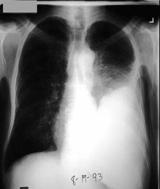
Left pleural effusion developed 4 days after antibiotic treatment for pneumococcal pneumonia. Patient developed fever, left-sided chest pain, and increasing dyspnea. During thoracentesis, purulent pleural fluid was removed, and the Gram stain showed gram-positive diplococci. The culture confirmed this to be Streptococcus pneumoniae.
Click to see larger pictureClick to see detailView Full Size Image
Media type:  X-RAY

Media file 2:  Empyema, pleuropulmonary. Left lateral chest radiograph shows a large, left pleural effusion.
Click to see larger pictureClick to see detailView Full Size Image
Media type:  X-RAY

Media file 3:  Empyema, pleuropulmonary. A right lateral decubitus chest radiograph shows a free-flowing pleural effusion, which should be sampled with thoracentesis for pH determination, Gram stain, and culture.
Click to see larger pictureClick to see detailView Full Size Image
Media type:  X-RAY

Media file 4:  Empyema, pleuropulmonary. CT scan of thorax shows loculated pleural effusion on left and contrast enhancement of visceral pleura, indicating the etiology is likely an empyema.
Click to see larger pictureClick to see detailView Full Size Image
Media type:  CT


  • Bartlett JG, Gorbach SL, Thadepalli H, Finegold SM. Bacteriology of empyema. Lancet. Mar 2 1974;1(7853):338-40. [Medline].
  • Bouros D, Schiza S, Tzanakis N, et al. Intrapleural urokinase versus normal saline in the treatment of complicated parapneumonic effusions and empyema. A randomized, double-blind study. Am J Respir Crit Care Med. Jan 1999;159(1):37-42. [Medline].
  • Cassina PC, Hauser M, Hillejan L, et al. Video-assisted thoracoscopy in the treatment of pleural empyema: stage-based management and outcome. J Thorac Cardiovasc Surg. Feb 1999;117(2):234-8. [Medline].
  • Davies CW, Lok S, Davies RJ. The systemic fibrinolytic activity of intrapleural streptokinase. Am J Respir Crit Care Med. Jan 1998;157(1):328-30. [Medline].
  • Davies RJ, Traill ZC, Gleeson FV. Randomised controlled trial of intrapleural streptokinase in community acquired pleural infection. Thorax. May 1997;52(5):416-21. [Medline].
  • Diacon AH, Theron J, Schuurmans MM. Intrapleural streptokinase for empyema and complicated parapneumonic effusions. Am J Respir Crit Care Med. Jul 1 2004;170(1):49-53.
  • Heffner JE, Brown LK, Barbieri C, DeLeo JM. Pleural fluid chemical analysis in parapneumonic effusions. A meta-analysis. Am J Respir Crit Care Med. Jun 1995;151(6):1700-8. [Medline].
  • Hope WW, Bolton WD, Stephenson JE. The utility and timing of surgical intervention for parapneumonic empyema in the era of video-assisted thoracoscopy. Am Surg. Jun 2005;71(6):512-4. [Medline].
  • Lawrence DR, Ohri SK, Moxon RE, et al. Thoracoscopic debridement of empyema thoracis. Ann Thorac Surg. Nov 1997;64(5):1448-50. [Medline].
  • LeMense GP, Strange C, Sahn SA. Empyema thoracis. Therapeutic management and outcome. Chest. Jun 1995;107(6):1532-7. [Medline].
  • Light RW, Girard WM, Jenkinson SG, George RB. Parapneumonic effusions. Am J Med. Oct 1980;69(4):507-12. [Medline].
  • Luh SP, Chou MC, Wang LS. Video-assisted thoracoscopic surgery in the treatment of complicated parapneumonic effusions or empyemas: outcome of 234 patients. Chest. Apr 2005;127(4):1427-32.
  • Maskell NA, Davies CW, Nunn AJ. U.K. Controlled trial of intrapleural streptokinase for pleural infection. N Engl J Med. Mar 3 2005;352(9):865-74.
  • Ng CS, Wan S, Lee TW. Post-pneumonectomy empyema: current management strategies. ANZ J Surg. Jul 2005;75(7):597-602.
  • Sahn SA, Light RW. The sun should never set on a parapneumonic effusion. Chest. May 1989;95(5):945-7. [Medline].
  • Sahn SA. State of the art. The pleura. Am Rev Respir Dis. Jul 1988;138(1):184-234. [Medline].
  • Schiza S, Siafakas NM. Clinical presentation and management of empyema, lung abscess and pleural effusion. Curr Opin Pulm Med. May 2006;12(3):205-11.
  • Striffeler H, Gugger M, Im Hof V, et al. Video-assisted thoracoscopic surgery for fibrinopurulent pleural empyema in 67 patients. Ann Thorac Surg. Feb 1998;65(2):319-23. [Medline].
  • Tokuda Y, Matsushima D, Stein GH. Intrapleural fibrinolytic agents for empyema and complicated parapneumonic effusions: a meta-analysis. Chest. Mar 2006;129(3):783-90.
  • Wait MA, Sharma S, Hohn J, Dal Nogare A. A randomized trial of empyema therapy. Chest. Jun 1997;111(6):1548-51. [Medline].

Empyema, Pleuropulmonary excerpt

Article Last Updated: Jun 23, 2006