Practice Essentials
Claudication, which is defined as reproducible ischemic muscle pain, is one of the most common manifestations of peripheral arterial occlusive disease (PAOD) caused by atherosclerosis. Claudication occurs during physical activity and is relieved after a short rest. Pain develops because of inadequate blood flow.
Angiography is the criterion standard arterial imaging study for the diagnosis of PAOD. The image below depicts a superficial femoral artery occlusion.
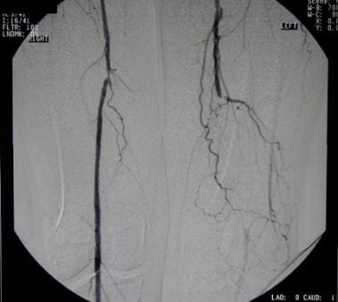 Peripheral arterial occlusive disease. Angiogram shows superficial femoral artery occlusion on one side (with reconstitution of suprageniculate popliteal artery) and superficial femoral artery stenosis on other side. This is most common area for peripheral vascular disease.
Signs and symptoms
Intermittent claudication typically causes pain that occurs with physical activity. Other signs and symptoms associated with peripheral arterial occlusive disease (PAOD) include the following:
-
Pain is reproducible within same muscle groups; pain ceases with a resting period of 2-5 minutes
-
The most common location of arterial lesions is the distal superficial femoral artery, which corresponds to claudication in the calf muscle area
-
Thigh/buttock muscle claudication predominates, with atherosclerosis distributed throughout the aortoiliac area
See Presentation for more detail.
Diagnosis
Examination of a patient with claudication should include a complete lower-extremity evaluation and pulse examination, including measuring segmental pressures. Attempt to palpate pulses from the abdominal aorta to the foot, with auscultation for bruits in the abdominal and pelvic regions. When palpable pulses are not present, a handheld Doppler device may be used to assess circulation.
A useful tool in assessing a patient with claudication is the ankle-brachial index (ABI), which is a noninvasive way of establishing the presence of PAOD and is calculated as the ratio of systolic blood pressure at the ankle to that in the arm (normal range, 0.9-1.1; PAOD, < 0.9).
Laboratory testing
A laboratory workup is helpful only for identifying accompanying silent alterations in renal function and elevated lipid profiles.
Imaging studies
The following radiologic studies may be used to evaluate suspected PAOD:
-
Angiography - The criterion standard for arterial imaging in the diagnosis of PAOD; usually reserved for when an intervention (either endovascular or traditional open surgery) is planned
-
Magnetic resonance angiography (MRA) - Useful for imaging large and small vessels
-
Computed tomography angiography (CTA) - Used to image arterial disease but requires large amount of contrast media and an upgraded CT scanner to reconstruct helpful images
-
Duplex ultrasonography - Evaluates status of a patient’s vascular disease and provides information about hemodynamics; is noninvasive and requires no contrast media but is highly technician-dependent
See Workup for more detail.
Management
Treatment of claudication is medical, with surgery reserved for severe cases. Medical management includes the following:
-
Tobacco cessation in patients who smoke
-
Regular exercise
-
Control of lipid profile, diabetes, and hypertension
Pharmacotherapy
The following medications are used in the management of PAOD:
-
Antiplatelet agents (eg, aspirin, clopidogrel, cilostazol, and pentoxifylline)
-
Antilipemic agents (eg, simvastatin)
Surgery
For patients in whom medical and exercise therapy fail or those who have claudication symptoms that are lifestyle-limiting, surgical treatment includes either open bypass surgery or endovascular therapy (eg, stents, balloons, or atherectomy devices).
See Treatment and Medication for more detail.
Background
Claudication, which is defined as reproducible ischemic muscle pain, is one of the most common manifestations of peripheral vascular disease caused by atherosclerosis (peripheral arterial occlusive disease [PAOD]). Claudication occurs during physical activity and is relieved after a short rest. Pain develops because of inadequate blood flow.
For patient education resources, see the Circulatory Problems Center and Cholesterol Center, as well as Peripheral Vascular Disease, High Cholesterol, and Cholesterol FAQs.
Pathophysiology
Single or multiple arterial stenoses produce impaired hemodynamics at the tissue level in patients with PAOD. Arterial stenoses lead to alterations in the distal perfusion pressures available to affected muscle groups.
Under resting conditions, normal blood flow to extremity muscle groups averages 300-400 mL/min. Once exercise begins, blood flow increases as much as 10-fold as a consequence of the increase in cardiac output and compensatory vasodilation at the tissue level. This allows the increase in oxygen demand to be met. When exercise ceases, blood flow returns to normal within minutes.
Resting blood flow in a person with PAOD is similar to that in a healthy person. In PAOD, however, blood flow cannot maximally increase in muscle tissue during exercise, because proximal arterial stenoses prevent compensatory vasodilation. When the metabolic demands of the muscle exceed blood flow, claudication symptoms ensue. At the same time, a longer recovery period is required for blood flow to return to baseline once exercise is terminated.
Similar abnormal alterations occur in distal perfusion pressure in affected extremities. In normal extremities, the mean blood pressure drop from the heart to the ankles is no more than a few millimeters of mercury. In fact, as pressure travels distally, the measured systolic pressure actually increases because of the higher resistance encountered in smaller-diameter vessels.
At baseline, a healthy person may have a higher measured ankle pressure than arm pressure. When exercise begins, no change in measured blood pressure occurs in the healthy extremity.
In the atherosclerotic limb, each stenotic segment acts to reduce the pressure head experienced by distal muscle groups. Correspondingly, at rest, the measured blood pressure at the ankle is less than that measured in a healthy person. Once physical activity starts, the reduction in pressure produced by the atherosclerotic lesion becomes more significant, and the distal pressure is greatly diminished.
The phenomenon of increased blood flow causing decreased pressure distally to an area of stenosis is a matter of physics. Poiseuille calculated energy losses across areas of resistance with varying flow rates by using the following equation:
-
Pressure difference = 8QvL/πr4
where Q is flow, v is viscosity, L is the length of the stenotic area, and r is the radius of the open area within the stenosis. In this equation, the pressure gradient is directly proportional to the flow and the length of the stenosis and inversely proportional to the fourth power of the radius. Thus, although increasing the flow rate directly increases the pressure gradient at any given radius, these effects are much less marked than those due to changes in the radius of the stenosis.
Because the radius is raised to the fourth power, it is the factor that has the most dramatic impact on a pressure gradient across a lesion. This impact is additive when two or more occlusive lesions are located sequentially within the same artery.
Epidemiology
United States and international statistics
Atherosclerosis affects up to 10% of the Western population older than 65 years. With the elderly population expected to increase 22% by the year 2040, atherosclerosis is expected to have a huge financial impact on medicine.
Estimated PAOD prevalence in the general US population, based on National Health and Nutrition Examination Survey (NHANES) data, was 4.3%. [1] Thus in 2000, about 5 million people in the US were affected by PAOD. That number increases with age; therefore, as the population ages the number of people affected by PAOD increases.
Age-, sex-, and race-related demographics
When claudication is used as an indicator, it is estimated that 2% of the population aged 40-60 years and 6% of the population older than 70 years are affected. Intermittent claudication most commonly manifests in men older than 50 years. Although younger patients may present with symptoms consistent with intermittent claudication, other etiologies of leg pain and claudication (eg, popliteal entrapment syndrome) must be strongly considered. There seems to be a higher prevalence of PAOD in non-Hispanic blacks.
Prognosis
Whether a patient progresses to limb amputation largely depends on the number and severity of cardiovascular risk factors (ie, smoking, hypertension, or diabetes). Continued smoking has been identified as the adverse risk factor most consistently associated with the progression of PAOD. Other factors are the severity of disease at the time of the initial patient encounter and, in some studies, the presence of diabetes.
In an effort to identify patients at highest risk for progression to critical limb ischemia (CLI), a simple risk score for PAOD was developed: the Graz CLI score. [2] Age and diabetes were among the most aggressive risk factors (respective odds ratios, 2.0 and 3.1).
As with most patients with vascular disease, survival is less than that of age-matched control groups. Coronary artery disease, with a subsequent myocardial event, is the major contributor to outcome. Predicted all-cause mortality for PAOD patients with claudication is approximately 30% at 5 years of follow-up, 50% at 10 years, and 70% at 15 years. [3]
In a double-blind trial (N = 6564), Bonaca et al randomly assigned patients with PAOD who had undergone revascularization to receive either rivaroxaban 2.5 mg bid plus aspirin (n = 3286) or placebo plus aspirin (n = 3278). [4] Compared with the patients in the aspirin-only group, those in the rivaroxaban-aspirin group had a significantly lower incidence of the composite outcome of acute limb ischemia, major amputation for vascular causes, myocardial infarction, ischemic stroke, or death from cardiovascular causes; however, they also had a significantly higher rate of major bleeding as defined by the International Society on Thrombosis and Haemostasis (ISTH).
-
Peripheral arterial occlusive disease. Measuring segmental pressures.
-
Peripheral arterial occlusive disease. Angiogram shows superficial femoral artery occlusion on one side (with reconstitution of suprageniculate popliteal artery) and superficial femoral artery stenosis on other side. This is most common area for peripheral vascular disease.
-
Peripheral arterial occlusive disease. Procedures performed during acute admission for peripheral arterial disease in US from 1996 to 2005. Reprinted from Journal of Vascular Surgery, Vol 49(4), Rowe VL et al, Patterns of treatment for peripheral arterial disease in the United States: 1996-2005, Pages 910-7, Apr 2009, with permission from Elsevier.
Tables

What would you like to print?
- Overview
- Presentation
- DDx
- Workup
- Treatment
- Guidelines
- DFA Guidelines on Management of Peripheral Arterial Disease in Diabetes-Related Foot Disease
- CCS Guidelines on Management of Peripheral Arterial Disease
- SVS/ESVS/WFVS Guidelines on Chronic Limb-Threatening Ischemia
- ESC/ESVS Guidelines on Lower-Extremity Arterial Disease
- AHA/ACC Guideline on Lower-Extremity Peripheral Arterial Disease
- SVS Guidelines on Atherosclerotic Occlusive Disease of Lower Extremities
- Show All
- Medication
- Questions & Answers
- Media Gallery
- References