Practice Essentials
Trauma is the leading cause of death, morbidity, hospitalization, and disability in Americans from the age of 1 year to the middle of the fifth decade of life. As such, it constitutes a major health care problem. According to the Centers for Disease Control and Prevention (CDC), 200,955 deaths occurred from unintentional injury in 2020. [1]
In particular, chest trauma is a significant source of morbidity and mortality in the United States. This article focuses on chest trauma caused by blunt mechanisms. Penetrating thoracic injuries are addressed in Penetrating Chest Trauma.
Blunt injury to the chest can affect any one or all components of the chest wall and thoracic cavity. [2] These components include the bony skeleton (ribs, clavicles, scapulae, and sternum), the lungs and pleurae, the tracheobronchial tree, the esophagus, the heart, the great vessels of the chest, and the diaphragm. In this article, each particular injury and injury pattern resulting from blunt mechanisms is discussed. The pathophysiology of these injuries is elucidated, and diagnostic and treatment measures are outlined.
Operative intervention is rarely necessary in blunt thoracic injuries. Most such injuries can be treated with supportive measures and simple interventional procedures such as tube thoracostomy.
Future directions for improving the diagnosis and management of blunt thoracic trauma involve diagnostic testing, endovascular techniques, and patient selection, as follows:
-
The use of thoracoscopy for the diagnosis and management of thoracic injuries will increase; the use of ultrasonography (US) for the diagnosis of conditions such as hemothorax and cardiac tamponade will become more widespread; spiral (helical) computed tomography (CT) techniques will be used more frequently for definitive diagnosis of major vascular lesions (eg, injuries to the thoracic aorta and its branches)
-
Endovascular techniques for the repair of great-vessel injuries will be developed further and applied more frequently
-
Patient selection and nonsurgical therapies for delayed operative management of thoracic aortic rupture will be refined
Anatomy
The thorax is bordered superiorly by the thoracic inlet, just cephalad to the clavicles. The major arterial blood supply to and the venous drainage from the head and neck pass through the thoracic inlet.
The thoracic outlets form the superolateral borders of the thorax and transmit branches of the thoracic great vessels that supply blood to the upper extremities. The nerves that make up the brachial plexus also access the upper extremities via the thoracic outlet. The veins that drain the arm (of which the most important is the axillary vein) empty into the subclavian vein, which returns to the chest via the thoracic outlet.
Inferiorly, the pleural cavities are separated from the peritoneal cavity by the hemidiaphragms. Communication routes between the thorax and abdomen are supplied by the diaphragmatic hiatuses, which allow egress of the aorta, esophagus, and vagal nerves into the abdomen and ingress of the vena cava and thoracic duct into the chest.
The chest wall is composed of layers of muscle, bony ribs, costal cartilages, sternum, clavicles, and scapulae. In addition, important neurovascular bundles course along each rib, containing an intercostal nerve, artery, and vein. The inner lining of the chest wall is the parietal pleura. The visceral pleura invests the lungs. Between the visceral and parietal pleurae is a potential space, which, under normal conditions, contains a small amount of fluid that serves mainly as a lubricant.
The lungs occupy most of the volume of each hemithorax. Each is divided into lobes. The right lung has three lobes, and the left lung has two lobes. Each lobe is further divided into segments.
The trachea enters through the thoracic inlet and descends to the carina at thoracic vertebral level 4, where it divides into the right and left mainstem bronchi. Each mainstem bronchus divides into lobar bronchi. The bronchi continue to arborize to supply the pulmonary segments and subsegments.
The heart is a mediastinal structure contained within the pericardium. The right atrium receives blood from the superior vena cava (SVC) and the inferior vena cava (IVC). Right atrial blood passes through the tricuspid valve into the right ventricle. Right ventricular contraction forces blood through the pulmonary valve and into the pulmonary arteries. Blood circulates through the lungs, where it acquires oxygen and releases carbon dioxide.
Oxygenated blood courses through the pulmonary veins to the left atrium. The left heart receives small amounts of nonoxygenated blood via the thebesian veins, which drain the heart, and the bronchial veins. Left atrial blood proceeds through the mitral valve into the left ventricle.
Left ventricular contraction propels blood through the aortic valve into the coronary circulation and the thoracic aorta, which exits the chest through the diaphragmatic hiatus into the abdomen. A ligamentous attachment (a remnant of the ductus arteriosus) exists between the descending thoracic aorta and the pulmonary artery just beyond the takeoff of the left subclavian artery.
The esophagus exits the neck to enter the posterior mediastinum. Through much of its course, it lies posterior to the trachea. In the upper thorax, it lies slightly to the right, with the aortic arch and descending thoracic aorta to its left. Inferiorly, the esophagus turns leftward and enters the abdomen through the esophageal diaphragmatic hiatus.
The thoracic duct arises primarily from the cisterna chyli in the abdomen. It traverses the diaphragm and runs cephalad through the posterior mediastinum in proximity to the spinal column. It enters the neck and veers to the left to empty into the left subclavian vein.
Pathophysiology
The major pathophysiologies encountered in blunt chest trauma involve derangements in the flow of air, blood, or both in combination. Sepsis due to leakage of alimentary tract contents, as in esophageal perforations, also must be considered.
Blunt trauma commonly results in chest-wall injuries (eg, rib fractures). The pain associated with these injuries can make breathing difficult, and this may compromise ventilation. Direct lung injuries, such as pulmonary contusions (see the image below), are frequently associated with major chest trauma and may impair ventilation by a similar mechanism. Shunting and dead-space ventilation produced by these injuries can also impair oxygenation.
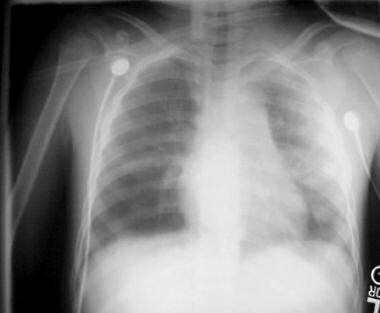 Left pulmonary contusion following a motor vehicle accident involving a pedestrian.
Space-occupying lesions (eg, pneumothorax, hemothorax, and hemopneumothorax) interfere with oxygenation and ventilation by compressing otherwise healthy lung parenchyma. A special concern is tension pneumothorax, in which pressure continues to build in the affected hemithorax as air leaks from the pulmonary parenchyma into the pleural space. This can push mediastinal contents toward the opposite hemithorax. Distortion of the SVC by this mediastinal shift can result in decreased blood return to the heart, circulatory compromise, and shock.
At the molecular level, animal experimentation supports a mediator-driven inflammatory process that further leads to respiratory insult after chest trauma. After blunt chest trauma, several blood-borne mediators are released, including interleukin (IL)-6, tumor necrosis factor (TNF), and prostanoids. These mediators are thought to induce secondary cardiopulmonary changes.
Blunt trauma that causes significant cardiac injuries (eg, chamber rupture) or severe great-vessel injuries (eg, thoracic aortic disruption) frequently results in death before adequate treatment can be instituted. This is due to immediate and devastating exsanguination or loss of cardiac pump function, which causes hypovolemic or cardiogenic shock and death.
Sternal fractures are rarely of any consequence, except when they result in blunt cardiac injuries.
Etiology
By far the most important cause of significant blunt chest trauma is motor vehicle accidents (MVAs). MVAs account for 70-80% of such injuries. As a result, preventive strategies to reduce MVAs have been instituted in the form of speed limit restriction and the use of restraints. Vehicles striking pedestrians, falls, and acts of violence are other causative mechanisms. Blast injuries can also result in significant blunt thoracic trauma.
Epidemiology
Trauma is responsible for more than 100,000 deaths annually in the United States. [1] Estimates of thoracic trauma frequency indicate that injuries occur in 12 persons per 1 million population per day. Approximately 33% of these injuries necessitate hospital admission. Overall, blunt thoracic injuries are directly responsible for 20-25% of all deaths, and chest trauma is a major contributor in another 50% of deaths.
Prognosis
For the great majority of patients with blunt chest trauma, outcome and prognosis are excellent. Most (>80%) require either no invasive therapy or, at most, a tube thoracostomy to effect resolution of their injuries. The most important determinant of outcome is the presence or absence of significant associated injuries of the central nervous system, abdomen, and pelvis.
Some injuries, such as cardiac chamber rupture, thoracic aortic rupture, injuries of the intrathoracic IVC and SVC, and delayed recognition of esophageal rupture, are associated with high morbidity and mortality.
A study using data from the TraumaRegister of the German Trauma Society (N = 50,519) found obesity to have a negative impact on outcomes after blunt chest trauma (ie, increased duration of mechanical ventilation, intensive care unit [ICU] stay, and hospital stay), though it did not document a comparable effect on mortality. [3]
A study by Beshay et al (N = 630) found that the presence of severe lung contusion, a higher Injury Severity Score (ISS), a higher Abbreviated Injury Scale (AIS) score in the thoracic region, and advanced age were independent risk factors directly related to higher mortality. [4]
Refaely et al analyzed clinical outcomes in patients with blunt and penetrating chest injuries who underwent urgent thoracotomy (ie, thoracotomy performed in the operating room within the first 48 hours of the patient's intensive care unit [ICU] stay) and found that mortality was higher in the blunt chest trauma group. [5] They suggested that in both penetrating and blunt chest trauma, urgent thoracotomy should be performed as quickly as possible and should be limited to damage control and that acidosis and hypothermia should be treated extremely aggressively before, during, and after the procedure.
-
Left pulmonary contusion following a motor vehicle accident involving a pedestrian.


