Practice Essentials
Essential features of ventricular septal rupture (VSR) may be summarized as follows:
-
The rupture typically occurs 3-8 days after an MI.
-
VSR is more likely to occur in the anterior septum than in the posterior septum (60% vs 40%).
-
The most consistent finding is a murmur (see Presentation).
-
The differential diagnosis includes ventricular septal rupture and mitral insufficiency secondary to papillary muscle rupture, papillary muscle dysfunction, or left ventricular dilatation.
-
In the differential diagnosis, exclude MR from papillary muscle rupture.
-
Diagnosis is confirmed with the aid of echocardiography and the presence of a left-to-right shunt (see Workup).
-
Catheterization results help determine the extent of coronary artery disease (CAD).
-
Emergent surgery is necessary but the condition still carries a very high mortality (see Treatment).
-
Today, percutaneous means of closure are available.
Background
Postinfarction ventricular septal rupture (VSR) is a rare but lethal complication of myocardial infarction (MI). The event occurs 2-8 days after an infarction and often precipitates cardiogenic shock. [1] The differential diagnosis of postinfarction cardiogenic shock should exclude free ventricular wall rupture and rupture of the papillary muscles. (See the image below.)
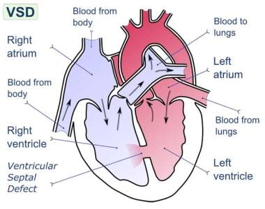 Postinfarction ventricular septal rupture. Ventricular septal rupture is defect in interventricular septum (wall dividing left and right ventricles of heart).
To avoid the high morbidity and mortality associated with this disorder, patients should undergo emergency surgical treatment. [2, 3, 4, 5] In current practice, postinfarction VSR is recognized as a surgical emergency, and the presence of cardiogenic shock is an indication for intervention. [6] Long-term survival can be achieved in patients who undergo prompt surgery. Concomitant coronary artery bypass grafting (CABG) may be required. The addition of CABG has helped improve long-term survival. [7, 8]
Surgery is performed via a transinfarction approach, and all reconstruction is performed with prosthetic materials to avoid tension. Developments in myocardial protection and improved prosthetic materials have contributed greatly to successful management of VSR. [9] Improved surgical techniques (eg, infarctectomy) and better perioperative mechanical and pharmacologic support have helped lower mortality. In addition, the development of surgical techniques to repair perforations in different areas of the septum has led to improved results. (should discuss the different techniques)
In current practice, patients undergoing VSR repair tend to be older and are more likely to have received thrombolytic agents, which may complicate repair. After successful repair, survival and quality of life are excellent, even in patients older than 70 years. [10]
For information, news, and CME activities on heart failure, see the Heart Failure Resource Center. For patient education resources, see the Heart Health Center, as well as Ventricular Septal Defect and Heart Attack.
Pathophysiology
The septal blood supply comes from branches of the left anterior descending coronary artery, the posterior descending branch of the right coronary artery, or the circumflex artery when it is dominant. Infarction associated with a ventricular septal rupture (VSR) is usually transmural and extensive. About 60% of VSRs occur with infarction of the anterior wall; 40% with infarction of the posterior or inferior wall (see the image below). Posterior VSR may be accompanied by mitral valve insufficiency secondary to papillary muscle infarction or dysfunction.
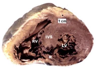 Postinfarction ventricular septal rupture. Heart sectioned transversely at level of middle left ventricle. Posterior ventricular septal defect is visible at site of recent acute myocardial infarction.
At autopsy, patients with VSR usually show complete coronary artery occlusion with little or no collateral flow. The lack of collateral flow may be secondary to associated arterial disease, anatomic anomalies, or myocardial edema. Sometimes, multiple septal perforations occur. These may occur simultaneously or within several days of each other. See the image below.
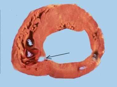 Postinfarction ventricular septal rupture. A section of the ventricle indicating location of the VSR (arrow).
VSR may be either anterior or inferior myocardial infarctions (MIs). Studies show that they occur with equal frequency. [11, 12] The anterior infarcts usually cause an apical VSR, which tend to be small and have a defined edge, making them a little simpler to repair. On the other hand, the inferior VSR usually have a thin and uneven edge which is often necrotic, making repair very difficult.
The natural history of postinfarction VSR is greatly influenced by hypertension, anticoagulation therapy, advanced age, and, possibly, thrombolytic therapy. The natural course in patients with postinfarction VSR is well documented and short. Most patients die within the first week, and almost 90% die within the first year; some reports indicate that fewer than 7% of patients are alive after 1 year.
This grim prognosis results from an acute volume overload exacted on both ventricles in a heart already compromised by a large MI and occasionally by extensive coronary artery disease (CAD) in sites other than that already infarcted. In addition, superimposed ischemic mitral valve regurgitation, a ventricular aneurysm, or a combination of these conditions may be present, further compromising heart function. The depressed left ventricular function commonly leads to impaired peripheral organ perfusion and death in most patients.
A few sporadic reports indicate that some patients with medically treated postinfarction VSR live for several years. Although many medical advances have been made in the nonsurgical treatment of these patients, including intra-aortic balloon counterpulsation (IABCP), these methods have not eliminated the need for surgery.
Etiology
Risk factors for postinfarction ventricular septal rupture (VSR) include the following:
-
Female gender
-
Advanced age
-
Higher Killip class
-
Myocardial infarction (MI) associated with ST segment elevation and elevated cardiac markers
-
Preoperative use of an intra-aortic balloon pump
-
Inferior wall MI
-
Renal failure with dialysis
Most studies show that the left anterior descending and right coronary arteries are chiefly involved in an MI that is associated with VSR.
Epidemiology
Rupture of the interventricular septum is an uncommon complication of MI. Although autopsy studies reveal an 11% incidence of myocardial free-wall rupture after MI, septal-wall perforation is much less common, occurring at a rate of approximately 1-2%.
Ventricular septal rupture (VSR) occurs in a zone of necrotic myocardial tissue, usually within the first 10-14 days. Clinical studies report an average time of 2.6 days from MI to VSR. However, some data suggest that initial treatment of MI with thrombolytics may affect both the time between infarction and VSR and the eventual outcome. Early use of thrombolytic agents may lead to reopening of the occluded vessels, thereby reducing the incidence of VSR.
The age range of patients who sustain a postinfarction VSR is wide, from 44 to 81 years. Men are affected more commonly than women are, though VSR is more common in women than would be predicted on the basis of the prevalence of CAD alone.
Prognosis
Operative mortality is directly related to the interval between MI and surgical repair. In a retrospective analysis of 41 patients treated for postinfarction ventricular septal defect (VSD), Serpytis et al confirmed that whereas female sex, advanced age, arterial hypertension, anterior-wall acute MI, absence of previous acute MI, and late arrival at hospital were associated with a higher risk of mortality from acute VSD, the time from the onset of acute MI to operation was the most important factor determining operative mortality and intrahospital survival. [13]
If repair of a postinfarction ventricular septal rupture (VSR) is performed 3 weeks or more after the infarction, mortality is approximately 20%; if it is performed before this time, mortality approaches 50%. The most obvious reason for this is that the greater the degree of myocardial damage and hemodynamic compromise, the more urgent the need for early intervention or the patients being repaired survived and were self selected.
With the use of an early operative approach, most studies show an overall mortality of less than 25%. Mortality tends to be lower for patients with anteriorly located VSRs and lowest for patients with apical VSRs. For anterior defects, mortality ranges from 10% to 15%; for posterior defects, mortality ranges from 30% to 35%.
More than 50% of deaths occurring after surgery for postinfarction VSR are due to cardiac failure. Sudden death is rare, and intractable heart failure can also occur. Other causes of death include cerebral embolism. Most patients who survive the hospital period have good functional status, with the majority falling into New York Heart Association (NYHA) class I or II. [14]
The most important risk factors for death in the early phase are poor hemodynamics and associated right ventricular dysfunction developing before the patient comes to the operating room. The amount and distribution of myocardial necrosis and scarring are responsible for both.
Right ventricular dysfunction results from ischemic damage or frank infarction of the right ventricle and is present when stenosis occurs in the right coronary artery system. The higher mortality observed after repair of defects located inferiorly in the septum is probably related to the higher prevalence of important right coronary artery stenosis.
The severity and distribution of CAD are also risk factors. Similarly, advanced age at operation, diabetes, and preinfarction hypertension are risk factors for death in the early phase.
Risk factors for death in patients with postinfarction VSR may be summarized as follows:
-
Posteriorly located VSRs are technically more difficult to repair and are associated with profound right ventricular dysfunction
-
The presence of multiple organ failure is a poor prognostic factor
-
The presence of cardiogenic shock does not bode well for the patient’s survival
In a retrospective analysis of 52 consecutive patients with surgically repaired postinfarction VSR over a 30-year period (mean follow-up, 7.8±7.7 years), Takahashi et al found that predictors of 30-day mortality on univariate analysis included the following [15] :
-
Renal insufficiency
-
Shock at surgery
-
Emergency surgery
-
Logistic EuroSCORE
-
Three-vessel disease
-
Significant left circumflex coronary arterial stenosis
-
Significant right coronary arterial stenosis
-
Incomplete revascularization
-
Surgical duration
-
Cardiopulmonary bypass time
On multivariate analysis, only incomplete coronary revascularization was an independent risk factor for 30-day mortality. [15]
Complications
Complications of postinfarction VSR include the following:
-
Of patients treated without surgery, 90% die
-
Surgical treatment must be carried out on an emergency basis, even if the patient is stable [3]
-
All VSRs are closed with a patch and associated coronary artery bypass grafting (CABG)
-
Operative mortality is 10-15% for anterior defects and 30-35% for posterior defects
-
Postinfarction ventricular septal rupture. Heart sectioned transversely at level of middle left ventricle. Posterior ventricular septal defect is visible at site of recent acute myocardial infarction.
-
Postinfarction ventricular septal rupture. Ventricular septal rupture is defect in interventricular septum (wall dividing left and right ventricles of heart).
-
Postinfarction ventricular septal rupture. Ventricular septal defect on echocardiography.
-
Postinfarction ventricular septal rupture. Acute anterior myocardial infarction on ECG.
-
Postinfarction ventricular septal rupture. An echocardiogram of a 69-year-old patient with a postinfarction VSR who decompensated very quickly.
-
Postinfarction ventricular septal rupture. A section of the ventricle indicating location of the VSR (arrow).


