Practice Essentials
Acute gastrointestinal (GI) bleeding is a potentially life-threatening abdominal emergency that remains a common cause of hospitalization. Upper GI bleeding (UGIB) is defined as bleeding derived from a source proximal to the ligament of Treitz.
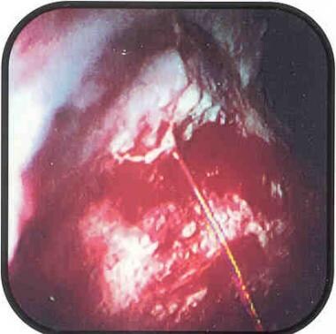
The image below depicts an ulcer with active bleeding.
Signs and symptoms
Signs and symptoms of acute UGIB [1] include the following:
-
Hematemesis
-
Melena
-
Hematochezia
-
Syncope
-
Presyncope
-
Dyspepsia
-
Epigastric pain
-
Heartburn
-
Diffuse abdominal pain
-
Dysphagia
-
Weight loss
-
Jaundice
See Presentation for more detail.
Diagnosis
Workup may include the following:
-
Orthostatic blood pressure (blood pressure in upright position)
-
Complete blood cell count with differential
-
Hemoglobin level
-
Type and crossmatch blood
-
Basic metabolic profile, blood urea nitrogen, and coagulation profile
-
Risk scoring assessment
-
Calcium level
-
Gastrin level
-
Endoscopy
-
Chest radiography
-
Nasogastric lavage
-
Computed tomography (CT) angiography (CTA)
-
Angiography (if bleeding persists and endoscopy fails to identify a bleeding site)
Standard CT scanning and ultrasonography may be indicated for the evaluation of the following [2] :
-
Liver disease with cirrhosis
-
Cholecystitis with hemorrhage
-
Pancreatitis with pseudocyst and hemorrhage
-
Aortoenteric fistula
See Workup for more detail.
Management
Treatment may include the following:
-
Secure the airway
-
Insert bilateral, 16-gauge (minimum), upper extremity, peripheral intravenous lines
-
Replace each milliliter of blood loss with 3 mL of crystalloid fluid
-
In patients with severe coexisting medical illnesses, pulmonary artery catheter insertion for monitoring hemodynamic cardiac performance
-
Foley catheter placement for continuous evaluation of urinary output as a guide to renal perfusion
-
Endoscopic hemostatic therapy for bleeding ulcers and varices
-
Surgical repair of perforated viscus
-
For high-risk peptic ulcer patients, high-dose intravenous proton pump inhibitors
-
Mesenteric angiography with embolization therapy
Indications for surgery in patients with bleeding peptic ulcers include the following:
-
Severe, life-threatening hemorrhage not responsive to resuscitative efforts
-
Failure of medical therapy and endoscopic hemostasis with persistent recurrent bleeding
-
A coexisting reason for surgery (eg, perforation, obstruction, malignancy)
-
Prolonged bleeding, with loss of 50% or more of the patient's blood volume
-
A second hospitalization for peptic ulcer hemorrhage
See Treatment and Medication for more detail.
Background
Acute gastrointestinal (GI) bleeding is a potentially life-threatening abdominal emergency that remains a common cause of hospitalization. [3, 4] Upper GI bleeding (UGIB) is defined as bleeding derived from a source proximal to the ligament of Treitz. [5]
The incidence of UGIB is approximately 100 cases per 100,000 population per year. [6] Bleeding from the upper GI tract is approximately 4 times more common than bleeding from the lower GI tract and is a major cause of morbidity and mortality. Mortality rates from UGIB are 6%-10% overall. [6] (See Epidemiology.)
The diagnosis of and therapy for nonvariceal UGIB has evolved since the late 20th century from passive diagnostic esophagogastroduodenoscopy with medical therapy until surgical intervention was needed to active intervention with endoscopic techniques followed by angiographic and surgical approaches if endoscopic therapy fails. [7] (See Workup and Treatment.)
Variceal hemorrhage is not discussed in this article because the underlying mechanisms of bleeding are different and require different therapies.
The underlying mechanisms of nonvariceal bleeding involve either arterial hemorrhage, such as in ulcer disease and mucosal deep tears, or low-pressure venous hemorrhage, as in telangiectasias and angioectasias. In variceal hemorrhage, the underlying pathophysiology is an elevated portal pressure transmitted to esophageal and gastric varices and resulting in portal gastropathy. A bleeding ulcer is seen below. (See Etiology.)
See Pediatric Gastrointestinal Bleeding for more information on this topic.
In patients with UGIB, comorbid illness, rather than the actual bleeding, is the major cause of death. Comorbid illness has been noted in 50.9% of patients, with similar occurrences in males (48.7%) and females (55.4%).
One or more comorbid illnesses have been noted in 98.3% of mortalities in UGIB; in 72.3% of patients, comorbid illnesses have been noted as the primary cause of death. [8, 9] (See Epidemiology and Prognosis.)
Significant comorbidities have become more prevalent as the patient population with UGIB has become progressively older. In a retrospective chart review by Yavorski et al, 73.2% of deaths occurred in patients older than 60 years. [9] (See Epidemiology and Prognosis.)
Rebleeding or continued bleeding is associated with increased mortality; therefore, differentiating the patient with a low probability of rebleeding and little comorbidity from the patient at high risk for rebleeding with serious comorbidities is imperative. (See Presentation and Workup.)
Peptic ulcer disease and UGIB
Peptic ulcer disease (PUD) remains the most common cause of UGIB. In a literature review involving more than 10,000 patients with UGIB, PUD was responsible for 27%-40% of all bleeding episodes. [10] High-risk patient populations at risk for PUD include those with a history of alcohol abuse, chronic renal failure, and/or nonsteroidal anti-inflammatory drug (NSAID) use. [11]
Peptic ulcer disease is strongly associated with Helicobacter pylori infection. The organism causes disruption of the mucous barrier and has a direct inflammatory effect on the gastric and duodenal mucosa, reducing mucosal defenses and increasing the back diffusion of acid by loosening the tight cellular junctions. (Rates of H pylori infection are reportedly lower in patients with complicated ulcer disease than in patients with uncomplicated ulcers. Hosking et al reported a 71% incidence of H pylori infection in patients with bleeding duodenal ulcers; patients with nonbleeding ulcers had an incidence of 93%.) This discrepancy may be due to the decrease in sensitivity of biopsy in patients with ulcer bleeding. [12]
Eradication of H pylori been demonstrated to reduce the risk of recurrent ulcers and, thus, recurrent ulcer hemorrhage after the initial episode. In fact, the proportion of UGIB cases caused by peptic ulcer disease has declined, [13] a phenomenon that is believed to be due to the use of proton pump inhibitors (PPIs) and anti-H pylori therapy.
Duodenal ulcers are more common than gastric ulcers, but the incidence of bleeding is identical for both. In most cases, the bleeding is caused by the erosion of an artery at the base of the ulcer. In approximately 80% of patients, bleeding from a peptic ulcer stops spontaneously. [10]
Initial endoscopic attempts to maintain hemostasis have a high success rate. Bleeding vessels larger than 1.5 mm in diameter are associated with an increased mortality rate due to the difficulty in producing adequate hemostasis with thermal probes.
A minority of patients experience recurrent bleeding after endoscopic therapy, and these cases are usually associated with risk factors for rebleeding. These factors include age older than 60 years, the presence of shock upon admission, coagulopathy, active pulsatile bleeding, and the presence of cardiovascular disease. (The appearance of the ulcer at the time of endoscopy provides important information regarding the risk of rebleeding.) These circumstances are associated with a poorer prognosis and a higher mortality rate. [14]
Despite the dangers associated with a bleeding peptic ulcer, a study by Sung et al of 10,428 cases of such bleeding (in 9,375 patients) found that most deaths were not caused by it. [15] Of the 577 deaths that occurred in the cohort, almost 80% resulted from other causes, including multiorgan failure, pulmonary conditions, and terminal malignancy. The authors concluded that the management of patients with peptic ulcers should focus not only on hemostasis but also on lowering the risk of multiorgan failure and cardiopulmonary death.
Recurrent bleeding risk in peptic ulcers
Forrest et al were the first to classify the stigmata of hemorrhage from peptic ulcers. Based on these classifications, the risk of recurrent bleeding can be predicted. The ulcers at highest risk for rebleeding are those that involve active arterial bleeding or those with a visible, protuberant, nonbleeding vessel at the base of the ulcer. The study not only correlated the incidence of rebleeding with the stigmata of recent bleeding and the endoscopic appearance of an ulcer, but also determined prognostic information regarding the need for surgery. Mortality was also correlated with these factors. [16]
In patients with H pylori infection, the rate of recurrent bleeding is increased. This is why documenting the presence of H pylori and aggressively treating the infection are important.
Patients who are not infected with H pylori may require a subsequent acid-lowering surgical procedure or long-term medical therapy for recurrent ulcer disease and bleeding.
Other causes of UGIB
Other major causes of UGIB are mucosal tears of the esophagus or fundus (Mallory-Weiss tear), erosive gastritis, erosive esophagitis, Dieulafoy lesion, gastric cancer, [17] and ulcerated gastric leiomyoma.
Patients with chronic liver disease and portal hypertension are at an increased risk for variceal hemorrhage and portal gastropathy in addition to ulcer hemorrhage.
Rare causes of UGIB include aortoenteric fistula, gastric antral vascular ectasia, angioectasia, and Osler-Weber-Rendu syndrome.
An aortoenteric fistula results from the erosion of the aortic graft into the bowel lumen, usually at the third or fourth portion of the duodenum. The result is a direct communication between the aortic graft lumen and the bowel lumen. Most aortoenteric fistulas involve the proximal aortic anastomotic suture line.
Acute stress-related mucosal disease (also known as stress ulcers), a disease process characterized by diffuse superficial mucosal erosions that appear as discrete areas of erythema, can also cause UGIB. [16] The bleeding is usually mild and self-limiting and rarely progresses to life-threatening hemorrhage. Stress ulcers can be detected endoscopically in as many as 75%-100% of critically ill patients, within 24 hours of admission to an intensive care unit (ICU).
In ICU patients, the incidence of clinically significant GI bleeding (eg, hypotension, transfusion) from acute stress ulcer was found to be 1.5%. [18] Although its incidence has fallen significantly over recent decades, likely due to better overall ICU care, mortality remains high in those with clinically important bleeding. [19] Stress ulcers are historically associated with (1) head injuries, with related elevations in intracranial pressure (Cushing ulcers) and (2) burn injuries (Curling ulcers).
Critically ill patients are at an increased risk of developing stress-related mucosal disease and subsequent stress-ulcer bleeding, most commonly with risk factors of respiratory failure and coagulopathy. [20] Other important factors include acute renal or hepatic failure, sepsis, hypotension, severe head or spinal cord injury, severe burns, acute lung injury, major or prolonged surgery, and a history of GI bleeding. [21]
Angiodysplasia of the upper GI tract accounts for 2%-4% of bleeding lesions. [10] The condition is also a cause of lower GI bleeding in 6% of cases. [16] The lesion is a vascular malformation that represents an abnormal dilation of mucosal and submucosal vessels.
Histologically, angiodysplasias are dilated, thin-walled vascular channels that appear macroscopically as a cluster of cherry spots. When located in the upper GI tract, they most commonly involve the stomach and duodenum. The lesions can be acquired or congenital, as in hereditary hemorrhagic telangiectasia and Osler-Weber-Rendu syndrome.
The acquired angiodysplasias are commonly found in patients with chronic renal failure requiring hemodialysis and with aortic valvular disease (especially aortic stenosis due to Heyde syndrome). Other diseases, such as cirrhosis and von Willebrand disease, are associated with a higher frequency of angiodysplasias. Most lesions are smaller than 1 cm in diameter and can be multiple in 66% of patients. [10]
For patient education information, see the Digestive Disorders Center and the Heartburn and GERD Center, as well as the patient education article Gastrointestinal Bleeding (GI Bleeding).
Etiology
Ulcer-related UGIB
Bleeding peptic ulcers account for the majority of patients presenting with acute upper gastrointestinal (GI) bleeding (UGIB). [3] As previously mentioned, peptic ulcer disease is strongly associated with H pylori infection. The organism causes disruption of the mucous barrier and has a direct inflammatory effect on the gastric and duodenal mucosa.
In cases of ulcer-associated UGIB, as the ulcer burrows deeper into the gastroduodenal mucosa, the process causes weakening and necrosis of the arterial wall, leading to the development of a pseudoaneurysm. The weakened wall ruptures, producing hemorrhage.
The flow through a vessel varies with the fourth power of the radius; thus, small increases in vessel size can mean much larger amounts of blood flow and bleeding, with more severe hypotension and more complications, especially in older patients.
Visible vessels usually range from 0.3-1.8 mm.
Exsanguinating hemorrhage has been reported from larger vessels. The larger vessels are located deeper in the gastric and duodenal submucosa and serosa. Larger branches of the left gastric artery are found high on the lesser curvature, while the pancreatoduodenal artery and its major branches are located posteroinferiorly in the duodenal bulb.
Vomiting-related UGIB
During vomiting, the lower esophagus and upper stomach are forcibly inverted. Vomiting attributable to any cause can lead to a mucosal tear of the lower esophagus or upper stomach. The depth of the tear determines the severity of the bleeding. Rarely, vomiting can result in esophageal rupture (Boerhaave syndrome), leading to bleeding, mediastinal air entry, left pleural effusion (salivary amylase can be present) or left pulmonary infiltrate, and subcutaneous emphysema.
Mallory-Weiss tears in UGIB
Mallory-Weiss tears account for 8%-15% of acute upper GI hemorrhage. [10, 22] Kenneth Mallory and Soma Weiss first described the syndrome in 1929. [23] The occasionally massive UGIB results from a tear in the mucosa of the gastric cardia. Like many upper GI tract lesions, the Mallory-Weiss tear may stop bleeding spontaneously 85%-90% of the time.
This linear mucosal laceration is the result of forceful vomiting, retching, coughing, or straining. These actions create a rapid increase in the gradient between intragastric and intrathoracic pressures, leading to a gastric mucosal tear from the forceful distention of the gastroesophageal junction. [24] In 80%-90% of cases, this is a single, 1.75- to 2.5-cm mucosal tear along the lesser curve of the stomach just distal to the gastroesophageal junction. [23]
See Mallory-Weiss Tear for more information on this topic.
Acute stress-related mucosal disease in UGIB
Acute stress-related mucosal disease (or stress ulcer) results from predisposing clinical conditions that have the potential to alter the local mucosal protective barriers, such as mucus, bicarbonate, blood flow, and prostaglandin synthesis. Any disease process that disrupts the balance of these factors results in diffuse gastric mucosal erosions.
This is most commonly observed in patients who have undergone episodes of shock, multiple trauma, acute respiratory distress syndrome, systemic respiratory distress syndrome, acute renal failure, and sepsis.
The principal mechanisms involved are decreased splanchnic mucosal blood flow and altered gastric luminal acidity.
Dieulafoy lesions in UGIB
The Dieulafoy lesion, first described in 1896, is a vascular malformation of the proximal stomach, usually within 6 cm of the gastroesophageal junction along the lesser curvature of the stomach. However, it can occur anywhere along the GI tract. This lesion accounts for 2%-5% of acute UGIB episodes. [25]
Endoscopically, the lesion appears as a large submucosal vessel that has become ulcerated. Because of the large size of the vessel, bleeding can be massive and brisk. The vessel rupture usually occurs in the setting of chronic gastritis, which may induce necrosis of the vessel wall. Alcohol consumption is reportedly associated with the Dieulafoy lesion.
In a review of 149 cases, the Dieulafoy lesion mostly occurred in men and mostly in those in their third to tenth decade. [26]
NSAIDs in UGIB
NSAIDs cause gastric and duodenal ulcers by inhibiting cyclooxygenase, which causes decreased mucosal prostaglandin synthesis and results in impaired mucosal defenses. Daily NSAID use causes an estimated 40-fold increase in gastric ulcer creation and an 8-fold increase in duodenal ulcer creation. [16]
Long-term NSAID use is associated with a 20% incidence in the development of mucosal ulceration. [27] Medical therapy includes avoiding the ulcerogenic drug and beginning a histamine-2 (H2)–receptor antagonist or a proton pump inhibitor that provides mucosal protection.
Epidemiology
US and international data
The incidence of acute upper gastrointestinal (GI) bleeding (UGIB) is about 100 per 100,000 adults per year. An estimated 15% of patients with UGIB present with hematochezia. In the United Kingdom, UGIB accounts for 70,000 hospital admissions each year, with the majority of cases nonvariceal in origin. [28] In a nationwide study from Spain, UGIB was six times more common than lower GI bleeding. [29]
UGIB is twice as common in men as in women and increases in prevalence with age (>60 y). However, the death rate is similar in both sexes. [9, 27]
Despite advances in therapy, in-hospital mortality remains high (13%), and rebleeding is common (15%). [30]
Prognosis
Recurrence of upper gastrointestinal (GI) bleeding (UGIB) is common. In a study to evaluate national 30-day readmissions after upper and lower gastrointestinal (GI) bleeding in US patients, of 82,290 patients admitted for UGIB, the all-cause 30-day readmission rate was 14.6% (vs 14.4% for LGIB). [31] The most common causes of readmission after UGIB were GI disease (33.9%), followed by cardiac (13.3%), infectious (10.4%), and respiratory (7.8%) etiologies. Significant predictors of 30-day readmission were metastatic disease, discharge against medical advice, and hospital stay for longer than 3 days. [31]
As previously mentioned, age older than 60 years is an independent marker for a poor outcome in UGIB, [32] with the mortality rate ranging from 12% to 25% in this group of patients.
The American Society for Gastrointestinal Endoscopy (ASGE) grouped patients with UGIB according to age and correlated age category to the risk of mortality. The ASGE found a mortality of 3.3% for patients aged 21-31 years, 10.1% for those aged 41-50 years, and 14.4% for those aged 71-80 years. [32]
The following risk factors are associated with an increased mortality, recurrent bleeding, the need for endoscopic hemostasis, or surgery [14, 33] :
-
Age older than 60 years
-
Severe comorbidity
-
Active bleeding (eg, witnessed hematemesis, red blood per nasogastric tube, fresh blood per rectum)
-
Hypotension
-
Red blood cell transfusion greater than or equal to 6 units
-
Inpatient at the time of bleed
-
Severe coagulopathy
Patients who present in hemorrhagic shock have a mortality rate of up to 30%.
-
Upper gastrointestinal bleeding (UGIB). Ulcer with active bleeding.
-
Upper gastrointestinal bleeding (UGIB). Ulcer with a clean base.
-
Upper gastrointestinal bleeding (UGIB). Diagram of an ulcer with a clean base.
-
Upper gastrointestinal bleeding (UGIB). Ulcer with an overlying clot.
-
Upper gastrointestinal bleeding (UGIB). Ulcer with a visible vessel.
-
Upper gastrointestinal bleeding (UGIB). Diagram of an ulcer with a visible vessel.
Tables
- Table 1. Probable Source of GI Bleeding Within the Gut
- Table 2. Estimated Fluid and Blood Losses in Shock
- Table 3. Effect of Number of Packed Erythrocyte Transfusions on Need for Surgery and Mortality from UGIB
- Table 4. Effect of the Color of the Nasogastric Aspirate and of the Stool on UGIB Mortality Rate
- Table 5. Ulcer Characteristics and Correlations
Clinical Indicator |
Probability of Upper GI Source |
Probability of Lower GI Source |
Hematemesis |
Almost certain |
Rare |
Melena |
Probable |
Possible |
Hematochezia |
Possible |
Probable |
Blood-streaked stool |
Rare |
Almost certain |
Occult blood in stool |
Possible |
Possible |
GI = gastrointestinal. |
||
Class 1 |
Class 2 |
Class 3 |
Class 4 |
|
Blood Loss, mL |
Up to 750 |
750-1500 |
1500-2000 |
>2000 |
Blood Loss, % blood volume |
Up to 15 |
15-30 |
30-40 |
>40 |
Pulse Rate, bpm |
< 100 |
>100 |
>120 |
>140 |
Blood Pressure |
Normal |
Normal |
Decreased |
Decreased |
Respiratory Rate |
Normal or Increased |
Decreased |
Decreased |
Decreased |
Urine Output, mL/h |
>35 |
30-40 |
20-30 |
14-20 |
CNS/Mental Status |
Slightly anxious |
Mildly anxious |
Anxious, confused |
Confused, lethargic |
Fluid Replacement, 3-for-1 rule |
Crystalloid |
Crystalloid |
Crystalloid and blood |
Crystalloid and blood |
bpm = beats per minute; CNS = central nervous system |
||||
Number of Units Transfused |
Need for Surgery, % |
Mortality, % |
0 |
4 |
4 |
1-3 |
6 |
14 |
4-5 |
17 |
28 |
>5 |
57 |
43 |
Nasogastric Aspirate Color |
Stool Color |
Mortality, % |
Clear |
Brown or red |
6 |
Coffee-ground |
Brown or black |
8.2 |
Red |
19.1 |
|
Red blood |
Black |
12.3 |
Brown |
19.4 |
|
Red |
28.7 |
|
UGIB = upper gastrointestinal bleeding. |
||
Ulcer Characteristics |
Prevalence Rate, % |
Rebleeding Rate, % |
Surgery Rate, % |
Mortality, % |
Clean base |
42 |
5 |
0.5 |
2 |
Flat spot |
20 |
10 |
6 |
3 |
Adherent clot |
17 |
22 |
10 |
7 |
Visible vessel |
17 |
43 |
34 |
11 |
Active bleeding |
18 |
55 |
35 |
11 |

What would you like to print?
- Overview
- Presentation
- DDx
- Workup
- Treatment
- Approach Considerations
- Proton-Pump Inhibitors
- Therapeutic Endoscopy
- Bleeding Peptic Ulcer Treatment
- Bleeding Gastric Ulcer Treatment
- Stress Ulcer Treatment
- Mallory-Weiss Syndrome Treatment
- Dieulafoy Lesion Treatment
- Angiodysplasia Treatment
- Aortoenteric Fistula
- Treatment Complications
- Posttreatment Monitoring and Care
- Deterrence and Prevention of UGIB
- Show All
- Guidelines
- Medication
- Questions & Answers
- Media Gallery
- Tables
- References