Overview
The success of a gynecologic procedure performed through an abdominal incision depends on careful selection of the incision site and proper closure of the wound. The surgeon needs to consider multiple factors before making an abdominal incision. These factors include the disease process, body habitus, operative exposure, simplicity, previous scars, cosmesis, and the need for quick entry into the abdominal cavity. The most important factor is adequate exposure to the operative field.
Complications during surgery can occur because of inadequate exposure, which is often due to the unwillingness of the surgeon to extend the incision. Incision location is particularly important when the patient has a gynecologic malignancy. These patients may need a colostomy, urinary diversion, or extraperitoneal lymph node dissection to satisfactorily manage the clinical situation. This article reviews pertinent abdominal wall anatomy, discusses various options for abdominal incisions, and examines various sutures available to surgeons.
Relevant Anatomy
A thorough understanding of abdominal wall anatomy is essential for choosing and making the proper surgical incision. The musculature of the abdominal wall is composed of 2 muscle groups. One group, the flat muscles, consists of the external oblique, internal oblique, and the transversus abdominis. The second group is composed of 2 muscles that run vertically, the rectus abdominis and the pyramidalis.
The external oblique muscle is the largest and most superficial of the flat muscles of the abdominal wall. Arising from the lower 8 ribs, the external oblique courses transversely to insert upon the iliac crests. The aponeurosis is a strong tendinous sheath that ends medially in the linea alba. The internal oblique muscle arises from the upper surface of the inguinal ligament, the iliac crest, and the thoracolumbar fascia. This muscle courses at a right angle to the fibers of the external oblique muscle. The aponeurosis of the internal oblique splits at the edge of the rectus muscle to envelope the rectus. The anterior layer blends with the aponeurosis of the external oblique. Posterior to the rectus muscle, this aponeurosis blends with the aponeurosis of the transversus abdominis to form a portion of the posterior rectus sheath.
The innermost of the flat muscles is the transversus abdominis. This muscle arises from the inguinal ligament, the iliac crest, the thoracolumbar fascia, and the lower costal cartilages. Coursing transversely to the midline, the upper three fourths of the transversus aponeurosis lies behind the rectus muscle. The lower one fourth of the aponeurosis passes in front of the rectus muscle. The aponeurosis of each flat muscle joins medial to the rectus muscle to form the linea alba.
Originating from the pubic crest, the rectus muscle runs vertically to insert into the xiphoid process and the fifth, sixth, and seventh costal cartilages. The muscle fibers contain 3 fibrous insertions known as the linea transversae. The rectus is surrounded by the rectus sheath, which consists of the aponeuroses of the oblique muscles and the transversus abdominis. A small, vestigial, triangular-shaped muscle, the pyramidalis, arises from the symphysis and inserts upon the linea alba. This muscle marks the midline and assists in the identification of the medial borders of the rectus muscle.
Two important surgical landmarks are formed by the aponeuroses of the abdominal wall muscles. The linea alba is in the midline between the 2 rectus muscles. Formed by the fusion of the aponeuroses of the external oblique, internal oblique, and transversus abdominis, identifying this structure during a midline incision is important. A second surgical landmark is the arcuate line that is found below the rectus muscle, approximately halfway between the umbilicus and the symphysis pubis. Above the arcuate line, the aponeuroses of the internal oblique and transversus abdominis fuse to form the posterior rectus sheath. Below the arcuate line, the posterior rectus sheath is absent. This anatomic finding occurs as the aponeuroses of the oblique muscles and the transversus pass in front of the rectus muscle.
Blood supply
The primary blood supply to the abdominal wall is from the superficial and deep vasculature. The superficial vasculature originates from branches of the femoral artery and includes the superficial epigastric, the superficial circumflex, and the superficial external pudendal arteries. These vessels course through the tissues anterior to the rectus sheath.
The deep vasculature is composed of vessels from the external iliac artery and the internal thoracic artery. The inferior epigastric artery originates from the external iliac artery and courses posterior to the lateral one third of the rectus muscle. Another branch of the external iliac is the deep circumflex artery, which courses cephalad lateral to the inferior epigastric artery. The superior epigastric artery is the terminal branch of the internal thoracic artery. This artery has multiple branches leading to the rectus muscle and has an anastomosis with the inferior epigastric artery. The internal thoracic artery is the source of the musculophrenic artery, which has an anastomosis with the deep circumflex artery. This large network of vascular anastomoses in the abdominal wall provides an excellent blood supply to all areas of the abdominal wall. See the image below.
 Location of deep and superficial vessels of the anterior abdominal wall. Blue circles indicate recommended locations for trocar placement.
Nerve supply
Innervation of the abdominal wall is by the thoracoabdominal nerves, the ilioinguinal nerves, and the iliohypogastric nerves. The thoracoabdominal nerves travel caudad between the transversus abdominis and the internal oblique. These nerves innervate the flat muscles of the abdominal wall and the rectus muscle. Innervating the lower abdominal wall are the iliohypogastric nerves and the ilioinguinal nerves. Both of these nerves arise primarily from the first lumbar nerve root. Damage to these nerves results in sensory changes in the mons pubis and the labia majora.
See Regions and Planes of the Abdomen for more information.
Types of Abdominal Incisions
Vertical incision
Several types of vertical abdominal incisions have been used in gynecologic surgery, including midline, paramedian, and wide paramedian incisions. A midline incision is almost exclusively the type of vertical incision used in gynecologic oncology surgery. The midline incision is the easiest and most versatile vertical incision for performing gynecologic cancer surgery. This incision allows quick entry into the abdominal cavity with little blood loss, and it is easily extended in length to accommodate the operative findings. The presumed disadvantages of a midline incision, compared with a transverse incision, include an increased risk of wound dehiscence and hernia formation. Most studies that support this idea are retrospective or lack proper statistical design. Recent studies have challenged this dictum and advocate that little difference exists in dehiscence rates between properly closed midline incisions and transverse incisions. [1, 2, 3, 4]
For a midline abdominal incision, the skin and subcutaneous fat are incised to the level of the fascia. The scalpel or electrocautery can be used to incise this tissue. Some surgeons believe the infection rate is higher with the use of electrocautery. Studies from the 1980s suggested a 2-fold increased risk of wound infection with electrocautery compared with a scalpel. However, more recent prospective studies indicate no increased wound complications with electrocautery compared with a scalpel in midline abdominal incisions. [5, 6, 7]
Using either instrument, the principle is to make long smooth strokes through the subcutaneous fat to the fascia. The subcutaneous fat should not be dissected from the fascia because this creates unnecessary dead space. Next, the fascia is incised, and the rectus muscles are separated vertically in the midline. The midline may not be evident in patients with previous abdominal surgery. Identifying where the rectus muscles diverge around the umbilicus or locating the pyramidalis muscles assists in identifying the midline. Once the rectus muscles are divided, the peritoneum is grasped between 2 hemostats, opened with a scalpel, and extended the length of the incision.
If the operative findings necessitate extending the incision above the umbilicus, avoid cutting through the umbilicus. Postoperative wound infections may be increased due to bacterial colonization of the umbilicus. Extension of the incision should pass to the left of the umbilicus to avoid cutting through the ligamentum teres.
Closure of the midline incision has evolved over the last two decades. [8, 9, 10] Layered closure using interrupted sutures was previously the choice of many surgeons. Today, most surgeons prefer to close the abdominal wall with a continuous running suture using delayed absorbable sutures. [11, 12]
The use of a continuous suture to close the fascia is faster, with dehiscence rates comparable to those of interrupted closures. [13, 14, 15, 16] Two basic techniques are used to close the abdomen with continuous suture, the single-layer mass closure and the internal mass closure. The single-layer mass closure involves using a heavy monofilament delayed-absorbable or permanent suture. Fascial closure involves penetrating the fascia 1.5 cm from the edge with the suture. The suture should also include the underlying muscle and peritoneum. [17, 18]
Some surgeons close the wound using the internal mass closure technique advocated by Smead-Jones. This is a far-far, near-near suturing technique. The anterior fascia is included in the near-near bite. The initial stitch is similar to the single-layer mass closure. The second bite only includes the anterior rectus fascia, approximately 0.5 cm from the fascial edge. Either technique requires starting from each end of the incision. Securing the suture with 5 knots at each end is sufficient. In patients who are slender, burying the knot is helpful. [19, 20]
A retrospective study by Spencer et al indicated that in patients with ovarian cancer who undergo primary laparotomy with midline incision, risk factors for incisional hernia development by 1 year postoperatively include poor nutritional status (with an albumin level below 3 g/dL) and less-than-ideal cytoreductive surgery results (with 1 cm or more of residual tumor remaining). Patient age of 65 years or above was associated with incisional hernia development by 2 years postoperatively. [21]
Transverse incision
Several useful transverse abdominal incisions are available to the surgeon performing gynecologic cancer surgery. Historically, the obstetrician-gynecologist has preferred this type of incision. Reported advantages include better cosmetic results, less pain, and low incidence of hernia formation. Gynecologic oncologists have embraced certain types of transverse incisions for specific gynecologic cancer operations. Several disadvantages of these incisions exist. Transverse incisions limit exploration of the upper abdomen, they are associated with greater blood loss, and they are more prone to hematoma formation when compared with a midline incision. Nerve injury, which can result in paresthesia of the overlying skin, is more frequent in a transverse incision compared with a midline incision.
Pfannenstiel incision
The Pfannenstiel incision results in good exposure to the central pelvis but limits exposure to the lateral pelvis and upper abdomen. These factors limit the usefulness of this incision for gynecologic cancer surgery. If the patient is thin and has a gynecoid or platypelloid pelvis, this incision can be used for a radical hysterectomy and pelvic lymph node dissection.
The incision is usually made 1-2 fingerbreadths above the pubic crest. Use of a marking pen is helpful to keep the incision symmetric. An incision length of 10-14 cm is sufficient. Increasing the length of the skin incision usually does not improve exposure due to the rectus muscles. The incision is made through the subcutaneous fat to the fascia. The superficial epigastric vessels are often near the lateral edges of the incision.
The anterior fascia is incised in the midline with a scalpel or electrocautery. Using curved scissors or electrocautery, the fascia is incised in a curvilinear fashion 1-2 cm lateral to the rectus muscle. The upper edge of the fascia is grasped with 2 Kocher clamps on either side of the midline. Using electrocautery, the rectus muscle is dissected free from the fascia. Electrocautery allows coagulation of multiple small vessels that perforate the rectus muscle to the fascia. The rectus muscles are mobilized off the fascia to the level of the umbilicus. Next, the lower fascial edge is grasped with Kocher clamps. Electrocautery is used again to dissect the rectus muscles and the pyramidalis muscle from the fascia. The rectus muscles are separated. The peritoneum is opened and incised vertically to complete a Pfannenstiel incision.
Closure of the Pfannenstiel incision is straightforward. The peritoneum does not need to be closed separately as re-epithelization occurs within 48 hours. Closure of the peritoneum does not add to the strength of the incision. A Cochrane review of peritoneal closure in nonobstetrical operations reaffirms of peritoneal closure with transverse incisions offers no short-term or long-term advantages. [22]
The rectus muscles should be thoroughly irrigated with water or saline, and any bleeding areas should be cauterized or ligated. Bleeding from small perforating vessels through the rectus muscle is the most common source of subfascial hematoma. The fascia is approximated with a delayed absorbable suture. Usually, a separate suture is started at each end of the fascial incision, and all layers of the anterior rectus sheath are incorporated. Unless a large area of dead space exists between the fascia and the skin, closure of the Scarpa fascia is not needed. Placement of a closed drainage system, like a Jackson-Pratt drain, may be needed if a large amount of fluid collection is anticipated. [23, 24]
Various studies have assessed the optimal closure technique of the skin after a Pfannenstiel incision with conflicting results. Most studies have not been in the specialty of obstetrics and gynecology. The advantage of staples compared with suture incision closure has yielded no conclusive advantages for either technique. Staple closure is faster, but rates of wound infection/disruption, cosmesis, pain, and cost-effectiveness appear to be no different between staples and suture. [25]
A 2013 Cochrane Review found no conclusive difference between outcomes of incisional closure with staples and sutures after cesarean delivery. The authors found that if staples are removed after 72 hours, incidence of skin separation increased. [26] Figueroa and colleagues reported their experience with incision closure after cesarean delivery in 400 patients. The primary outcome was the incidence of wound disruption and wound infection 4-6 weeks after surgery. The authors reported a 14.5% rate of wound disruption/infection with staples compared with 5.9% in the suture group. Suture closure added 10 minutes to the surgery compared to staples. Interesting, staples were removed on postoperatively day 3 or 4. The evidence suggests wound closure with either staples or suture is acceptable, with similar outcomes. [27]
Maylard incision
In an effort to improve surgical exposure to the lateral pelvic sidewall with a transverse incision, Maylard proposed a transverse muscle-splitting incision. This incision usually refers to a subumbilical transverse incision. For gynecologic surgery, the incision is made 3-8 cm superior to the pubis symphysis. The anterior rectus sheath is cut transversely. The inferior epigastric vessels are identified under the lateral edge of each rectus muscle and then are ligated. Patients with significant peripheral arterial disease may experience ischemia from ligation of the inferior epigastric vessels. These patients may have collateral flow from the epigastric vessels to the lower extremities. After ligation of the inferior epigastric vessels, electrocautery is used to transversely cut the rectus muscle. The peritoneum is opened and cut laterally. [28, 29]
To facilitate closure of a Maylard incision, flex the operating table. Close the peritoneum with an absorbable suture. Next, inspect the ties placed on each inferior epigastric vessel, and irrigate with water. Examine the cut edges of the rectus muscles for any bleeding areas. The fascia and underlying rectus muscle can be closed with a monofilament absorbable suture.
Cherney incision
Cherney described a transverse incision that allows excellent surgical exposure to the space of Retzius and the pelvic sidewall. The skin and fascia are cut in a manner similar to a Maylard incision. The rectus muscles are separated to the pubis symphysis and separated from the pyramidalis muscles. A plane is developed between the fibrous tendons of the rectus muscle and the underlying transversalis fascia. Using electrocautery, the rectus tendons are cut from the pubic bone. The rectus muscles are retracted and the peritoneum opened.
Closing a Cherney incision begins with closure of the peritoneum. Attach the cut ends of the rectus muscle to the distal end of the anterior rectus sheath with interrupted nonabsorbable sutures. Fixing the rectus muscle to the pubis symphysis can result in osteomyelitis. Next, the fascia is closed with 2, running, continuous, delayed-absorbable sutures. [30]
Several types of incisions facilitate extraperitoneal para-aortic lymph node dissection. An upper abdominal transverse incision, which is a high Maylard incision, is made approximately 2 cm above the umbilicus. The incision is extended laterally and caudad to the anterior superior iliac spines. The fascia and rectus muscles are incised transversely, usually requiring ligation of the inferior and superior epigastric vessels. Next, the transversus abdominis muscle is cut, exposing the peritoneum. Using blunt dissection, the peritoneal sac is dissected caudad to cephalad to expose the psoas muscle, the aorta, and the common iliac vessels. Often, a drain needs to be placed in the area of the lymph node dissection. [31]
Modified Gibson incision
Some gynecologic oncologists perform an extraperitoneal lymph node dissection using a modification of the Gibson incision. This incision can be made on each side of the midline, but often, the skin is cut only on the left. The incision is started 3 cm superior and parallel to the inguinal ligament. Extension is made vertically 3 cm medial to the anterior superior iliac spine to the level of the umbilicus. The fascia is cut and the peritoneum bluntly dissected, as described above. The round ligament and the inferior epigastric vessels are ligated to facilitate surgical exposure. Care is needed when exposing the lymph nodes using only a left-sided incision. Too much traction on the peritoneum can result in avulsion of the inferior mesenteric vessels.
Incisions in Patients Who Are Obese
Surgery in patients who are obese and morbidly obese represents a challenge for every surgeon. Wound complication rates are uniformly higher in patients who are obese, regardless of the type of incision. [32] Obtaining adequate surgical exposure requires patience, understanding of changes in anatomical landmarks, and proper surgical equipment.
The abdominal wall landmarks are distorted in patients who are obese, particularly in the presence of a large panniculus. The umbilicus is located caudad to its normal position. If a vertical incision is needed, first pull the panniculus downward. A periumbilical incision is made and the fascia incised to the symphysis. Care is needed not to buttonhole the skin under the panniculus. Use of a ring retractor, such as the Bookwalter, optimizes surgical exposure. [33]
The site of a transverse incision in patients who are obese should never be made under the fold of the panniculus. Wound complications are invariably higher compared with an incision made away from the panniculus. Ideally, a paraumbilical midline incision should be made. In some patients, this will not allow for adequate exposure to the pelvic organs. The surgeon may find the distance to the pelvic structures exceeds the length of the surgical instruments and the retractors. In this scenario, a panniculectomy should be performed. A panniculectomy allows the fascial incision to be within several centimeters of the pubis symphysis, allowing easier access to the pelvic organs. Large suction drains should be placed above the fascial closure with a panniculectomy, and kept in place until the drainage is less than 25 mL in 24 hours. [34, 35]
Sutures
A suture is any strand of material used to approximate tissue or ligate vessels. Various materials have been used for sutures throughout history. Materials incorporated into sutures include horsehair, linen, silk, animal intestines, and wire. The ideal suture has yet to be created. Qualities important in a suture include uniform tensile strength, knot security, nonallergenic properties, and high tensile-strength retention during wound healing. Choosing the correct suture requires knowledge of the healing characteristics of tissues and understanding of the physical properties of various suture materials.
Absorbable sutures
Today, sutures are classified based on their absorptive properties. Absorbable sutures are prepared from the collagen of animals or synthetic polymers. These sutures are removed from the body by enzymatic action or hydrolysis. The ability of the suture to retain tensile strength dictates where the suture should be used in wound closure. Do not confuse the loss of tensile strength with the rate of absorption. Sutures can maintain adequate tensile strength until wound healing is complete, followed by rapid absorption. Conversely, some sutures may lose tensile strength rapidly and undergo slow absorption. All absorbable sutures eventually completely dissolve.
Absorbable sutures have some limitations. For patients with fever, infection, or poor nutritional status, absorption of absorbable suture may accelerate and lead to premature diminution of tensile strength. If these sutures are exposed to significant moisture, such as ascites, absorption rates are accelerated. The common absorbable sutures used in gynecologic surgery are as follows:
-
Surgical gut
Plain
Chromic
Fast absorbing
-
Polyglactin 910 (Vicryl)
Uncoated
Coated
-
Polyglycolic acid (Dexon)
-
Poliglecaprone (Monocryl)
-
Polydioxanone (PDS) (Quill)
-
Polyglyconate (Maxon)
Surgical gut sutures can be used to reapproximate mucosal surfaces or peritoneal edges, but they lack the tensile strength for use in fascial closure. Poliglecaprone 25 (Monocryl) is an absorbable suture that retains 50% of its tensile strength after 2 weeks. This suture should not be used to reapproximate the abdominal wall fascia.
Synthetic absorbable sutures are used extensively in many gynecologic surgeries. Polyglactin (Vicryl) and polyglycolic acid (Dexon) are frequently used to ligate pedicles during a hysterectomy. These sutures can be used to close a transverse incision in a healthy patient, although monofilament sutures are preferred by many surgeons for fascial closure of a transverse incision. [36, 37, 38]
Two monofilament delayed absorbable sutures useful in fascial closure are polyglyconate (Maxon) and polydioxanone (PDS). Both of these sutures invoke little tissue reaction and maintain 50% of their tensile strength at 4 weeks. These sutures are often used with midline incision closures in gynecologic surgeries. Studies indicate that using a delayed absorbable suture in a mass closure of all layers of the abdominal wall is efficient and safe. [39, 40]
The Quill is a longer-lasting absorbable polydioxanone suture that is typically used for deeper tissue closures and subcuticular closure. This suture has a helical barbed design that does not require tying knots to secure the suture.
Nonabsorbable sutures
Enzymatic activity or hydrolysis does not digest nonabsorbable sutures. These sutures are composed of multiple filaments of metal, synthetic fibers, and organic fibers fashioned into a strand by twisting, braiding, or spinning. The commonly used nonabsorbable sutures are as follows:
-
Natural
Silk
Cotton
Stainless-steel wire (Flexon)
Nylon (Dermalon, Surgilon)
Polypropylene (Prolene, Novafil)
Braided synthetics (Dacron, Tevdek)
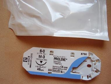
Some investigators recommend the use of nonabsorbable sutures, polypropylene (Prolene) (see the image below), or polybutester (Novafil), to close the fascia in a midline abdominal incision. A meta-analysis of 32 trials published in 2000 compared the closure techniques of the abdominal fascia. This study found a 32% decreased risk of incisional hernia when the fascia was approximated with nonabsorbable sutures compared with absorbable sutures. This study may have included patients with fascial closure using rapidly absorbed sutures such as Vicryl or Dexon. [41] A study by van't Riet in 2002 found no difference in incisional hernia rates between delayed absorbable sutures and nonabsorbable sutures. [14]
Laparoscopy
Operative laparoscopy is commonly used in the surgical treatment of gynecologic malignancies. Numerous studies have demonstrated minimally invasive surgery, compared to laparotomy, results in reduced operative blood loss, decreased number of hospital days, and improved patient quality of life.
Various techniques are used to insert trocars into the abdominal cavity for minimally invasive surgery (see the image below). A Verress needle can be inserted at the subumbilical site or in the left upper quadrant at the midclavicular line just below the ribs to create a pneumoperitoneum. A trocar is bluntly inserted at the subumbilical site into the abdominal cavity after an adequate peritoneum is established. This method requires a blind insertion of the trocar into the abdomen. Many surgeons prefer to visualize the trocar entering the abdominal cavity to decrease injury to the intestines or vascular structures.
Location of deep and superficial vessels of the anterior abdominal wall. Blue circles indicate recommended locations for trocar placement.
Visualization of the trocar into the abdominal cavity is performed by making a subumbilical incision. The fascia is grasped with Kocher clamps and a 10-12 mm incision is created in the fascia. The peritoneum is then incised and a blunt trocar is inserted into the abdomen with direct visualization. An alternative method is to insert a 2 mm Verress needle into the left upper quadrant. A 2 mm laparoscope is inserted through the needle after a pneumoperitoneum is created. This technique allows for larger trocars to be inserted under direct visualization.
Closure of trocar incision sites has not been standardized. The incidence of an incisional hernia at trocar sites has been estimated to be 21 per 100,000. Most incisional hernias after laparoscopy occur with fascial incisions greater than 10 mm, which prompts many surgeons to close the fascia in this situation. Case reports have described hernias at 5 mm trocar sites but fascial incisions less than 10 mm are not usually repaired. [42] Skin closure techniques include subcuticular closure with a 4-0 absorbable suture or octylcyanoacrylate (Dermabond, Ethicon, Sommerville, NJ). A randomized trial from 2005 demonstrated skin closure with octylcyanoacrylate yielded cost savings and decreased operative time compared to skin closure with a 4-0 absorbable suture. [43, 44, 45]
-
Location of deep and superficial vessels of the anterior abdominal wall. Blue circles indicate recommended locations for trocar placement.
-
Polypropylene sutures. Image courtesy of Wikimedia Commons.