History of the Procedure
Clark performed the first radical hysterectomy for cervical cancer at Johns Hopkins Hospital in 1895. In 1898, Wertheim, a Viennese physician, developed the radical total hysterectomy with removal of the pelvic lymph nodes and the parametrium. In 1905, Wertheim reported the outcomes of his first 270 patients. The operative mortality rate was 18%, and the major morbidity rate was 31%.
In 1901, Schauta described the radical vaginal hysterectomy and reported a lower operative mortality rate than the abdominal approach. In the late 20th century, radiation therapy became the favored approach because of the high mortality and morbidity of the surgical approach.
In 1944, Meigs repopularized the surgical approach when he developed a modified Wertheim operation with removal of all pelvic nodes (the Wertheim-Clark plus Taussig operation). Meigs reported a survival rate of 75% for patients with stage I disease and demonstrated an operative mortality rate of 1% when these procedures were performed by a specially trained gynecologist. Throughout the remainder of the 20th century, various modifications have been made for this radical procedure, especially in light of improvements in the areas of anesthesia, intensive care, antibiotics, and blood product transfusion science. Finally, the concurrent decrease in the incidence of invasive cervical cancer, the most common rationale for this procedure, has declined over the past several decades and has led to more conservative procedures (ie, conization for early-stage disease) or nonsurgical modalities (ie, radiotherapy). [1]
Problem
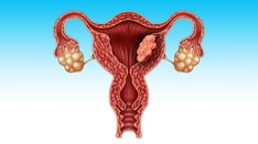
Radical hysterectomy was initially developed as a surgical treatment for cervical cancer due to the absence of other modalities for treatment. Squamous cell carcinoma and adenocarcinoma are the most common variants that arise in the cervix. The uterine cervix comprises the distal third of the uterus. The cervix projects into the vagina and continues up to the lower uterine segment. The portion of the cervix exposed to the vagina is most commonly covered with squamous epithelium. The squamous epithelium transitions to columnar epithelium at the squamocolumnar junction, which is also known as the transformation zone. It is this vulnerable area of the cervix, where columnar cells are actively undergoing metaplastic change to squamous epithelium, in which the majority of cervical malignancies occur.
Epidemiology
In the United States, approximately 14,100 new cases of cervical cancer are predicted to occur in 2022. In addition, about 4280 deaths caused by cervical cancer are expected in 2022. [2]
The death rate from cervical cancer has decreased dramatically since the American Cancer Society recommended the use of the Papanicolaou test (Pap test) for cervical cancer screening in the mid 1940s. Over the next 40 years, the death rate from cervical cancer decreased by more than 70% because preinvasive lesions and cervical cancers were detected at an earlier stage.
Worldwide, cervical cancer is the third most common cancer in women. More than 85% of the global burden occurs in developing countries. [3] The lack of a screening cytology program (ie, Pap test) has resulted in this significant problem in the area of women's health. Thus, the most effective strategy of prevention of this malignancy due to the detection of a preinvasive phase is negated and most cases of cervical cancer are not diagnosed until they are advanced in stage and the patient becomes symptomatic.
Etiology
Multiple factors have been associated with the development of cervical cancer. This malignancy most commonly arises at the squamocolumnar junction, where cells are most actively undergoing metaplastic change from columnar epithelium to squamous epithelium.
Infection with human papillomavirus (HPV) is detected in more than 99% of cervical cancers. Although more than 70 different subtypes of HPV have been identified, women infected with high-risk subtypes have an increased risk of developing dysplasia and a subsequent malignancy. The most common high-risk subtypes are HPV-16 and HPV-18, which account for 70% of cervical cancers in the United States. [4] The E6 protein product of these high-risk HPVs binds to the tumor suppressor protein p53, which is thought to disrupt the p53-dependent control of the cell cycle. [5] The E7 protein causes an inactivation of the tumor suppressor retinoblastoma gene (Rb) via its interaction with the Rb protein, whose normal function is seen with the negative control of cell growth. [6]
Risk factors associated with HPV infection include multiple sexual partners, history of other sexually transmitted infections, high parity, immunosuppression, and cigarette smoking. [7]
Cigarette smoking has been associated with an increased severity of dysplasia and squamous cell carcinoma in women with underlying HPV infection. [8] Nicotine, co-nicotine, hydrocarbons, and tars, carcinogenic breakdown products of cigarette smoke, have been seen concentrated in cervical secretions. [9]
The effect of oral contraceptive use on the risk of cervical cancer is controversial because it is difficult separate sexual behavior from contraceptive use in studies. However, several studies have demonstrated that long-term oral contraceptive use resulted in an increased incidence of cervical cancer. [7, 10, 11] To adequately demonstrate an association, such studies must control for sexual behavior and for the interval of last cervical screening in all study groups. Finally, there are no proven benefits from the cessation of oral contraceptives in the clinical management of cervical dysplasia.
Immunosuppression, either induced or acquired (eg, from HIV infection), is a risk factor for the development of significant preinvasive disease for cervical cancer. [12] In the era of organ transplantation and chronic diseases that require systemic immunosuppression, there exists a cohort of patients with increased risk for the development of cervical cancer.
Sexually transmitted diseases, such as those caused by Chlamydia trachomatis, Neisseria gonorrhoeae, herpes simplex virus, and Trichomonas vaginalis, may be associated with preinvasive disease of the cervix and ultimately a risk for malignancy. [13] With the evidence of HPV as an etiologic agent, such diseases may represent more than a co-infective process, and, in fact, they may be a cofactor in the ability for the establishment of the viral infection via disruption of epithelial integrity.
Pathophysiology
Squamous cell carcinoma is the most common histologic variant of cervical cancer. HPV is now known to be definitively associated with cervical carcinogenesis and its precancerous precursors, low-grade squamous intraepithelial lesions (LSIL) and high-grade squamous intraepithelial lesions (HSIL). The molecular basis for the malignant potential of these viruses has been determined in the dysregulation of the cell cycle by the viral oncogenes E6 and E7. [14]
The progression rate of mild dysplasia to a severe dysplasia or worse is approximately 1% per year; high-grade lesions (moderate and severe dysplasia) have demonstrated a progression to a worsening lesion in approximately 16-36% of cases. Therefore, the treatment strategy for a high-grade lesion usually involves removal of the lesion, but a colposcopically confirmed low-grade lesion can be conservatively managed. The progression time to an invasive malignancy is variable and can span a period of 1 year to several decades.
Adenocarcinoma, the second most common histologic type of cervical cancer, arises from the subcolumnar reserve cells of the columnar endocervical epithelium. A strong association has been demonstrated between cervical adenocarcinoma and HPV-18. The overall incidence for this variant has increased and is associated with women younger than 35 years. Approximately 15% will exhibit no visible lesion due to its endocervical point of origin. Adenocarcinoma in situ of the cervix is strongly associated with an underlying squamous dysplastic lesion and/or cancer in more than 50% of cases, thus making this a high-risk cytologic finding.
Other histologic findings of malignancy involving the cervix include minimal-deviation adenocarcinoma, papillary villoglandular adenocarcinoma, endometrioid adenocarcinoma, serous adenocarcinoma, mesonephric adenocarcinoma, glassy cell carcinoma, adenoid basal carcinoma, basal cell carcinoma, verrucous carcinoma, clear cell adenocarcinoma, adenosquamous carcinoma, adenoid cystic carcinoma, adenoid basal epithelioma, and neuroendocrine tumors. [15] Rarely, cervical lesions result from direct invasion by advanced endometrial, vaginal, bladder, urethra, or colon cancers.
Presentation
Patients with early-stage cervical cancer are relatively asymptomatic; these cases are usually detected via cytologic screening. With the advancement of the disease, signs and symptoms of abnormal bleeding and vaginal discharge may occur. Postcoital bleeding may be the first reported sign in sexually active women; in women who are not sexually active, cervical cancer may not produce clinical manifestations, such as postmenopausal or abnormal uterine bleeding, until the malignancy is in an advanced stage.
As tumors enlarge and outgrow their blood supply, they may become necrotic and produce a malodorous discharge. Larger tumors may cause size-related symptoms such as urinary frequency or retention, rectal pressure, constipation, neurologic symptoms (ie, sciatic pain due to local extension), lower extremity pain, and swelling. Urinary or fecal incontinence due to a local tumor eroding into the bowel or bladder may be the symptom that prompts patients to seek care. Symptomatic anemia may be encountered due to persistent bleeding of the cervical lesion.
The most common sign of cervical cancer is a grossly visible lesion upon a vaginal speculum examination. An exophytic or ulcerative lesion may be obvious during the clinical examination, but an endocervical lesion may remain occult and demonstrate a normal-appearing ectocervical mucosa in the presence of a firm, enlarged cervix. With microinvasive cervical cancer, colposcopic evaluation may provide the means of detection. Colposcopic detection of atypical vessels that demonstrate irregular distribution, unusual caliber, and acute angles are associated with an early invasive tumor of the ectocervix. Presence of any ulcerative or erosive lesion warrants a histologic evaluation by biopsy despite a normal cytologic (Pap test) antecedent result.
The size of the cervix is most accurately determined via a rectal examination, which can also determine the involvement of the adjacent parametrial tissue and/or pelvic side wall. Endocervical lesions expanding or prolapsing through the cervical os can be mistaken for cervical or prolapsing leiomyomata. Biopsy of any abnormally firm or grossly abnormal lesions of the cervix should be undertaken.
The remainder of the physical examination for a patient suspected of a diagnosis of cervical cancer should include a careful evaluation of the vagina, vulva, and rectum for the presence of locally advanced disease. In addition, surveillance of the inguinal, femoral, and supraclavicular lymph nodes by careful palpation should be performed in search of overt evidence of advanced distal disease.
Indications
Radical hysterectomy is indicated for patients with International Federation of Gynecology and Obstetrics (FIGO) stage IA2-IIA cervical cancer who are medically fit enough to tolerate an aggressive surgical approach and wish to avoid the long-term adverse effects of radiation therapy. Prospective randomized trials have validated equal curative rates from radical surgery and radiotherapy (overall survival similar at 83%). [16]
Increased complication rates are noted with combined radical therapies (ie, requirement for adjuvant radiotherapy). However, adjuvant radiotherapy is recommended after radical hysterectomy if there is parametrial involvement, positive surgical margins, or pelvic lymph node metastases, and it should be considered if there is a combination of lymphovascular space invasion, tumor size greater than 2 cm, and deep invasion. [17, 18]
Currently, with stage IB disease, approximately 54% of patients with tumors 4 cm or less in size (stage IB1) and 84% with tumors greater than 4 cm (stage IB2) will require postoperative adjuvant radiotherapy. Recent encouraging data for improved outcomes with combined chemoradiation therapy and the increased morbidity noted with the combined surgical and adjuvant radiotherapy has brought into question the role of radical surgery with stage IB2 and stage IIA. However, a review of survival in 4,885 women with stage IB1-IIA cervical cancer in the Surveillance, Epidemiology, and End Results database showed that radical hysterectomy is superior to primary radiation for the treatment of cervical cancer lesions smaller than 6 cm, and especially for those smaller than 4 cm. [19]
Young patients who desire ovarian preservation and retention of a functional, nonirradiated vagina are ideal candidates for this procedure. Patients who have relative or absolute contraindications to radiation therapy, such as a pelvic kidney or a history of pelvic abscess or pelvic irradiation, should be afforded surgical treatment. In the setting of recurrence, radical hysterectomy has been performed for very small, centrally recurrent or persistent cancers after radiation therapy. Radical hysterectomy is also indicated for other disease processes that involve the cervix (eg, primary upper vaginal carcinoma, endometrial cancer with involvement of the lower uterine segment or cervix).
Relevant Anatomy
Knowledge of the relevant anatomy of the pelvis is important. Radical hysterectomy includes removal of the uterus with parametrial and paracervical tissue, proximal vagina, and proximal uterosacral ligaments. The uterine artery is transected at its origin, lateral to the ureter. In order to complete this dissection, the ureter is unroofed from the paracervical tunnel until the point of entry into the bladder. In order to resect the parametrial and paracervical tissue and unroof the ureter, the paravesical and pararectal spaces must be developed. The pertinent boundaries are the paravesical space and the pararectal space.
The paravesical space is bordered as follows:
-
Medially by the obliterated umbilical artery
-
Laterally by the obturator internus muscle
-
Posteriorly by the cardinal ligament
-
Anteriorly by the pubic symphysis
The pararectal space is bordered as follows:
-
Medially by the rectum
-
Laterally by the hypogastric artery (the internal iliac artery)
-
Posteriorly by the sacrum
-
Anteriorly by the cardinal ligament
The pelvic lymphadenectomy is performed in a systematic fashion. The anatomy of this procedure involves removal of all nodal tissue and skeletonization of all vessels from the mid portion of the common iliac vessels and the internal and external iliac vessels to the level of the circumflex iliac vein distally, from the mid portion of the psoas muscle laterally to the ureter medially, with preservation of the genitofemoral nerve on the psoas muscle. The nodal tissue in the obturator fossa is removed from above the obturator nerve to the external iliac vein superiorly and laterally to the pelvic sidewall. Care must be taken in the obturator fossa to avoid injury to the obturator nerve or to an accessory obturator vein, which is present in approximately 20% of patients.
Contraindications
Contraindications to radical hysterectomy include patients who are medically infirm and those who refuse surgical treatment. Because between one third and two thirds of surgical patients require transfusion, radiation therapy should be considered for patients whose religious or personal beliefs prohibit blood product transfusion. As with any other surgery, careful preoperative risk assessment must be performed. A relative contraindication concerns the possible requirements for adjuvant radiotherapy (ie, stage IB2/IIA or intraoperative findings of locally advanced disease with overt parametrial involvement or grossly positive pelvic or para-aortic lymph nodes).
-
Radical hysterectomy specimen demonstrating the vaginal margin.
-
Radical hysterectomy specimen demonstrating the parametrial margins.