Practice Essentials
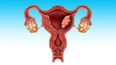
Endometrial hyperplasia involves the proliferation of endometrial glands that results in a greater than normal gland-to-stroma ratio. This results in varying degrees of architectural complexity and cytologic atypia. The clinical significance of this diagnosis is progression to or concurrent endometrioid endometrial adenocarcinoma.
Endometrial carcinoma is the most common gynecologic malignancy and the fourth most common cancer in women in the United States. In 2021, the number of projected cases is 66,570 patients, leading to 12,940 deaths. [1] Significant morbidity or mortality can occur if endometrial hyperplasia is untreated with progression to cancer. Accurate classification and diagnoses are key to guiding appropriate treatment.
Endometrial hyperplasia is most frequently diagnosed in postmenopausal women, but women of any age can be at risk if they are exposed to a source of unopposed estrogen. Endometrial hyperplasia is increasingly seen in young women with chronic anovulation due to polycystic ovary syndrome (PCOS) or obesity.
This article reviews the classification, pathophysiology, clinical features, and treatment of endometrial hyperplasia.
For other information, see Medscape's Ob/Gyn & Women's Health Specialty Center.
Background / Classification
Endometrial hyperplasia is believed to produce lesions that may be the precursor to endometrial carcinoma of endometrioid histology. [2] The classification of endometrial hyperplasia has had numerous terminologies. The classification below was the most commonly used system historically and was used by the World Health Organization (WHO) and the International Society of Gynecologic Pathologists since 1994. This system characterizes the glandular architectural pattern as simple or complex and describes the presence or absence of nuclear atypia.
WHO 1994
- Simple hyperplasia - Increased number of glands but regular glandular architecture
- Complex hyperplasia - Crowded irregular glands
- Simple hyperplasia with atypia - Simple hyperplasia with presence of cytologic atypia (prominent nucleoli and nuclear pleomorphism)
- Complex hyperplasia with atypia - Complex hyperplasia with cytologic atypia
This is based on the original retrospective study of 170 patients by Kurman et al found that lesions with varying degrees of complexity and presence of atypia, when left untreated for a mean of 13 years, progressed to adenocarcinoma at different rates. Simple hyperplasia was associated with a 1% rate of progression to cancer, complex hyperplasia 3% rate of progression, simple atypical hyperplasia 8% rate of progression, whereas complex atypical hyperplasia had a 29% rate of progression to cancer. [3]
Not only does the concern exist for atypical hyperplasia progressing to invasive cancer if untreated, but numerous studies found coexisting carcinoma at rates ranging from 17-56%. [4, 5, 6, 7, 8, 9, 10] A prospective Gynecologic Oncology Group study found that 306 patients with preoperative biopsies that diagnosed atypical endometrial hyperplasia had concurrent invasive adenocarcinoma in 42.6% of hysterectomy specimen. [11] Part of the difficulty in diagnosing concurrent carcinoma is due to lack of reproducibility in diagnosing hyperplasia, especially atypical hyperplasia versus adenocarcinoma among even expert gynecologic pathologists. Studies report only 40-69% interobserver agreement for hyperplasia or cancer. [12, 13, 14, 11]
Due to the poor reproducibility of diagnosis, and confusion regarding optimal clinical management, gynecologic pathologists proposed a simpler classification of endometrial hyperplasia (EH) versus endometrial intraepithelial neoplasia (EIN) using a computerized morphometric analysis. Endometrial Hyperplasia is benign hyperplasia and correlates closely to simple hyperplasia, whereas EIN is a pre-malignant condition. EIN is defined as when the volume of glandular crowding is greater than the stromal volume, the presence of cytologic alterations, a lesion larger than 1 mm, and exclusion of mimics or carcinoma. Classification as complex atypical hyperplasia (WHO’94) or as EIN had similar sensitivities and negative predictive values for coexisting endometrial cancer. [15] Others found the EIN classification to be better at predicting progression to cancer. Thus the WHO formally adopted the simplified classification of endometrial hyperplasia into 2 categories.
WHO 2014
1. Benign endometrial hyperplasia
2. Endometrial Intraepithelial Neoplasia (EIN)/well differentiated carcinoma.
The ACOG and SGO Committee opinion 2015 also recommend the use of EIN schema for a more clear terminology to distinguish the two clinicopathologic categories. [2, 16] [17] Not only does this classification reflect clinical outcome, but also underlying genetic and molecular changes. [18, 19, 20]
Pathophysiology
Endometrial hyperplasia results from continuous estrogen stimulation that is unopposed by progesterone. This can be due to endogenous estrogen or exogenous estrogenic sources. Endogenous estrogen may be caused by chronic anovulation associated with PCOS. Obesity also contributes to unopposed estrogen exposure due to chronic high levels of estradiol that result from aromatization of androgens in adipose tissue and conversion of androstenedione to estrone. Endometrial hyperplasia and cancer can also result from estradiol-secreting ovarian tumors such as granulosa cell tumors.
Exogenous unopposed estrogen without progesterone has been associated with increased endometrial hyperplasia and adenocarcinoma. [21] The Postmenopausal Estrogen/Progestin Interventions (PEPI) trial found that unopposed estrogen exposure with 0.625 mg of conjugated equine estrogen replacement therapy increased the risk of complex hyperplasia by 22.7% and atypical hyperplasia by 11.8% over 3 years of use compared with a less than 1% increase in placebo controls. [22] The risk of endometrial cancer was not increased when 2.5 mg of medroxyprogesterone acetate was used in combination with 0.625 mg of conjugated equine estrogens in 8506 women in the Women's Health Initiative (WHI) study. [23] Tamoxifen, with its estrogenic effect on the endometrium, increases the risk of endometrial hyperplasia and endometrial cancer. The risk of progression to cancer is associated with an increased duration of use. [24] While unopposed estrogen in oral contraceptive pills or estrogen replacement therapy increases the risk of hyperplasia and cancer, combination oral contraceptive pills and combination hormone replacement therapy does not increase and may decrease the risk of hyperplasia and cancer.
Other risk factors for endometrial hyperplasia are the same as those for type I endometrial adenocarcinoma, including obesity, nulliparity, early menarche, and late menopause. The independent risk factors for predicting when endometrial hyperplasia coexists with cancer include age older than 53 years, postmenopausal status, diabetes, abnormal bleeding, body mass index (BMI) of 27 or more, and atypical hyperplasia. [8]
The exact mechanism of estrogen's role in the transformation of normal endometrium to hyperplasia and cancer is unknown. Genetic alterations are known to be associated with hyperplasia and type I endometrial cancers. Benign Hyperplasia is associated with low levels of somatic mutations, whereas EIN is associated with genetic alternations similar to endometrioid endometrial cancer such as microsatellite instability, and mutations in PTEN and KRAS. [25, 19, 20] PTEN tumor suppressor gene mutations have also been found in 55% of hyperplasia cases and 83% of hyperplasia cases once it has progressed to endometrial cancer. [26]
Clinical Features
Clinical presentation
The most common clinical presentation of patients with endometrial hyperplasia is abnormal uterine bleeding, whether in the form of menorrhagia, metrorrhagia, or postmenopausal bleeding. It can be associated with uterine hemorrhage, requiring emergent medical or surgical interventions, loss of fertility, and blood transfusion therapy. Others present with abnormal vaginal discharge or Pap smear results showing glandular abnormalities. The abnormal Pap smear result may be atypical glandular cells or presence of atypical endometrial cells.
When abnormal bleeding is present, a full history and physical examination is warranted with careful examination of the lower genital tract for lesions of the vulva, vagina, cervix, and palpation of uterus and ovaries. The source of vaginal discharge or bleeding, the size of the uterus and endometrial cavity, and any pelvic masses should be noted. If the patient is obese and a pelvic examination is inadequate, pelvic ultrasonography may be helpful to assess for ovarian masses. A diagnostic procedure is then needed to rule out hyperplasia or cancer if the patient is symptomatic or has abnormal cytology.
Diagnosis
Diagnosis of endometrial hyperplasia is usually made by sampling the endometrial cavity with an endometrial biopsy in the office. Tissue sampling should be performed in women with risk factors who present with symptoms of abnormal vaginal bleeding or discharge. This includes women older than 35 years with abnormal bleeding, women younger than 35 years with bleeding and risk factors, women with persistent bleeding, and women with unopposed estrogen replacement, tamoxifen therapy, and HNPCC cancer syndrome.
In addition, a biopsy should be performed in women with atypical glandular cells (AGC) Pap smear or endometrial cells in Pap smears of women older than 40 years when out of synch with menstrual cycle. [27, 28] While no evidence of improved survival has been documented, some also advocate routine screening by endometrial biopsy in asymptomatic women with hereditary nonpolyposis colorectal cancer (HNPCC) syndrome or those on tamoxifen therapy. However, most of this high-risk population present with abnormal vaginal bleeding; thus, other experts recommend work-up only when symptoms are present.
If a patient does not tolerate an office biopsy or has cervical stenosis, endovaginal ultrasonography is an effective method to assess thickness of the endometrial echo complex and to evaluate uterine bleeding. The American College of Obstetricians and Gynecologists recommends transvaginal ultrasonography as a reasonable alternative to endometrial sampling for the evaluation of an initial episode of bleeding in a postmenopausal woman. [29]
Endovaginal ultrasonography has a sensitivity of greater than 96% for ruling out endometrial carcinoma if the endometrial echo complex is less than 5 mm. Persistent bleeding despite a thin stripe still warrants tissue biopsy because of the risk of missing a type 2 cancer that is not associated with hyperplasia and thickening of the endometrial echo complex. [30] If hyperplasia is diagnosed by office biopsy, one should consider D&C and hysteroscopy to more definitely rule out atypia or cancer prior to conservative medical management. This is because blind D&C and Pipelle endometrial biopsies both sample only 50-60% of the endometrial cavity, and can miss focal lesions. [31]
Treatment
The accurate diagnosis of hyperplasia type is vital for appropriate treatment based on risk of cancer without over or under treatment. Once a tissue diagnosis of endometrial hyperplasia is made, treatment depends on the type of hyperplasia, the patient's symptoms such as severity of bleeding, surgical risks, and wish for future childbearing. Progestins can effectively treat endometrial hyperplasia to control bleeding and prevent progression to cancer. They can serve as prevention of recurrence in those with continued risk factors. Benign hyperplasia responds well to progestins. More than 98% of women with hyperplasia treated with cyclic progestins experienced regression of the disease in 3-6 months. [32] However, atypical hyperplasia or EIN lesions should undergo hysterectomy for definitive diagnosis and treatment if they have completed childbearing and are reasonable surgical candidates due to a 42% risk of concurrent endometrial cancer. [11]
The PEPI trial showed a 94% normalization of complex or atypical hyperplasia in 45 women treated with progestins. [22] One cohort study found that 115 patients with complex atypical hyperplasia had approximately 30% persistence or progression of disease whether treated with progestins or not. However, of 70 patients with atypical hyperplasia, 67% vs 27% had persistence or progression when not treated with progestins. [33]
Multiple regimens of progestin therapy have been found effective in reversing endometrial hyperplasia, including the following [34] :
-
Medroxyprogesterone acetate - MPA (Provera), 10-20 mg qd, or cyclic 12-14 days per month
-
Depot Medroxyprogesterone (Depo-provera) 150 mg IM q 3 months
-
Micronized vaginal progesterone (Prometrium), 100-200 mg qd or cyclic 12-14 days per month
-
Levonorgestrel-containing IUD (Mirena), 20 mcg/day [35]
-
Megestrol acetate (Megace), 40-200 mg per day, usually reserved for women with atypical hyperplasia
If the patient has not completed childbearing or is not a surgical candidate, then concurrent cancer must first be ruled out by D&C with hysteroscopy prior to medical management. We recommend megestrol acetate for EIN, with or without levonorgestrel IUD for patients wishing to preserve fertility or for those too ill for surgical management. Any progestin should be adequate for treatment of benign hyperplasia or for maintenance after resolution of EIN. Patient should be sampled to assess for response every 3 to 6 months for regression to normal endometrium. If there is inadequate response in 6 months, consider increasing dose or changing progestins. There is no proven protocol for selection or dosing. Continued surveillance after regression of the lesion is recommended every 6-12 months if risk factors persist. Repeat biopsy is also indicated for recurrent abnormal bleeding or discharge. Prevention of recurrence include use of daily or cyclic progesterone, indwelling levonorgestrel IUD, along with weight loss for obese patients.
Due to the large number of young anovulatory women diagnosed with atypical hyperplasia/EIN or early endometrial cancer, numerous studies have examined the outcome of fertility-sparing hormonal therapy. A meta-analysis of 24 observational studies that included a total of 151 women with atypical hyperplasia found that those who had fertility-sparing treatments had 86% regression, 26% relapse, and live birth rate of 26%. [36]
Another review of complex atypical hyperplasia/EIN patients who underwent progestin therapy found a complete response rate of 66%. Median time to response was 6 months. Recurrence rate after initial response was 23%. During study follow-up time of 39 months, 41% of patients with atypical hyperplasia became pregnant. [37] Interest is increasing in the use of levonorgestrel-releasing intrauterine system (LNG-IUS) as a treatment option for endometrial hyperplasia to deliver local effect with less systemic side-effects. [38] A 15-year study using the LNG-IUS for 34 patients with endometrial hyperplasia found regression to atrophic endometrium in 94%. However, this study had only 4 (12%) patients with atypical hyperplasia. [39] A meta-analysis of 24 observational studies, including 1,001 cases, indicate that progestins appeared to induce a lower disease regression rate than LNG-IUS in the treatment of complex and atypical endometrial hyperplasia. However, a controlled clinical trial is necessary to confirm the observational findings. The authors feel data using the LNG-IUD as the only treatment modality for atypical hyperplasia or endometrial cancer are still limited. [40]
As mentioned above, the Gynecologic Oncology Group pathologic study with biopsy diagnosis of atypical hyperplasia found 42.6% concurrent endometrial carcinoma on hysterectomy specimen; 31% of these had myometrial invasion, including 10.6% with outer half myometrial invasion. [11] Others also found atypical endometrial hyperplasia on office biopsy or D&C was associated with a 48-56% cancer rate on permanent pathology. Thus, if a hysterectomy is planned for treatment of atypical hyperplasia based on office endometrial biopsy, the authors recommend having a gynecologic oncologist be primary surgeon, or be available for surgical staging if needed based on frozen section of uterine specimen. [10, 31] Patients should be counseled and consented for washings, hysterectomy, possible BSO and lymph node dissection depending on intra-op exam or frozen pathology findings. If a D&C with hysteroscopy had been performed to rule out concurrent carcinoma more definitively, an oncologic surgeon is likely not needed. Due to the risk of cancer, supracervical, morcellation, or endometrial ablation is not recommended. While a simple hysterectomy is adequate for definitive treatment of hyperplasia, one can consider bilateral salpingo-oophorectomy in perimenopausal or postmenopausal women due to possibility of cancer on permanent section. Ovaries should only be removed if cancer is diagnosed in premenopausal women. They should be counseled a second surgery may be required to remove ovaries and perform lymph node staging if cancer if found on final pathology.
The need for hysterectomy to exclude concurrent myoinvasive endometrioid adenocarcinoma presents a barrier to nonsurgical management of endometrial hyperplasia. A Gynecologic Oncology Group study examined the histomorphometric 4-class rule (4C), which measures epithelial abundance, thickness, and nuclear variation as applied to diagnostic biopsies to predict myoinvasive cancer outcomes at hysterectomy. [41] Women with endometrial biopsies or curettages with a community diagnosis of atypical endometrial hyperplasia were enrolled in a clinical trial in which subsequent hysterectomy was scored for endometrial adenocarcinoma, and 4C rule ability to predict cancer outcomes was measured. Qualifying biopsies were stratified into high-risk and low-risk histomorphometric subgroups. Assignment to a high-risk category by 4C rule was highly sensitive in predicting any (71%) or deeply (92%) myoinvasive adenocarcinoma at hysterectomy, and assignment to a low-risk group had a high negative predictive value for absence of any (90%) or deeply (99%) myoinvasive disease. At present, this use of histomorphometry is most suited to a centralized reference laboratory performing histomorphometry for a variety of diagnostic applications. However, in the future, formal histomorphometry of endometrial biopsies using the 4C rule may become a more common method to identify a subset of women with premalignant disease who are unlikely to have concurrent myoinvasive adenocarcinoma and therefore may qualify for nonsurgical therapy.