Practice Essentials
Benign vulvar disorders are a significant issue for patients. These disorders include vulvar atrophy, benign tumors, hamartomas and cysts, infectious disorders, and nonneoplastic epithelial disorders. [1, 2] Infectious disorders include diseases caused by known transmissible agents, such as viruses, bacteria, fungi, and protozoa. They may first be seen by physicians of various specialties, including dermatologists and gynecologists, and often require a multidisciplinary approach. In an Australian study, the most common final dermatologic diagnoses made at a tertiary dermatology center for referrals for vulvar complaints were predominately for lichen sclerosus and much less so for dermatitis and psoriasis. [3]
Developmental abnormalities of vulva are generally rare. Vulvar atrophy may be related to advanced age or other disorders, but these abnormalities often represent an almost physiologic finding in the elderly. [4]
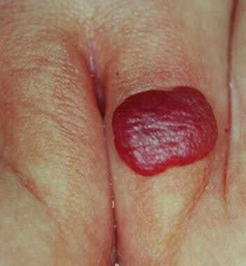
Benign tumors of the vulva are relatively uncommon and may show nonspecific clinical features. Therefore, a biopsy is often needed to exclude a malignant neoplasm and to indicate proper treatment. Vascular neoplasms may also occur in the vulva and are similar to such lesions found elsewhere. See the image below.
Nonneoplastic epithelial disorders include several inflammatory, ulcerative, and blistering disorders, as well as pigmentary changes involving the vulvar region.
Inflammatory diseases involving the vulvar region include the following:
-
Lichen sclerosus
-
Squamous cell hyperplasia (with and without atypia)
-
Lichen simplex chronicus (localized neurodermatitis)
-
Primary irritant dermatitis
-
Fixed drug eruption
-
Reiter disease
-
Lupus erythematosus
-
Darier disease
-
Aphthosis and Behçet disease
-
Pyoderma gangrenosum
-
Plasma cell vulvitis
-
Vulvar vestibulitis
Blistering diseases involving the vulvar region include the following:
-
Familial benign chronic pemphigus (Hailey-Hailey disease)
Pigmentary changes involving the vulvar region include the following:
-
Lentigo, lentiginosis, and benign vulvar melanosis
-
Postinflammatory hypopigmentation
-
Vulvar melanosis [5]
Benign tumors, hamartomas, and cysts involving the vulvar region include the following [6] :
-
Bartholin and Skene duct cysts
-
Acrochordon (fibroepithelial polyp)
-
Fibroma, fibromyoma, and dermatofibroma
-
Hidradenoma
-
Syringoma
-
Hemangioma
-
Angiokeratoma
-
Heterotopic sebaceous glands and sebaceous gland hyperplasia
-
Papillomatosis (papillary vulvar hirsutism)
-
Schwannoma [7]
-
Angiomyxoma: a benign and aggressive mesenchymal tumor [8]
Congenital malformations involving the vulvar region include the following:
-
Ambiguous external genitalia
-
Congenital labial hypertrophy
Atrophy of the vulva may also occur.
Background
In the last few years, interest in vulvar disease has greatly increased. However, the relevant material has been scattered throughout the literature of various specialties, including dermatology, genitourinary medicine, gynecology, and pathology. The spectrum of involved specialties reflects the complexity of vulvar diseases and the necessity of a multidisciplinary approach to the study of the vulva. [9] In response to the various approaches of the specialists faced with treating vulvar disease, the World Health Organization, the International Society for the Study of Vulvar Disease, the International Federation of Gynecology and Obstetrics, and the International Society of Gynecological Pathologists have made an effort to standardize the nomenclature.
Some general anatomic, embryologic, and histologic findings of the vulva merit review. The vulva is the part of the female genital tract located between the genitocrural folds laterally, the mons pubis anteriorly, and the anus posteriorly. Embryologically, it is the result of the junction of the cloacal endoderm, urogenital ectoderm, and paramesonephric mesodermal layers. This hollow structure contains the labia majora, labia minora, clitoris, vestibule, urinary meatus, vaginal orifice, hymen, Bartholin glands, and Skene ducts. Different epithelia, from keratinized squamous epithelium to squamous mucosa, cover the vulva. The labia minora are rich with sebaceous glands but have few sweat glands and no hair follicles. The epithelium of the vestibule is neither pigmented nor keratinized and contains eccrine glands.
Vulvar pruritus is an important symptom. It is most often caused by vulvovaginal candidiasis, followed by chronic dermatoses, such as lichen sclerosus and vulvar dermatitis. [10] If the pruritus is unresponsive to therapy, an invasive or preinvasive squamous cell carcinoma should be considered.
Etiology and Pathophysiology
Nonneoplastic epithelial disorders are discussed below.
Inflammatory diseases
Lichen sclerosus
The etiology of this condition is unknown. [11] A higher prevalence of the disease in postmenopausal women suggests hormonal factors, but this has not been confirmed. [12, 13] A study demonstrated that oral contraceptives with antiandrogenic properties might trigger the early onset of lichen sclerosus in susceptible young women. [14]
Investigative studies aimed toward identifying an infection-causing agent (eg, spirochetes, viruses) have yielded inconclusive results.
Lichen sclerosus has been weakly linked to autoimmune diseases and genetic factors. Approximately 21% of patients have an autoimmune disease, most commonly a thyroid disorder. Familial occurrence is also well recognized. Forty-four percent of patients have one or more autoantibodies, and 22% percent have a positive family history. [13, 14, 15, 16, 17]
The role of local factors (eg, trauma, friction, chronic infection, and irritation) is well recognized, and recurrence near vulvectomy scars has been observed.
Squamous cell hyperplasia
Repetitive scratching or rubbing from irritants can result in squamous cell hyperplasia. Because this condition is often thought to be equivalent to lichen simplex chronicus, some diagnostic confusion exists.
Ambiguous and even inexplicable terms, such as atypical epithelial hyperplasia (dysplasia), vulvar dystrophy, vulvar atypia, atrophic dystrophy, mixed dystrophy, and vulvar intraepithelial neoplasia, inhibit effective communication between clinicians and pathologists. These terms have different meanings to dermatologists, pathologists, and gynecologists, further complicating the problem.
Lichen simplex chronicus (localized neurodermatitis)
Lichen simplex chronicus of the vulva is the end stage of the itch-scratch-itch cycle. The initial stimulus to itch may be underlying seborrheic dermatitis, intertrigo, tinea, or psoriasis; however, in most cases, the underlying cause is not evident and may have been transient vulvitis or vaginal discharge. [18]
Any itching disease of the vulva may become secondarily lichenified.
Primary irritant dermatitis
In the absence of any immune reactivity, this condition is a common cause of vulvar burning and pruritus due to irritation.
Chemical agents that remove surface lipids, denature epidermal keratins, or damage cell membranes with a direct cytotoxic effect on cells may cause irritation. Common irritants include perfumed soaps and detergents, fabric softeners, feminine hygiene products (eg, tampons, pads, diapers, wipes, deodorant sprays), bubble baths, bath oils, colored or scented toilet paper, physically abrasive contactants (eg, face cloths, sponges), and body secretions (eg, urine, semen).
Obsessive cleaning of the vulvar area may also cause serious irritation.
Irritation may eventually arise as a result of friction from tight clothing, trapping of moisture due to lack of ventilation (eg, from wearing synthetic fabrics), or activities such as bicycling and horseback riding.
Intertrigo
Intertrigo is a nonspecific inflammatory eruption of skin folds that can be precipitated by sweating, obesity, and occlusion.
Allergic contact dermatitis
Vulvar allergic dermatitis may occur as a result of a delayed, cell-mediated, type IV hypersensitivity reaction. An inflammatory disorder originating from local contact with an agent to which the patient has previously been sensitized, vulvar allergic dermatitis develops 12-48 hours after exposure. Less commonly, vulvar pruritus may coincide with more generalized allergic symptoms. Known allergens include topical medications and various chemicals in products such as perfumes, preservatives, and latex. Nickel sensitivity from snaps or zippers in denim jeans or undergarments may also occur. [19, 20, 21]
Topical allergens in vulvar allergic dermatitis include the following:
-
Antibiotics - Neomycin, clindamycin, tetracycline, sulfonamides, nifuratel
-
Anesthetics - Benzocaine, lidocaine, prilocaine, pramoxine
-
Antihistamines - Promethazine, diphenylenediamine hydrochloride, p-phenylenediamine, ethylenediamine dihydrochloride
-
Antiseptics - Hexachlorophene
-
Clothing dyes
-
Moisturizers - Lanolin
-
Nail polish
-
Nickel
-
Plants - Poison ivy
-
Preservatives - Paraben, imidazolidinyl urea
-
Perfumes - Balsam of Peru
-
Rubber - Gloves, condoms, diaphragms
Fixed drug eruption
A fixed drug eruption is a cell-mediated allergic drug reaction typically recurring in the same site upon reexposure. Common causative agents include oral drugs such as nonsteroidal anti-inflammatory agents, acetaminophen, sulfonamides, tetracycline, and barbiturates. Fluconazole as a possible causative agent has also been recently reported. [22]
Atopic dermatitis
Atopic dermatitis usually occurs in patients with a personal or family history of asthma, hay fever, childhood eczema, or dry skin and is related to a cutaneous hypersensitivity associated with defective cell-mediated immunity and immunoglobulin E overproduction. [23]
Although airborne and food allergens may generally play a role, because of their skin hypersensitivity, atopic individuals sometimes show vulvar symptoms as a result of irritation by personal hygiene products (eg, soaps, cleansers, lotions, perfumes, sanitary napkins).
Seborrheic dermatitis
The cause of seborrheic dermatitis is unknown. Seborrhea is evidently a substantial predisposing factor because lesions occur in areas of the skin where sebaceous glands are most active, such as the face, scalp, body folds, and, less commonly, the genitalia.
Seborrheic dermatitis is commonly observed in neonates during the early months of life (as a result of sebaceous gland activation by maternal androgens) or after puberty. An association with Pityrosporum yeasts has been noted.
Neurologic factors have also been thought to play a role; emotional stress, Parkinson disease, nerve injury, and syringomyelia have been related to onset or worsening of the disease.
Seasonal factors, zinc deficiency, and HIV infection have been linked to this disorder.
Psoriasis
Psoriasis is a hereditary disorder of the skin that affects approximately 2% of the population in the Western world. The characteristic silver-white scales on erythematous plaques are caused by rapid cell turnover and primarily occur in sites of repetitive trauma, such as the scalp, elbows, forearms, knees, hands, and feet.
Rarely, vulvar involvement occurs and is sometimes triggered or worsened by local factors such as irritation from scratching, irritant soaps, bacterial or yeast superinfections, or increases in heat and humidity secondary to the use of tight synthetic clothing and sanitary napkins.
Reiter disease
Reiter disease is a sterile oligoarthritis typically associated with conjunctivitis and with a distant infection (eg, Chlamydia trachomatis, Ureaplasma urealyticum, Shigella, Salmonella), causing nongonococcal urethritis or enteritis. Additional findings include palmoplantar keratoderma, circinate balanitis, and, less frequently, vulvar involvement.
About 80% of patients with Reiter disease are HLA-B27 positive. However, the exact role of this major histocompatibility class I antigen in the development of Reiter disease remains unclear.
Lichen planus
Lichen planus can be an acute or chronic dermatosis affecting the skin, mucous membranes, or both. Its cause is unknown, but evidence suggests that it is an immunologically mediated disorder. [17] Some drugs have been found to induce lichen planus–type eruptions. [24]
Vulvar lesions may be more common than generally considered; a report found genital involvement in 51% of women with cutaneous disease. [25] Involvement may sometimes be hypertrophic. [26]
Lupus erythematosus
Lupus erythematosus is an idiopathic autoimmune disorder that can affect many organ systems. According to the degree of systemic involvement, the disease is classified into systemic, subacute, chronic, or discoid forms.
Genital involvement is uncommon, and vulvar manifestations have seldom been described. In one study, vulvar lesions were found in 2 (5%) of 42 women affected by the chronic form.
Darier disease
Darier disease is a heritable disorder of keratinization transmitted as an autosomal dominant trait. Altered keratinization is a result of the disruption of desmosomal proteins with consequent tonofilament detachment and acantholysis because of the mutation of a gene located on band 12q23-24.1. [27]
Genital involvement is frequent and is precipitated by heat, humidity, sweat, and friction.
Aphthosis
Aphthosis is a condition of unknown etiology, but it may be associated with autoimmune disease. Premenstrual exacerbations are also common. Aphthosis is characterized by single or multiple painful canker sores on the oral and genital mucosa. It may be associated with COVID-19 infection. [28] Synonyms include aphthous ulcers, canker sores (on the vulva), Lipschütz ulcers, and ulcus vulvae acutum. [29]
Behçet disease
Behçet disease is a chronic multisystemic inflammatory disorder characterized by oral and genital aphthous ulcers and ocular involvement. [30] Its etiology is unknown but probably involves altered immunity in genetically predisposed subjects because an association with the expression of some HLAs (B51, DR5, and DQw3) and a racial predilection for individuals of Asian or Middle Eastern descent have been shown. [31]
Pyoderma gangrenosum
The etiology of pyoderma gangrenosum is still unknown, although evidence of its etiologic and pathogenic background may be found in its frequent association with systemic autoimmune diseases (eg, Crohn disease, arthritis, monoclonal gammopathy). Moreover, evidence suggests that altered immunity occurs in some patients with pyoderma gangrenosum.
Vulvar involvement occurs, albeit rarely, with approximately 8 cases reported in the literature. [32]
Crohn disease
Crohn disease is a chronic granulomatous inflammatory bowel disease that may primarily or secondarily involve the vulvar (2%) and inguinal regions. [33] Vulvar Crohn disease is most often evident with vulval edema, less so with ulceration and fissures. [34] Proposed causes include an unrecognized infectious agent or a disturbed immunologic reaction to an intestinal organism in a genetically predisposed individual.
Findings from the largest case series of vulvar Crohn disease suggest that patients with Crohn disease should have regular gynecologic checkups. [35]
Hidradenitis suppurativa
Hidradenitis suppurativa is a chronic suppurative inflammatory disorder of the apocrine glands resulting—as with acne vulgaris and acne mechanica—from acute and chronic follicular occlusion.
Several factors, including onset at puberty, female predilection (female-to-male ratio, 3:1), typical flares with menstruation, spontaneous regression at menopause, and occasional association with the use of an oral estroprogestinic, suggest a role for hormonal factors. [36]
Familial cases and racial predilection (ie, more common in Blacks) indicate that genetic factors may also play a role.
Obesity, profuse sweating, and overheating are other precipitating factors.
Fox-Fordyce disease
Fox-Fordyce disease is an uncommon condition of the axillary and anogenital regions related to apocrine sweat duct occlusion and is typically exacerbated by factors that stimulate apocrine secretion. [37] Its cause is unknown. Improvement is observed at menopause, with pregnancy, or following oral contraceptive intake. [38]
Plasma cell vulvitis
Plasma cell vulvitis, corresponding to Zoon plasma cell balanitis, has also been reported on the vulva. The etiology is unknown.
Vulvar vestibulitis
The pathogenesis of vulvar vestibulitis is obscure. Studies suggesting a relative inability to down-regulate proinflammatory interleukin-1 beta activity by interleukin-1 receptors need further confirmation. [39]
Sexual activity, nulligravidity, bacterial infection, or candidal infections seem to be unlikely factors. [40, 41] Association with sensitization to seminal fluid [42] or with interstitial cystitis may occasionally be found. [43]
Localized pain of vulvar vestibulitis may result from regionally elevated cytokines produced by vulvar vestibule-specific fibroblasts. [44] Altered density of nerve endings [45] and estrogen receptors has been reported. [44, 46, 47]
A study demonstrated that vulvar vestibulitis is more frequent in patients who use oral contraceptives with low-dose estrogen than those who use oral contraceptive with high-dose estrogen. [48] The use of oral contraceptives might be a contributing factor by increasing the sensitivity of the vestibular mucosa. [49]
Blistering diseases
Familial benign chronic pemphigus (Hailey-Hailey disease)
Hailey-Hailey disease is a rare autosomal dominant acantholytic disorder due to a mutation on band 3q21-q24 and is characterized from late adolescence or adulthood by recurrent eruptions of vesicles and blisters typically located on the neck, axillae, and groin.
Intrinsic desmosomal fragility may only become evident when trigger factors (eg, friction, infections, irritants, increased temperature and humidity, acute UV exposure) precipitate acantholysis. Because most of these conditions frequently occur in the anogenital area, vulvar problems may be common.
Bullous pemphigoid
Bullous pemphigoid is a blistering autoimmune disorder that usually affects elderly patients. The disorder is caused by antibodies binding 2 different antigens, BPAg1 (230 kd) and BPAg2 (a hemidesmosomal antigen of 180 kd now thought to be collagen XVII), located in the basement membrane of the skin. [50]
Mucosal lesions of the vulva occur less frequently and are less severe than other blistering disorders. [51]
A juvenile localized subtype of immunoglobulin G (IgG)–mediated bullous pemphigoid occurring in children in the first year of life and characterized by a self-limited nonscarring bullous pemphigoid–like process involving only the vulva has been described. [52]
Cicatricial pemphigoid
Cicatricial pemphigoid is an autoimmune condition with autoantibodies directed against collagen XVII and laminin-5 located in the basement membrane zone. Resulting inflammation and subepidermal splitting leads to typical mucosal scarring. Genital involvement is common and reportedly occurs in at least 50% of patients.
Pemphigus vulgaris
Pemphigus vulgaris is an autoimmune blistering disorder that affects the skin and mucosa. This is due to circulating autoantibodies directed toward desmoglein III (130 kd), a desmosomal cadherin that mediates cell-to-cell adhesion in the epidermis. The vulva is affected in approximately 10% of cases.
Contact pemphigus induced by topical imiquimod may occur. [53]
Erythema multiforme (minor/major)
Erythema multiforme is a cutaneous hypersensitivity reaction that may be triggered by herpesvirus infection or drug intake. Sulfonamides, beta-lactam antibiotics, and nonsteroidal anti-inflammatory drugs are the offending agents in most patients.
Vulvar involvement is frequently observed in the more severe forms of the disease, along with lesions located on other mucosal (eg, eye, mouth, throat) and skin sites.
Epidermolysis bullosa
The term epidermolysis bullosa indicates a group of congenital disorders characterized by skin fragility due to an inherited defect in the epidermal, junctional, or dermal components underlying the physiologic mechanical properties of the skin. Mucous membranes, including those of the genitalia, may be involved.
Epidermolytic hyperkeratosis
Vulvar epidermolytic hyperkeratosis is an uncommon benign genodermatosis. A causative role of keratin 1 and 10 mutations has been considered. Mechanical trauma may act as a precipitating factor. [54]
Pigmentary changes
Acanthosis nigricans
Acanthosis nigricans is a diffuse pigmentary change typically observed in intertriginous areas and skin folds. It may be hereditary, drug induced, or associated with obesity or endocrine diseases. [55] It sometimes represents a paraneoplastic syndrome, revealing associated malignant epithelial tumors or lymphoproliferative disorders.
Lentigo, lentiginosis, and benign vulvar melanosis
Lentigo, lentiginosis, and benign vulvar melanosis are characterized by benign epidermal melanocytic hyperplasia. These are the most common pigmented lesions reported to occur in the vulva. [56]
Hyperpigmented macules of less than 4 mm in diameter define lentigo, whereas larger macules are usually vulvar melanosis.
Lentiginosis is characterized by multiple spots of hyperpigmentation and may sometimes be an expression of a genetic disorder such as Peutz-Jeghers syndrome, LEOPARD syndrome (ie, lentigines, electrocardiographic [conduction abnormalities], ocular [hypertelorism], pulmonary [stenosis], abnormal [genitalia], retardation [of growth], and deafness syndrome), or somatic mosaicism.
Melanocytic nevus
Vulvar nevi are fairly common (0.1% of nevi have this location). The etiology of nevi at other skin sites is still a matter of debate. Nevus cells deriving from the neural crest migrate into the skin during embryogenesis and collect in the basal cell layer of the epidermis, where they proliferate in small nests.
Postinflammatory hyperpigmentation
Postinflammatory hyperpigmentation is due to melanin deposition in the dermis following a previous inflammatory process caused by drug intake (end stage of a fixed drug eruption) or other dermatologic disorders localized to the vulva (eg, lichen planus, discoid lupus erythematosus, psoriasis).
Postinflammatory hypopigmentation
Postinflammatory hypopigmentation is due to temporary or permanent melanocyte damage of different causes, including skin injury with scar formation and chronic inflammatory skin diseases (eg, lichen sclerosus, discoid lupus erythematosus).
Vitiligo
Vitiligo is an acquired loss of pigmentation secondary to possible immunologically mediated melanocyte damage. Genital involvement is common.
Benign tumors, hamartomas, and cysts
Mucous cysts
Mucous cysts are dysontogenetic cysts arising from the minor vestibular glands or from mesonephric duct remnants.
A cyst of the canal of Nuck (processus vaginalis peritonei) that fills with peritoneal fluid may also occur.
Bartholin cyst and Skene duct cyst
Bartholin and Skene duct cysts develop as a result of ductal occlusion. [57, 58]
Epidermal inclusion cyst
Epidermal inclusion cyst is a common cystic lesion that arises from obstruction of a sebaceous gland.
Seborrheic keratosis
The etiology of seborrheic keratosis, a common pigmented epithelial proliferation, is unknown, although it may be inherited.
Acrochordon
A fibroepithelial polyp (acrochordon) is a skin tag commonly observed in areas subject to irritation. They can be rather large, rarely up to 20 cm. [59]
Fibroma, fibromyoma, and dermatofibroma
Fibromas and related fibromyomas are the most common benign solid vulvar tumors. Their cause is unknown.
Lipoma
Lipomas, or fatty tumors, are the second most common solid tumors found in the vulvar area. The etiologies of other benign connective tumors in this area are unknown.
Hidradenoma
Hidradenomas are now considered benign tumors arising from anogenital mammary-like glands. [60]
Syringoma
Syringomas are common benign adnexal tumors of eccrine origin. They may rarely be familial. Because their onset and aggravation may be related with puberty, menstruation, or pregnancy, a hormonal influence has been suggested, but results of immunostaining for estrogen and progesterone receptors are controversial. [61, 62, 63, 64] They may become large in size. [65]
Vascular neoplasms
The hormonally sensitive vulva may display numerous vascular tumors. Vulvar vascular lesions were classified in 85 patients identified over a quarter century. The breakdown was as follows [66] :
-
Benign vascular tumors (32, 38%)
-
Dilatations of preexisting vessels (31, 36%)
-
Hyperplasia/reactive (7, 8%)
-
Tumors with significant vascular component (11, 13%)
-
Malformations (3, 4%)
-
Malignant vascular tumors (1, 1%)
Hemangioma
Hemangiomas are benign tumors of the vascular endothelium that occur in infants. They enlarge by active proliferation of endothelial cells due to still unknown factors.
Lymphangioma
Lymphangiomas are benign tumors of the lymphatic vessels that only rarely occur on the vulva. They may be congenital or acquired, [67] and in such cases they may represent a complication of radiation therapy. [68] It may be present as verrucous plaques. [69]
Angiokeratoma
Angiokeratomas of Fordyce are small benign vascular proliferations of unknown cause. They are the result of dilation of ectatic subdermal vessels and congested capillaries. [70] They are common on hair-bearing skin of the external genitalia (eg, vulva, scrotum). The clitoris is an extremely rare location. [70] Association with radiotherapy has sometimes been reported. [71] Recently, the first case of angiokeratoma of the vulva following chronic human papillomavirus (HPV) infection was reported. [72]
Pyogenic granuloma
This type of granuloma is a vascular proliferation of unknown cause; occasionally, pyogenic granulomas are associated with a history of trauma.
Endometriosis
Endometriosis is an uncommon benign neoplasm in the vulva produced by implantation of endometrial fragments, often following trauma or surgical procedures (eg, endometrial curettage, cesarean delivery). [73]
Chocolate cysts of this benign metastasizing neoplasm may also be found near the umbilicus.
Heterotopic sebaceous glands and sebaceous gland hyperplasia
Heterotopic sebaceous glands are normal variants and represent the equivalents of Fordyce spots on the oral mucosa.
Sebaceous gland hyperplasia is a benign hamartomatous condition commonly observed on the face in elderly patients, but it has also been reported on the vulva. [74]
Papillomatosis (papillary vulvar hirsutism)
Similar to pearly penile papules occurring over the corona and penile sulcus, these papillary growths of vestibular mucosa are normal variants of female anatomy. Their origin is unknown. No association with human papillomavirus infection has been detected so far.
Congenital malformations
Ambiguous external genitalia
Sexual ambiguity is a term used to describe situations in which the external genital organs are not clearly female or male at birth. Abnormalities of the external genitalia occur in 1 in 4500 births. [75] The 3 main etiologic categories include (1) female pseudohermaphroditism, accounting for 80% of ambiguous genitalia; (2) male pseudohermaphroditism, occurring in approximately 15% of cases; and (3) disorders of differentiation.
The preponderance of female pseudohermaphroditism is due to a recessive congenital enzymatic defect of adrenal steroid biosynthesis (most commonly, 21-hydroxylase deficiency), resulting in androgen overproduction that virilizes the external genitalia of a female 46,XX fetus with normal ovaries. Although rare, maternal factors can also virilize a female fetus.
In male pseudohermaphroditism, healthy 46,XY infants have a partial or complete block in the masculinization process during development as a result of a lack of gonadotropin, an enzyme defect in testosterone biosynthesis, or a defect in androgen-dependent target tissue response.
Differentiation disorders arise from an abnormality of the number or structure of the X and Y chromosomes or of a male-specific transplant antigen that interacts with the Y chromosome to induce testicular differentiation.
Congenital labial hypertrophy
Noted at puberty, hypertrophy of the labia minora is not an uncommon developmental anomaly. The condition is an anatomic variant rather than a malformation. Rarely, it occurs because of lymphostasis or chronic physical pulling on the labia.
Labial adhesions
Labial adhesions result from fusion of the labia minora and occur in 1.4% of prepubertal girls. The etiology is unknown. Contributing factors include local irritation, poor hygiene, and lack of estrogen, resulting in a mild inflammatory reaction.
Atrophy of the vulva
Genital atrophy
Sometimes termed senile atrophy, genital atrophy is quite common in postmenopausal women and is a physiologic condition related to aging and reduction of sexual hormones levels.
Clinical History and Physical Findings
Nonneoplastic epithelial disorders are discussed below.
Inflammatory diseases
Lichen sclerosus
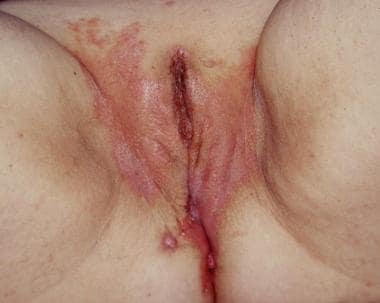
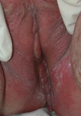
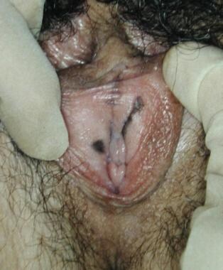
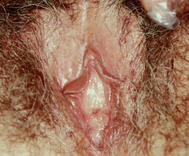
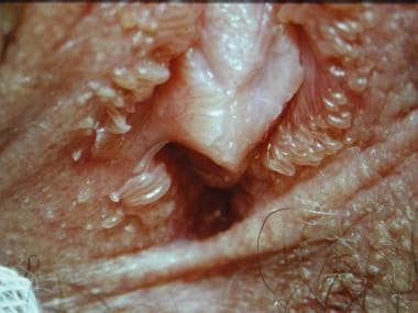
Lichen sclerosus is commonly characterized by whitish lesions of the vulva. It is asymptomatic, but intractable pruritus can sometimes be present. Burning and pain are less likely manifestations. Clinically, the lesions are characterized by a wrinkled ("cigarette-paper") or parchmentlike (shiny, delicate, pale) appearance of the skin that commonly extends around the anal area in a figure-8 or keyhole configuration. [12, 13]
In late stages of the disease, normal architecture may be lost. [8] Additionally, atrophy and fusion of the labia minora, constriction of the vaginal orifice (kraurosis), synechiae, ecchymoses, fissures, and telangiectases may be noted. Squamous cell carcinoma develops in 3-6% of women affected by vulvar lichen sclerosus, which is therefore now regarded as a preneoplastic condition. [11, 76, 77] The presence and the duration of symptoms and the loss of vulvar architecture are not useful indicators of potential cancer risk.
Squamous cell hyperplasia
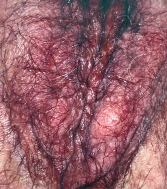
Squamous cell hyperplasia appears as ill-defined, single or scattered, asymmetrical, whitish, thickened, and sometimes verrucous plaques that may be accompanied by excoriations or fissurations that cause pain and soreness.
Itching is a common symptom. If hyperkeratosis is not prominent, lesions may appear as reddish plaques. The clitoris, labia minora, and inner aspects of the labia majora are more commonly affected.
Extensive lesions may result in stenosis of the vaginal introitus.
Lichen simplex chronicus (localized neurodermatitis)
Patients with lichen simplex chronicus present with a hyperkeratotic, usually ill-defined, grayish, thickened, and sometimes excoriated lesion, usually located over the labia majora and merging with normal skin.
Hyperpigmentation is common, and prominent skin markings are evident when the skin is involved. Itching is always present and may be intense.
Primary irritant dermatitis
Typical physical findings of primary irritant dermatitis include diffuse reddening of the involved skin with areas of excoriation. Secondary infection may occur.
An association with regional intertrigo, especially following mechanical irritation, may be observed.
Chronic irritant dermatitis may lead to squamous cell hyperplasia.
Intertrigo
Typically, intertrigo is characterized by erythema, local edema, oozing, maceration, and fissuring of the inguinal fold, sometimes accompanied by considerable odor. It may be associated with similar findings in other skin folds.
The surrounding skin may show reactive postinflammatory hyperpigmentation. Soreness and itching are common symptoms. Secondary candidosis may exacerbate intertrigo.
Allergic contact dermatitis
Physical examination often reveals dryness, scaling, excoriations, and, at times, ulceration. Itching is usually intense.
The clinical pattern may be subacute, with weeping and oozing, especially when bacterial superinfection occurs. Without treatment, allergic dermatitis can progress to squamous cell hyperplasia.
Fixed drug eruption
Fixed drug eruption appears as an erythematous and edematous plaque that frequently involves the genital area and typically resolves, leaving postinflammatory hyperpigmentation. [78]
The lesions are usually single at first presentation, but following reingestion of the drug, development of new elements may occur in addition to recurrence of the primary lesion.
The main complaint is burning, but some patients are asymptomatic or have mild pruritus.
Atopic dermatitis
Atopic dermatitis consists of a subacute or chronic, symmetric, and ill-defined eczematous rash, usually involving the labia majora and, less frequently, the labia minora and inner thighs.
The eruption is characterized by mild erythema, dryness, and fine scaling. Itching and burning are common symptoms.
Excoriation secondary to repeated scratching may cause bacterial superinfection with honey-colored crusting and, in chronic forms, lichenification. In some patients, the itch-scratch-itch cycle may gradually lead to development of lichen simplex chronicus.
Seborrheic dermatitis
When seborrheic dermatitis involves the vulva, the labia majora and mons pubis are primarily affected.
The lesions appear as dry-to-greasy scales superimposed on red-to-yellow brownish plaques and are often pruritic, extending to the gluteal cleft and thighs.
Psoriasis
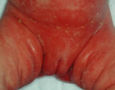
Occasionally, psoriasis may manifest in infancy as a bright-red, glazed, and well-demarcated eruption in the napkin area (napkin psoriasis).
In adults, vulvar psoriasis usually involves the genitocrural areas and the lateral aspects of the labia majora, sparing the mucosa. It may range in severity from scattered, nonscaling erythematous patches to thick, confluent erythematous plaques with silvery-white adherent scales covering all of the labia majora and the mons pubis.
Symptoms are highly variable and range from intense pruritus to minimal discomfort. Secondary changes, such as excoriations and lichenification or oozing and crusting from bacterial and yeast colonization, may occur.
Reiter disease
In approximately half of patients affected with Reiter disease, cutaneous lesions occur as psoriasiform, crusting, and sometimes pustular papules and plaques over the hands and feet. Circinate genital erosions or oral mucosa involvement are often associated. Conjunctivitis, arthritis, and low back pain may also occur.
Reiter disease is unusual in women; therefore, vulvar lesions are poorly described. [79] Red crusted plaques associated with vaginal discharge, circinate erosions, or linear ulcers associated with verrucous lesions and pustules have been observed. [80]
Lichen planus
In the vulvar area, the disease may occur in 3 patterns: papulosquamous, erosive, and hypertrophic. [81] Note the following:
-
The papulosquamous form, occurring as part of a generalized disease, is the most common and is characterized by flat-topped, polyhedral, violaceous, shiny, and itchy papules located on keratinized skin of the labia and mons pubis. Delicate and whitish reticulated papules may be present on the mucosa, but no atrophy or scarring is observed.
-
The erosive form involves the mucous membranes of the mouth and vulvovaginal area and may be locally destructive, leading to atrophy and scarring. [82] (Synonyms include erosive vaginal lichen planus, desquamative inflammatory vaginitis, vulvovaginal-gingival syndrome and ulcerative lichen planus. [8] ) Itching is rare, but pain, burning, and irritation occur and may be responsible for dyspareunia and dysuria. [79]
-
The rare hypertrophic form, clinically resembling lichen sclerosus, manifests with extensive white scarring of the periclitoral area with variable degrees of hyperkeratosis. [26] It may be very itchy. Extensive vaginal involvement may result in a malodorous discharge. Large denuded areas may become adherent, causing stenosis of the vaginal introitus and dyspareunia. Marked atrophy may develop with time.
Malignancy is possible in long-standing and ulcerative lichen planus.
Lupus erythematosus
Vulvar manifestations may be different in each clinical subset of the disease. [83] In chronic discoid lupus erythematosus, the skin is mostly involved (rarely, the mucous membranes). Vulvar involvement may occur as a scarred plaque of variable size with or without central ulceration and with marked peripheral hyperpigmentation located anywhere on the vulva or perineum.
In subacute and systemic lupus erythematosus, the patient is usually asymptomatic and a lichen planus–type reticulated pattern may sometimes be observed. Occasionally, tender, punched-out mucosal ulcers with possible scarring occur in the vestibule or vagina.
Darier disease
The onset of Darier disease is usually in late childhood, with keratotic, crusted, skin-colored, yellow, or brown papules located mainly on the neck, upper thorax, and flexural areas. These papules grow and multiply and tend to become more widespread and verrucous with age.
The vulva is often involved. [1] As the condition becomes more pronounced, hygiene becomes progressively difficult to maintain, and the buildup of keratotic debris causes secondary infection and a foul smell.
Aphthosis
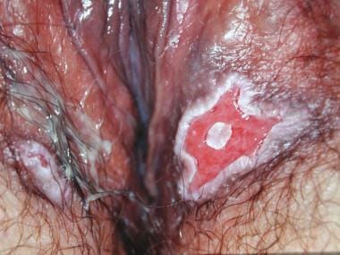
Patients with aphthosis report single or multiple, sharply demarcated, punched-out, and shallow vulvar ulcers with fibrinous bases and erythematous borders. The lesions are very painful and are sometimes accompanied by systemic symptoms, such as fever and malaise, but they are usually self-limited, clearing spontaneously in a few weeks.
In some cases, especially in older women, relapsing episodes akin to partial Behçet disease may occur. In time, older patients with recurrent ulcers may develop Behçet disease or inflammatory bowel disease.
Behçet disease
Behçet disease is a rare multisystem disorder characterized by the triad of oral ulcers, genital ulcers, and posterior uveitis; however, almost any organ system may be involved. [30]
Oral ulcers are usually multiple and recurrent and may sometimes be extensive, showing a pseudomembranous coating.
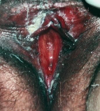
Vulvar ulcers, ranging in diameter from a few millimeters to 3 cm, often appear as multiple crops of well-defined and very tender ulcers with fibrinous bases and considerable undermining. Fistulae, with partial or complete destruction of the labia, may develop.
Other manifestations of the disease include fever, malaise, acneiform lesions or cutaneous nodules on the skin, arthritis, synovitis, and thrombophlebitis. Associated erythema nodosum and erythema multiforme have been reported.
Pyoderma gangrenosum
Pyoderma gangrenosum often appears as a deep, painful nodule or pustule that breaks down, draining a purulent discharge and forming an irregular ulcer with distinct undermined and purplish edges. The lesion extends peripherally as the inflammatory process spreads within the dermis. [32]
Satellite pustules may be observed, eventually coalescing and forming a multicentric ulceration.
The pain is intense, and the course of the disease is unpredictable. The lesion may heal spontaneously, remain quiescent for months (even years), or worsen again after minimal trauma, surgery, or an inapparent triggering cause.
Crohn disease
Vulvar involvement in Crohn disease is uncommon. [84, 85]
Cutaneous changes may occur before the onset of bowel symptoms. The area of involvement may extend to the perineal and perianal area. Localized or generalized labial edema, with erosions and multiple painful ulcers of variable severity, may be observed. Ulcers may be solitary, deep, and necrotic, possibly leading to formation of fistulae. [86] Perianal and rectovaginal fistulae are common complications. [87] Malignant degeneration has occurred in some such cases. [88]
Unilateral labial hypertrophy, coalescing pustules, and vegetating lesions mimicking anogenital warts (pyostomatitis vegetans) have also been described. [89]
Hidradenitis suppurativa
In the vulvar area, hidradenitis suppurativa primarily affects the labia majora and intercrural folds, with erythematous acneiform papules, nodules, and cysts scattered among multiheaded comedones, but it may also involve the mons pubis, labia minora, and clitoris.
In some cases, deep, painful subcutaneous nodules may ulcerate and ooze a purulent yellow discharge, leading to open sinuses and extensive scarring; in other cases, nodules may coalesce, thus forming conglobate plaques interconnected by sinus tracts. Over time, pseudoepitheliomatous hyperplasia may develop. Occasionally, vulval squamous cell carcinoma has been observed arising in chronic hidradenitis suppurativa. [90]
Fox-Fordyce disease
Fox-Fordyce disease is an itchy papular eruption of the axillary and anogenital regions that appears on the vulva as multiple, monomorphous, skin-colored or slightly hyperpigmented, dome-shaped follicular lesions, mainly involving the mons pubis and labia majora.
Plasma cell vulvitis
Plasma cell vulvitis appears as an erythematous, well-demarcated, smooth, and shiny plaque that may be either asymptomatic or cause mild itching, burning, or soreness. [91, 92]
Vulvar vestibulitis
Vulvar vestibulitis is characterized by severe burning and pain with vestibular touch or attempted vaginal entry. Typically, it follows a chronic course and may show variable numbers of minute spots of vestibular erythema, ranging in diameter from 2-7 mm. Rarely, small ulcerations are detectable. [8, 93]
The pain is enough to make intercourse uncomfortable or completely impossible. [94] Associated deep pain from secondary vaginismus may occur. Understandably, varying degrees of sexual dysfunction may cause depression and anxiety. [95, 96]
Frequently, orofacial pain is associated. [97]
Blistering diseases
Familial benign chronic pemphigus (Hailey-Hailey disease)
Hailey-Hailey disease is characterized by recurrent eruptions of vesicles and blisters that easily erode and develop crusting. Painful deep fissures may also occur. The lesions typically involve the inguinal fold and may extend along the edge of the labia majora and on the inner aspect of the thighs. Bacterial and fungal superinfections often occur.

Bullous pemphigoid
The course of bullous pemphigoid is chronic and benign. Often, a prodromal phase of fixed urticarial plaques occurs with itching and irritation, which can be generalized.
On the vulva, blisters arising on the labia easily erode, leaving erosions that may cause variable degrees of discomfort.
Cicatricial pemphigoid
In cicatricial pemphigoid, blisters typically develop on mucosal sites, gradually leading to the development of disabling scarring adhesions.
Common symptoms include vulvar itching, soreness, and pain, along with the presence of mucosal erosions and vaginal discharge. [8] Vulvar involvement may cause synechiae of the labia and/or vagina, with consequent dyspareunia.
Pemphigus vulgaris
The erosions of pemphigus vulgaris, either arising on the mucosa of the inner labia and vestibule or on vulvar skin cause considerable burning and pain. Long-term disease may result in vulvar scarring, vaginal scarring, or both.
Erythema multiforme (minor/major)
In erythema multiforme, vesicular and bullous lesions with a typical iris pattern may show variable symmetrical extension according to the severity of the disease.
In the major form, blistering of the mucous membranes is extensive and may cause the formation of synechiae in later stages of the disease.
Epidermolysis bullosa
Genital involvement has been reported in dystrophic forms of epidermolysis bullosa and may cause painful blistering in the vulvar area with consequent scarring, vaginal obstruction, and obstructive uropathy. [98]
Epidermolytic hyperkeratosis
It is often evident as multiple pruritic and persistent well-defined sessile grayish papules, with confluence into plaques and sometimes exophytic hyperkeratotic nodules. Epidermolytic hyperkeratosis may resemble other benign inflammatory and malignant vulvar entities clinically and histologically. [54]
Pigmentary changes
Acanthosis nigricans
In acanthosis nigricans, the skin of the inguinal and axillary regions appears diffusely hyperpigmented with a velvety or warty surface.
Patients with this condition are usually asymptomatic, but some report local irritation and pruritus.
The malignant form is associated with a malignant tumor, most commonly a gastric carcinoma, a lymphoma, or a sarcoma.
Lentigo, lentiginosis, and benign vulvar melanosis
Lentigo appears as a small (< 4 mm), hyperpigmented, brownish macule that may be found anywhere on the vulvar skin or mucosa. [99]
Lentiginosis is characterized by a circumscribed grouping of pigmented small macules with normal background pigmentation. [99]
In vulvar melanosis, larger brown-to-black macules (≤10 cm in diameter), often showing irregular margins, are observed. [99]
Melanocytic nevus
Melanocytic nevi appear as small, circumscribed, variably pigmented macules or raised papules that may be congenital or acquired.
Some authors have suggested that nevi occurring on the vulva are more likely to undergo malignant transformation; therefore, careful examination is recommended. [99]
Postinflammatory hyperpigmentation
Varying degrees of macular or patchy hyperpigmentation may occur with postinflammatory hyperpigmentation.
The color can range from brown to black, although it is usually irregular and can also show scattered patches of hypopigmentation.
Postinflammatory hypopigmentation
Postinflammatory hypopigmentation is characterized by diffuse depigmentation or patches of hypomelanosis that may be single or multiple and variably involve vulvar skin.
Vitiligo
Patients with vitiligo may develop asymptomatic progressively enlarging white patches on the vulvar skin and mucosae that, in fair-skinned individuals, may be barely appreciable upon clinical observation using a natural light source. Vitiligo may be confused with lichen sclerosus, but the skin in this condition is not atrophic.
Benign tumors, hamartomas, and cysts
Mucous cysts
Mucous cysts usually cause no symptoms and appear as a lump or mass that may be found at the introitus and labia minora.
Cysts of the canal of Nuck can give rise to a hydrocele located high in the labia majora and are associated with a concurrent inguinal hernia in 30% of cases.
Bartholin cyst and Skene duct cyst
Bartholin cysts are the most common vulvar cystic growths. They usually occur in the lower and lateral portion of the labia majora, although lesions expanding anteriorly have also been described, and, if large, they may cause variable discomfort, hampering sexual intercourse and micturition. [57]
Skene duct cysts arise adjacent to the urethral meatus and, if large enough, may cause urinary obstruction.
In both conditions, acute infection with abscess formation may occur, thus causing considerable pain.
Epidermal inclusion cyst
Epidermal inclusion cysts are most commonly observed in the vagina, but they can also be found on the vulva. Such cysts are subcutaneous and generally asymptomatic unless they become infected. Spontaneous rupture often occurs.
 Benign vulvar lesions. Epidermal inclusion cyst located in the middle portion of the labium majus.
Seborrheic keratosis
Seborrheic keratoses appear as single or multiple verrucous, roundish, yellowish-brown, sharply circumscribed papules ranging in diameter from 2-10 mm and covered with a greasy friable scale. [100, 101] The lesions often have a "stuck-on" appearance.
Acrochordon
Acrochordon lesions, often multiple and appearing as soft, pedunculated, brown, tan, or skin-colored lesions (0.2-1.5 cm in diameter), can particularly be found in the inguinal folds of obese and/or diabetic patients.
Fibroma, fibromyoma, dermatofibroma, and trichoblastoma
Fibromas, fibromyomas, and dermatofibromas usually appear as solitary, slightly raised, gray-brown, mobile indurated lesions (3-8 mm in diameter) developing along the insertion of the round ligament into the labia majora.
Fibromas may be pedunculated and may rarely reach a considerable size.
In dermatofibromas, lateral compression produces a slight indentation known as the dimple sign, which is characteristic of these tumors. These lesions usually cause no symptoms until they reach a larger size and/or are located near the introitus or urethra.
A trichoblastoma may occur too, a benign vulvar neoplasm that may have potential for malignant transformation and, in addition, needs to be distinguished from a basal cell carcinoma, which it resembles histologically. [102]
Lipoma
On the labia majora, lipomas may appear as soft sessile or pedunculated masses varying in diameter from 1 cm to several centimeters.
Large lesions may gradually ulcerate.
Hidradenoma
Hidradenomas usually occur in postpuberty as single mobile nodules (~1-1.5 cm in diameter) arising in the interlabial sulcus.
Ulceration may occur, and in these cases, the lesions may show an exophytic proliferation clinically resembling a malignant neoplasm.
Syringoma
Vulvar syringomas manifest as small, multiple, bilateral, skin-colored to yellowish or brownish pruritic papules over the labia majora. Typical syringomas on the eyelids may coexist in one third of cases. [62]
Hemangioma
Most genital hemangiomas involve the labia majora, but the labia minora, the perineal area, and the perianal area may also be involved to varying degrees. They appear as red macules that rapidly progress to well-circumscribed, raised, red, and soft lesions of variable size.
Over time, regression occurs, with involution and fibrosis. Possible complications include ulceration, bleeding, urethral obstruction, and Kasabach-Merritt syndrome (a consumptive coagulopathy mainly described in association with large hemangiomas).
Lymphangioma
This condition is usually detected early in infancy on the labia minora or majora as an asymptomatic, raised, compressible, doughy mass, sometimes showing multiple clustered, superficial, thin-walled, translucent, and persistent pseudovesicles filled with clear fluid that may progressively grow over time. [67]
Angiokeratoma
Angiokeratomas manifest as 1-3 mm, dark, red-to-purple, and sometimes hyperkeratotic papules. Patients are usually asymptomatic.
Occasionally, patients become symptomatic, with vulvar itch, discomfort, or pain. The lesions may bleed as a result of trauma. When they appear in teenagers and are associated with angiokeratomas of the lower abdomen, Fabry disease should be excluded.
Pyogenic granuloma
Pyogenic granuloma appears as a bright red papule or nodule of no more than 1-2 cm in diameter; erosion and bleeding may occur.
It may persist indefinitely unless destroyed.
Endometriosis
Often painful, vulvar endometriosis manifests as an ill-defined, dark red, brown, or blue-black cystic papule or nodule, usually located on the posterior fourchette. A case of endometriosis infiltrating the Bartholin gland has been observed.
It sometimes shows a cyclical variation in size and symptoms according to menses. More widespread involvement is a significant cause of pain and distress.
Heterotopic sebaceous glands and sebaceous gland hyperplasia
Heterotopic sebaceous glands often arise on the labia minora and inner aspects of the labia majora as multiple superficial yellow papules (1-3 mm in diameter) that can be clearly seen when the mucosa is stretched.
The clinical features of sebaceous gland hyperplasia differ from those of typical lesions on the face and have been described as polypoid tumors no greater than 2.5 cm in diameter on the labia majora, covered by normal-appearing skin. These lesions may regress, and they have no malignant potential. [74]
Papillomatosis (papillary vulvar hirsutism)
Papillomatosis lesions are found distal to the hymenal ring. They consist of raised, fleshy, skin-colored, soft, asymptomatic micropapillae of the inner labia minora, usually 1-3 mm in diameter and symmetric, that occur singly or become confluent, forming a fimbriated fringe.
Congenital malformations
Ambiguous external genitalia
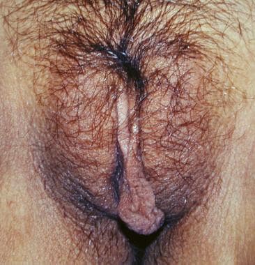
Infants with female pseudohermaphroditism usually present with an enlarged phallus, alone or associated with some degree of labioscrotal fusion.
Reduced levels of cortisol and consequent sodium depletion, which can be life-threatening in neonates, may be associated in forms resulting from 21-hydroxylase deficiency.
Underdevelopment of male genitalia can yield a female phenotype in the most extreme cases of male pseudohermaphroditism.
Congenital labial hypertrophy
Patients with congenital labial hypertrophy are asymptomatic except for nuisances regarding hygiene, physical activity, or sexual intercourse. The large labia may be unilateral or bilateral. If hygienic care is not maintained, erythema and irritation may result.
Labial adhesions
The extent of fusion varies and often causes no symptoms. The natural history is one of spontaneous resolution with pubertal estrogenization.
Atrophy of the vulva
The mucosal surface is dry, and mild atrophy of the labia minora, clitoris, and inner aspects of the labia majora occurs.
The clinical appearance may resemble that of the later stages of lichen sclerosus. These patients have relatively few symptoms, and hyperkeratosis is not evident.
Histologic Findings
Nonneoplastic epithelial disorders are discussed below.
Inflammatory diseases
Histologic findings in lichen sclerosus include hyperkeratosis, epithelial thinning with flattening of the rete pegs, cytoplasmic vacuolation of basal keratinocytes, follicular plugging, homogenization of the subepithelial layer, and inflammatory cell infiltration consisting of lymphocytes with few plasma cells. [103]
Squamous cell hyperplasia
Histologic examination reveals thickening of the keratin layer (hyperkeratosis) greater than that seen with lichen sclerosus, and epithelial hyperplasia with elongation, widening, and distortion (acanthosis) of the rete pegs. Retention of nuclei in the keratin layer (parakeratosis) is a common finding.
Cellular elements of the epithelium proliferate, but maturation is usually normal. An inflammatory response in the dermis usually occurs, consisting of lymphocytic and plasma cell infiltration. Varying degrees of cellular atypia with increased mitotic activity and loss of polarity may be observed in the epidermis.
This vulval squamous epithelial hyperplasia with atypia corresponds to the entity formerly indicated as leukoplakia, which has a malignant potential. It appears to be related to conditions that approximate vulvar intraepithelial neoplasms (VINs) and has been found to progress to invasive carcinoma in 10% of cases.
Lichen simplex chronicus (localized neurodermatitis)
Histologically, epidermal and epithelial hyperplasia, hyperkeratosis, and fibrotic vertical streaks of collagen between the hyperplastic rete are present. A superficial perivenular infiltrate is also present.
Primary irritant dermatitis
Histologic features are highly variable, from extensive ulceration to diffuse parakeratosis with vascular congestion and ectasia to a spongiotic pattern essentially identical to allergic contact dermatitis. In some instances, a significant individual keratinocyte necrosis with nuclear karyorrhexis and cytoplasmic pallor occurs.
Intertrigo
Histopathologic findings are nonspecific.
Allergic contact dermatitis
In the epidermis, variable degrees of intercellular edema and spongiosis are present, which may eventually lead to the development of an intraepidermal vesicle. Lymphocytic infiltration of the epidermis is always present.
Concomitant with these changes are varying degrees of epithelial proliferation ranging from mild acanthosis in early acute dermatitis to a psoriasiform epidermal hyperplasia in chronic variants. The dermis is often congested, and edema is usually marked in active lesions.
Fixed drug eruption
Histologic changes resemble those of erythema multiforme. Necrosis of keratinocytes in the stratum malpighii occurs.
Scattered dyskeratotic keratinocytes with eosinophilic cytoplasm and pyknotic nuclei are frequently seen in the epidermis and represent apoptosis.
Atopic dermatitis
Mild spongiosis, exocytosis of lymphocytes, and parakeratosis are present in the epidermis. Hyperkeratosis and wedge-shaped hypergranulosis may also be observed. [104]
A perivascular lymphocytic infiltrate with scattered histiocytes is present in the superficial dermis. [23]
Seborrheic dermatitis
The histopathologic features are a combination of those observed in psoriasis and spongiotic dermatitis. Moderate acanthosis with focal areas of parakeratosis, regular elongation of the rete ridges, mild spongiosis, and focal exocytosis of lymphocytes are noted. The dermis contains a sparse mononuclear cell infiltrate.
Psoriasis
The histologic picture varies considerably with the stage of the lesion and is usually diagnostic only in early scaling papules and near the margin of advancing plaques, ie, acanthosis with regular elongation of the rete ridges, thinning of suprapapillary epidermis with occasional small spongiform pustules, diminished or absent granular layer, confluent parakeratosis, elongation and edema of the dermal papillae, and dilated and tortuous capillaries.
Reiter disease
Early pustular lesions show a spongiform macropustule in the upper epidermis that is indistinguishable from the spongiform pattern observed in pustular psoriasis. Parakeratosis and elongation of the rete ridges are also noted.
Lichen planus
Vulvar lichen planus shows a dense dermal inflammatory infiltrate extending to the dermoepidermal junction in conjunction with a prominent granular cell layer, hyperkeratosis, and acanthosis.
Lupus erythematosus
Histologic findings alone may not be sufficient to allow correct classification of the subtype of eruption.
In well-developed lesions, hydropic degeneration of the basal cell layer occurs in association with edema of the upper dermis and extravasation of erythrocytes.
Fibrinoid deposits in the connective tissue of the skin are not specific but are often observed in erythematous, edematous lesions, especially in patients with systemic lupus erythematosus.
Subcutaneous fat is often involved in systemic disease.
Darier disease
The typical histologic hallmarks of Darier disease are hyperkeratosis, with a peculiar form of dyskeratosis resulting in the formation of corps ronds and grains and suprabasal acantholysis, leading to the formation of suprabasal clefts or lacunae with irregular upward proliferation of papillae lined with a single layer of basal cells.
A chronic inflammatory infiltrate is present in the dermis.
Aphthosis
Histologic features include necrotizing vasculitis of the superficial postcapillary venules with associated fibrinoid necrosis, endothelial swelling, and lymphocytic perivascular infiltrate with sometimes abundant neutrophils.
Behçet disease
Vulvar lesions show features similar to those of minor aphthosis.
Pyoderma gangrenosum
Although suggestive, histopathologic features alone are not diagnostic. Features include dermal edema; dense, diffuse neutrophilic infiltrate; engorgement and thrombosis of small-to-medium–sized vessels; necrosis; and hemorrhage.
Crohn disease
Discrete noncaseating granulomas with isolated multinucleated giant cells are present throughout the superficial and deep dermis, with extension into subcutaneous tissue.
Occasionally, the granulomatous infiltrate is perivascular and may create secondary vascular changes.
Hidradenitis suppurativa
Histology shows chronic dermal inflammation with fibrosis and foreign body giant cells and the presence of neutrophils and bacteria in apocrine gland ducts.
Fox-Fordyce disease
Histologic examination shows enlarged apocrine sweat glands surrounded by a dermal inflammatory infiltrate.
Plasma cell vulvitis
Below an atrophic epidermis showing no signs of keratinocyte atypia, a dense lichenoid infiltrate with a large number of plasma cells and occasional dilated blood vessels and hemosiderin deposition is evident in the upper and mid dermis.
Vulvar vestibulitis
Histologic examination reveals a nonspecific, chronic lymphocytic inflammatory infiltrate in the dermis, with foci of squamous metaplasia of the minor vestibular glands that may show nodular hyperplasia in some cases. [105]
Some patients are found to have koilocytosis after biopsy and/or human papillomavirus infection after in situ hybridization testing.
Blistering diseases
Familial benign chronic pemphigus (Hailey-Hailey disease)
Epidermal parakeratosis and dyskeratotic suprabasal acantholysis, with the typical appearance of a dilapidated brick wall, are common findings.
Bullous pemphigoid
A typical histopathologic finding on direct immunofluorescence is the presence of a subepidermal blister with deposition of immunoglobulins along the basement membrane.
Cicatricial pemphigoid
In addition to subepidermal splitting, lamellar fibrosis beneath the epidermis is a hallmark of this condition, but it may not be present in the initial lesions. Neutrophils and lymphocytes predominate in the inflammatory infiltrate.
Pemphigus vulgaris
Rarely, the earliest recognized change may be eosinophilic spongiosis; more commonly, the earliest noted change is spongiosis in the lower epidermis.
Acantholysis first leads to the formation of clefts and then to blisters in a predominantly suprabasal location.
Erythema multiforme (minor/major)
Erythema multiforme is considered the prototype of the vacuolar form of interface dermatitis. Because of its acute nature, an orthokeratotic stratum corneum is formed.
Mild spongiosis and exocytosis are observed. Necrosis of keratinocytes in the stratum malpighii is typical.
Epidermolysis bullosa
Dermoepidermal fissuring is a common feature.
Different histopathologic, ultrastructural, and laboratory findings may be observed according to the clinical subset of disease.
Epidermolytic hyperkeratosis
Vulvar epidermolytic hyperkeratosis tends to have a histologic pattern of distinctive hyperkeratosis, papillomatosis, acanthosis, and hypergranulosis, with a variable epidermal reticular degeneration. This last finding along with prominent cytolysis may easily be confused with the viral cytopathic effect and/or the cytological atypia of HPV infections and squamous cell carcinomas, respectively. [54]
Pigmentary changes
Acanthosis nigricans
Histologic examination reveals hyperkeratosis and papillomatosis but only slight, irregular acanthosis and, usually, no hyperpigmentation.
Lentigo, lentiginosis, and benign vulvar melanosis
A slight or moderate elongation of the rete ridges with an increase in the concentration of melanocytes in the basal layer is observed.
Melanocytic nevus
Typical findings are nests of round melanocytic cells without dendrites and no sign of atypia. Histopathology allows identification of all the evolutional steps of these lesions, which typically begin as junctional nevi and, after having become intradermal nevi, undergo involution.
Postinflammatory hyperpigmentation
Epidermal melanin is increased. Melanophages are present in the superficial dermis, along with a variably dense lymphohistiocytic infiltrate around superficial blood vessels and in dermal papillae.
Postinflammatory hypopigmentation
Epidermal melanin is decreased. A superficial and perivascular lymphohistiocytic infiltrate may be observed in the dermis.
Vitiligo
The central process is the destruction of melanocytes at the dermoepidermal junction.
Benign tumors, hamartomas, and cysts
Mucous cysts
Histologically, mucous cysts show a fibrous wall lined by epithelial cubical cells and filled with mucin or, in the case of canal of Nuck cysts, a clear fluid.
Bartholin cysts and Skene duct cysts
These cysts show a fibrous wall lined by a flattened epithelium. [57, 58]
Epidermal inclusion cysts
Epidermal cysts have a wall composed of true epidermis and are filled with horny material arranged in laminated layers.
Seborrheic keratosis
This is an exophytic and papillomatous proliferation of basaloid epidermal cells containing horn cysts and often showing marked basal and suprabasal intracellular melanin pigmentation.
Acrochordon
Loose connective tissue rich in vessels covered by normal epidermis is observed.
Fibroma, fibromyoma, and dermatofibroma
Fibroma, fibromyoma, and dermatofibroma show a well-demarcated area of interwoven collagen fiber bundles without elastic fibers covered by normal or hyperplastic epidermis.
In fibromyoma, muscle fiber bundles are evident. A dermatofibroma is a variant of the fibromyoma group that, because of its vascularity, can present a confusing histologic picture.
Lipoma
By definition, the principal component of lipomas is mature adipocytes. [106]
Hidradenoma
Histopathologic features may be confusing; for example, a complex papillary-adenomatous pattern arranged in an aggressive fashion with absent mitoses may be observed. [107]
Syringoma
Histology shows sweatlike glandular structures and solid epithelial nests embedded within a sclerosing stroma; miliumlike epithelial cysts may coexist. [62]
Hemangioma
Early lesions are very cellular with few vascular channels; mitotic figures and mast cells may be prominent. Later, vessel lumina become apparent, producing a cavernous pattern.
Lymphangioma
Cavernous dilated lymphatic channels of different sizes are evident in the dermis or subcutaneous fat and sometimes extend into the overlying epidermis.
Angiokeratoma
Histopathologic features consist of hyperkeratosis and epidermal acanthosis overlying a dermis that contains dilated capillary vessels in proximity to the epidermis. [106]
Pyogenic granuloma
This is a lobular vascular proliferation with distinctive plump epithelioid endothelial cells admixed with a varying lymphocytic and eosinophilic infiltrate that tends to obscure the vessels.
Endometriosis
Typical glandular and stromal tissue of the endometrium lying within the dermis are observed.
Heterotopic sebaceous glands and sebaceous gland hyperplasia
Histologic examination shows sebaceous gland hyperplasia. [74]
Papillomatosis (papillary vulvar hirsutism)
Histologically, these lesions are angiofibromas.
Congenital malformations
Ambiguous external genitalia, congenital labial hypertrophy, and labial adhesions: No definitive histologic changes occur.
Atrophy of the vulva
No definitive histologic changes occur.
Differential Diagnosis and Workup
Workup and procedures
If the diagnosis is not readily apparent, unaided (ie, naked-eye) or colposcopic examination of the vulva may define areas of abnormality that may warrant biopsy using a Keyes punch or biopsy forceps under local anesthesia.
The mainstay of diagnosis is vulvar biopsy. Furthermore, all patients with a nonneoplastic vulvar epithelial disorder should be checked at regular intervals. Areas of ulceration or foci of granulation or nodularity that develop should be biopsied to exclude malignant change. The formation of hyperkeratotic plaques or erosions that do not respond to treatment should arouse suspicions of malignancy. Multiple biopsies may be necessary.
Biopsy is indicated when the diagnosis is in doubt or if management strategies would be influenced by more information. An outpatient procedure with local anesthesia is almost always feasible. The request form should indicate the area from which the biopsy will be taken. Excisional biopsy is feasible for small lesions, but larger areas require sampling by punch biopsy.
Preliminary application of lidocaine and prilocaine (EMLA Cream) that is left on for about 10 minutes is helpful. Lignocaine 1% is infiltrated in the areas to be biopsied. Disposable 2- to 6-mm punches are used (eg, Keyes punch biopsy instruments). The 6-mm punch is used for larger lesions. A rotary motion of the instrument removes a core of tissue, which is removed by snipping off at the base with scissors. Hemostasis is usually satisfactorily achieved with pressure, chemicals such as silver nitrate or Monsel solution, or electrocautery. With larger biopsies, the use of absorbable sutures, such as 4-0 Vicryl, achieves hemostasis. As a rule, late bleeding is rare and healing is rapid.
Nonneoplastic epithelial disorders are discussed below.
Inflammatory diseases
Lichen sclerosus
A skin biopsy is necessary to confirm the diagnosis and to exclude the presence of malignant degeneration.
The differential diagnosis includes lichen planus, vitiligo, postmenopausal atrophy, cicatricial pemphigoid, extramammary Paget disease, and sexual abuse. [12, 8, 108]
Squamous cell hyperplasia
The diagnosis is one of exclusion after psoriasis, lichen sclerosus, lichen planus, and chronic eczematous dermatitis have been ruled out. In doubtful cases, a biopsy is suggested. This also helps identify cases of squamous cell hyperplasia with atypia that may have a propensity to develop carcinoma.
Lichen simplex chronicus (localized neurodermatitis)
Primary irritant dermatitis, chronic eczematous dermatitis, squamous cell hyperplasia with or without atypia, and lichen planus should be excluded. [18]
Primary irritant dermatitis
Skin swabs and patch testing are useful to exclude superimposed bacterial or fungal infections and allergic contact dermatitis, respectively.
The differential diagnosis includes candidal vulvitis and allergic contact dermatitis.
Intertrigo
The clinical presentation is often diagnostic. The differential diagnosis includes candidosis and other conditions that may be found in intertriginous areas, such as familial benign chronic pemphigus (Hailey-Hailey disease), psoriasis, and seborrheic dermatitis.
Bacterial and mycologic investigations may be useful to detect secondary infections.
Skin biopsy is indicated when treatment fails or when an underlying disorder (eg, Hailey-Hailey disease) is considered possible.
Allergic contact dermatitis
The diagnosis is usually made on the basis of history findings, although less obvious cases may require patch tests. Because vulvar epithelium is more permeable than exposed skin, standard clinical patch tests may not sufficiently mimic vulvar exposures.
The differential diagnosis includes atopic dermatitis psoriasis, intertrigo, and tinea.
Fixed drug eruption
The clinical history and typical morphologic features usually confirm the diagnosis.
The differential diagnosis includes recurrent herpes simplex virus (HSV) infection, lichen planus, intertrigo, [109] and bullous pemphigoid.
Atopic dermatitis
The diagnosis is made based on personal and family history and on clinical detection of typical lesions elsewhere in the body. Histology is seldom necessary.
The differential diagnosis includes psoriasis, seborrheic dermatitis, contact dermatitis, and eczematous candidiasis.
Seborrheic dermatitis
The presence of characteristic lesions elsewhere on the body may indicate the diagnosis.
The differential diagnosis includes tinea, psoriasis, and other scaling disorders, and it may sometimes be clinically difficult to confirm.
Psoriasis
Biopsy is confirmatory but is seldom needed because the diagnosis is often clinical.
The differential diagnosis includes seborrheic dermatitis, candidal or dermatophyte infection, lichen simplex chronicus, and contact dermatitis.
Reiter disease
The diagnostic dilemma is differentiating Reiter syndrome from pustular psoriasis. [110] Acrodermatitis enteropathica and lymphogranuloma venereum should also be considered in the differential diagnosis.
Discrimination points include cervicitis, which is common in Reiter syndrome but not reported in psoriasis; the greater prevalence of HLA-B27 positivity; iritis; conjunctivitis; and other mucous membrane lesions in Reiter syndrome.
Biopsy, radiographs, and laboratory investigations are usually necessary to confirm the diagnosis and to assess the extent and severity of the disease. [110]
Lichen planus
Although demonstration of typical oral changes carries more diagnostic weight than biopsy, histopathology is often necessary to confirm the diagnosis. [81, 111]
The differential diagnosis includes psoriasis, dermatophyte infection, lichen simplex chronicus, lichen sclerosus, cicatricial pemphigoid, pemphigus, lupus erythematous, and bullous pemphigoid. [8]
Lupus erythematosus
To confirm the clinical diagnosis, histopathologic, immunohistopathologic, and serologic investigations are required. The latter include indirect immunofluorescence for antinuclear antibodies.
The differential diagnosis includes all causes of genital ulcers and lichen planus in its clinical variants.
Darier disease
The clinical diagnosis necessitates histopathologic confirmation.
The differential diagnosis of lesions located in the genital and perineal area includes acanthosis nigricans, benign familial pemphigus, and impetigo.
Aphthosis
The diagnosis is essentially clinical and one of exclusion, following appropriate cultures, serologic testing, and biopsies to rule out other conditions. Histologic features alone are not diagnostic.
The differential diagnosis includes HSV infection, chancroid, granuloma inguinale, tuberculosis, syphilis, lymphogranuloma venereum, and Crohn disease. [29] Idiopathic vulvar ulceration should also be considered in the premenarchal age. [112]
Behçet disease
The diagnosis of systemic disease is based on a history of recurrent oral ulcers in conjunction with genital ulcers, eye findings, or skin lesions. [113]
The differential diagnosis for vulvar lesions is the same as that for aphthosis.
Pyoderma gangrenosum
Bacterial and mycobacterial infections, tropical ulcers, tertiary syphilis, chronic ulcerative HSV infection, deep mycoses, ecthyma gangrenosum, and postoperative progressive gangrene are excluded based on history findings, clinical features, and laboratory investigations. [32]
Crohn disease
Biopsy findings show the typical granulomatous changes.
The differential diagnosis includes hidradenitis suppurativa, Behçet disease, [114] lymphogranuloma venereum, cutaneous sarcoidosis, and genitourinary tuberculosis. [115] The latter may be evident as vulvar tuberculosis cutis orificialis. [116]
Hidradenitis suppurativa
The diagnosis is usually made based on clinical findings rather than histologic findings. [90]
The differential diagnosis includes bacterial infections, Crohn disease, and lymphogranuloma venereum. [36]
Fox-Fordyce disease
The clinical presentation and biopsy findings define the diagnosis.
The differential diagnosis includes syringomas and folliculopapular lichen simplex chronicus.
Plasma cell vulvitis
Clinically, plasma cell vulvitis may mimic low-grade VINs and in situ carcinomas involving the same mucosal area.
Histopathologic examination is necessary to exclude intraepithelial carcinoma and to confirm the diagnosis. [91, 92]
Vulvar vestibulitis
The diagnosis is based on the history, the physical findings [94, 96] and the lack of another satisfactory etiology. Biopsy is not diagnostic.
The differential diagnosis includes cyclic monilial vulvovaginitis and dysesthetic vulvodynia. [40, 8]
Blistering diseases
Familial benign chronic pemphigus (Hailey-Hailey disease)
The diagnosis is easily confirmed on the basis of clinical features, family history, and histopathologic findings.
The differential diagnosis includes intertrigo, other autoimmune blistering disorders, HSV infection, and psoriasis.
Bullous pemphigoid
Direct immunofluorescence is necessary to confirm the diagnosis. Circulating antibasement membrane antibodies can be found in the sera of 70% of patients and may be identified by immunoblotting techniques. [52]
The differential diagnosis includes pemphigus vulgaris, bullous drug eruptions, cicatricial pemphigoid, and erythema multiforme.
Cicatricial pemphigoid
A biopsy for routine and immunofluorescent histopathology, showing linear deposits of immunoglobulins along the basement membrane, is usually confirmatory. Immunoblotting shows the presence of circulating autoantibodies in 30-50% of cases.
The differential diagnosis includes erosive lichen planus and lichen sclerosus. [8]
Pemphigus vulgaris
The diagnosis is made on the basis of clinical presentation and is confirmed by histology and immunofluorescence test results.
The differential diagnosis includes bullous and cicatricial pemphigoid, erosive lichen planus, erythema multiforme, fixed drug eruption, and paraneoplastic pemphigus.
Erythema multiforme (minor/major)
The diagnosis is made based on the clinical pattern and is confirmed by histologic examination.
The differential diagnosis includes pemphigus vulgaris, bullous pemphigoid, drug reaction, and toxic epidermal necrolysis.
Epidermolysis bullosa
Familial and clinical histories are useful to suggest the diagnosis. Ultrastructural and immunohistochemical evaluation and genetic investigations (DNA mutation analysis) are required for final confirmation and identification of the disorder.
Epidermolytic hyperkeratosis
Chronic dermatitis, white sponge nevi, HPV infections, and squamous cell carcinoma should be considered in the differential diagnosis.
Pigmentary changes
Acanthosis nigricans
The malignant type differs from the benign types by showing more extensive and more pronounced lesions.
Endocrinologic investigations are recommended.
Lentigo, lentiginosis, and benign vulvar melanosis
The role of dermoscopy in the diagnosis of pigmented mucosal lesions is currently still under evaluation. [56, 117, 5] Histopathologic examination may be necessary to confirm the diagnosis.
In cases of lentiginosis, one should rule out Peutz-Jeghers syndrome, LEOPARD syndrome, and inguinal freckling associated with neurofibromatosis.
The differential diagnosis of lentigo and vulvar melanosis includes postinflammatory hyperpigmentation, melanocytic nevi, malignant melanoma, and pigmented basal cell carcinoma.
Melanocytic nevus
The diagnosis is confirmed by histopathology findings. [99]
The differential diagnosis includes lentigo, melanoma, seborrheic keratosis, and pigmented basal cell carcinoma.
Postinflammatory hyperpigmentation
The clinical picture and confirmatory skin biopsy define the diagnosis.
The differential diagnosis includes benign vulvar melanosis and vulvar intraepithelial neoplasia.
Postinflammatory hypopigmentation
The differential diagnosis includes vitiligo and lichen sclerosus.
Vitiligo
The diagnosis is clinical. Wood lamp examination may allow a better assessment of the extent of disease in fair-skinned individuals. Histopathology is often unnecessary.
The differential diagnosis includes postinflammatory hypopigmentation, piebaldism, and lichen sclerosus.
Benign tumors, hamartomas, and cysts
Mucous cyst, Bartholin cyst, Skene duct cyst, and epidermal inclusion cyst
The diagnosis is usually clinical. In case of nonspecific clinical findings, excisional biopsy is recommended.
The differential diagnosis includes cystic and solid lesions of the vulva, such as hidradenoma papilliferum, lipoma, fibroma, leiomyoma, endometriosis.
Seborrheic keratosis
The differential diagnosis includes bowenoid papulosis, melanocytic nevus, melanoma, pigmented basal cell carcinoma, and verruciform xanthoma.
Dermoscopy may be helpful to rule out other pigmented vulvar lesions.
Acrochordon
The diagnosis of acrochordon is clinical.
The differential diagnosis includes melanocytic nevus, neurofibroma, molluscum, and neuroma.
Fibroma, fibromyoma, and dermatofibroma
The diagnoses of fibroma, fibromyoma, and dermatofibromas are usually made on a clinical basis.
The differential diagnosis includes dermal melanocytic nevus, histiocytoma, leiomyoma, neurofibroma, and keloid.
Lipoma
Diagnosis is clinical and confirmed by biopsy.
Hidradenoma
The diagnosis of hidradenoma requires histopathologic examination. If ulceration is present, malignant adenocarcinoma must be excluded.
Syringoma
Histology is mandatory.
Hemangioma
The diagnosis is clinical. Biopsy samples are seldom taken from hemangiomas.
Lymphangioma
The diagnosis is usually clinical. The lesion may sometimes be clinically misdiagnosed as anogenital warts, molluscum contagiosum, or other noninfectious conditions, and in such cases diagnosis is made by biopsy. [118, 119]
Angiokeratoma
The diagnosis of angiokeratoma is based on clinical and histopathologic features.
The differential diagnosis includes melanoma, vulvar warts, and nevi.
Pyogenic granuloma
This growth may closely resemble a nodular melanoma, but the short history, the pedunculated growth, and the epithelial collar are typical.
Endometriosis
The lesions of endometriosis may be clinically suggestive of a melanoma. Typical histology is confirmatory.
Papillomatosis (papillary vulvar hirsutism)
The diagnosis of papillomatosis is made based on clinical findings showing monomorphous papules with a symmetric distribution.
The differential diagnosis includes human papillomavirus infection.
Heterotopic sebaceous glands and sebaceous gland hyperplasia
The diagnosis of heterotopic sebaceous glands is clinical, and no biopsy is needed.
The diagnosis of a solitary raised tumor usually requires histologic confirmation.
Congenital malformations
Ambiguous external genitalia, congenital labial hypertrophy, and labial adhesions
Infants with ambiguous genitalia must be immediately assessed by a pediatrician, gynecologist, geneticist, and endocrinologist. [120]
The diagnosis of congenital labial hypertrophy and labial adhesions is essentially clinical.
Atrophy of the vulva
The diagnosis relies on careful clinical and histologic evaluation in order to differentiate the condition from lichen sclerosus and other problems that may cause vulvar atrophy.
Treatment, Follow-up, and Prevention
Nonneoplastic epithelial disorders are discussed below.
Inflammatory diseases
Lichen sclerosus
Patients with lichen sclerosus typically present with thin, parchmentlike skin, which is a poor barrier to the loss of moisture. Patients should avoid excessive drying of this skin after bathing. Bland emollients should be used to improve moisture retention. For instance, a thin layer of petrolatum (eg, Vaseline) may be helpful. Aqueous creams or emulsifying ointments are safe and cheap. Many proprietary preparations of moisturizing lotions, creams, or ointments are available.
Note the following:
-
Careful hygiene, avoidance of irritants and allergens, use of cotton underwear, and avoidance of constricting and heat-inducing clothing are sensible adjuncts of local care. The condition is independent of whether the patient is taking hormone replacement therapy.
-
Currently, potent topical corticosteroids provide the best outcomes. [13, 121, 122] Clobetasol propionate 0.05% ointment, applied twice daily for 1-3 months (with the dose gradually tapered) provides short-term relief and long-term control in most patients. [123] Maintenance therapy with 1-2 applications per week may be useful. [124, 125] In using a potent corticosteroid, the amount used should be monitored, with 30 g over 3 months providing a dosage level below which few local or systemic adverse effects are likely to occur. Once daily application of mometasone furoate 0.1% cream may be an option, but its efficacy compared to ultrapotent topical steroids has not been assessed in double-blind comparison trials. [11, 126]
-
Long-term maintenance therapy of vulvar lichen sclerosus with a moisturizing cream (both safe and inexpensive) can maintain the symptom relief induced by topical corticosteroids. This treatment may also be associated with a reduction in topical corticosteroid use. [128]
-
Estrogen and testosterone creams have little or no role in the treatment of lichen sclerosus. Testosterone propionate 2% in petrolatum applied 2-3 times a day for up to 6 months has been used, but it is only slightly more effective than placebo and has many adverse effects (eg, clitoral hypertrophy, increased libido, hirsutism, voice alterations). [129] Moreover, testosterone is contraindicated in children because it is systemically absorbed, causing androgenic adverse effects. Topical progesterone has been used for adults who did not respond to steroids or testosterone and for children. This agent is prepared by mixing 400 mg of progesterone in oil with 4 oz of Aquaphor and is applied twice daily. As with testosterone, pruritus must first be controlled with steroid cream before use of the progesterone cream.
-
Alternative treatments include intralesional steroid injections and/or cryosurgery, focal ultrasonography, photodynamic therapy, [138, 139, 140, 141] and surgery. For pruritus unresponsive to topical steroids, triamcinolone (Kenalog-10) may be injected locally at 1-cm grids. Surgery may occasionally be necessary to excise hyperplastic or fissured areas of lichen sclerosus unresponsive to medical therapy, but patients must realize that recurrence rates after excision are high. This applies even after skin grafting, when lichen sclerosus may recur in the grafted skin. Surgery is reserved for patients in whom biopsy has identified associated vulvar intraepithelial neoplasia or invasive SCC. In such cases, referral to a gynecologist oncologist is in order. When introital stenosis is causing symptoms, consultation with a plastic surgeon and vaginoplasty may be indicated. [13, 142, 143, 144]
-
Clinical efficacy of 5-aminolevulinic acid photodynamic therapy for vulvar lichen sclerosus has been documented in a retrospective study that included 42 patients. [145]
-
Platelet-rich plasma and adipose-derived stem cell therapy have also been advocated to treat vulvar lichen sclerosus. [146]
Squamous cell hyperplasia
Treatment of squamous cell hyperplasia is the same as that for lichen sclerosus and is aimed at halting the itch-scratch-itch cycle. General attention to proper hygiene is suggested.
If the skin is moist or macerated, aluminum acetate 5% (Burow) solution applied 3-4 times daily for 30-60 minutes is beneficial.
Systemic antihistamines or tricyclic antidepressants, especially when taken at bedtime, may help. For lichen sclerosus, the treatment of choice is a potent corticosteroid cream. In refractory lesions, intralesional injections of triamcinolone acetonide may be an alternative. [121]
Lichen simplex chronicus (localized neurodermatitis)
Treatment includes removal of irritants and/or allergens (if identified), followed by topical application of mild-to-high–potency corticosteroids. Do not use high-potency topical steroids for prolonged periods because of adverse effects.
Avoid soaps and cleansing agents other than aqueous cream. Discourage excessive cleaning of the genital area; use of hot water; overheating; and wearing of synthetic, rough, and/or tight clothing.
Review all cases after lichenification has resolved because lichen simplex chronicus may be associated with underlying dermatoses (eg, Paget disease, Bowen disease).
Topical use of calcineurin inhibitors has recently been advocated. [147]
Primary irritant dermatitis
Identification and avoidance of the offending irritants is crucial. Symptomatic relief may be obtained with cool sitz baths and an application of Burow solution.
Corticosteroid ointments may also be used for short-term treatment.
Discourage excessive cleaning with inadequate or aggressive soaps and wearing of tight and/or synthetic fabrics that may cause mechanical irritation and occlusion.
Intertrigo
Gentle cleansing is often enough to elicit considerable improvement or healing.
The use of antiseptic solutions (eg, triclosan, chlorhexidine) and/or absorbent powders may also be helpful. Sparingly applied mild topical corticosteroids (eg, hydrocortisone 1%) may be necessary to promptly relieve local symptoms.
Instruct patients to avoid tight, hot, synthetic clothing and to keep the area cool and dry in order to stop friction and prevent relapses.
Allergic dermatitis
Treatment starts with identification of the offending agent, followed by environmental control. [21]
Oral antihistamines and short-term use of a topical corticosteroid ointment (eg, betamethasone-17-valerate 0.1%, triamcinolone acetonide 0.1%) are usually effective.
Fixed drug eruption
Identification and elimination of the offending agent is the mainstay of treatment. [78]
Atopic dermatitis
In cases of acute rash, a sitz bath or compresses with Burow solution once or twice daily are helpful.
Topical steroids, such as betamethasone-17-valerate 0.1% cream, and systemic antihistamines, such as hydroxyzine, are indicated to relieve symptoms. [23]
Antibiotics are recommended in cases of secondary infection.
Advise patients to avoid using irritating detergents, lotions, or perfumed products, and encourage wearing of cotton undergarments.
Seborrheic dermatitis
Treatment includes Burow solution and short-term, low-potency steroid ointments.
Topical antifungals may be helpful because Pityrosporum organisms are thought to play a role in causing the disorder.
Psoriasis
Treatment is aimed at symptom relief, thereby minimizing the scratching and rubbing that stimulate cell turnover.
Instruct patients to avoid chemical or mechanical traumas, including use of irritating detergents and tight systemic clothing, in order to minimize symptoms.
The treatment of choice is calcipotriene ointment, a topical vitamin D-3 preparation that is effective without the risk of skin atrophy.
Low-potency corticosteroid ointments may be used but are seldom effective as monotherapy.
Advise patients to avoid tar preparations, which are irritating to the vulvar skin.
In severe cases, systemic antipsoriatic agents (eg, cyclosporin, acitretin, methotrexate) may be used.
Reiter disease
Methotrexate is the treatment of choice. Systemic retinoids or cyclosporine and topical corticosteroids may provide some relief.
Lichen planus
The first-line treatment consists of topical high-potency corticosteroid ointments. [24] Intralesional corticosteroid injections may also be used. Short courses of systemic corticosteroids may be needed for severe symptoms or flares of the disease. [24]
Encouraging results have been reported in the erosive form with tacrolimus 0.1% ointment or topical cyclosporine. [148]
Systemic administration of cyclosporine, retinoids (eg, etretinate, acitretin), or oral methotrexate may be curative in steroid-resistant cases. [149]
Lupus erythematosus
A team consisting of a dermatologist, rheumatologist, and internal medicine expert is needed for successful management in cases of systemic involvement.
Chronic disease may be addressed with current pharmacologic treatments for discoid lupus, including high-potency steroids (eg, halobetasol, clobetasol), antimalarials (eg, chloroquine, hydroxychloroquine), and intralesional triamcinolone.
Darier disease
Tazarotene gel is a new-generation topical retinoid that provides a useful and safe alternative for patients with mild disease in whom the use of systemic retinoids or other drugs is contraindicated. [150]
Consider systemic treatment with oral retinoids or high-dose vitamin A for severe debilitating disease.
Daily washing with triclosan solution and use of absorbent powders are recommended.
Aphthosis
Treatment is symptomatic and mainly consists of topical fluorinated corticosteroid ointments. Recommend gentle cleansing, sitz baths, and avoidance of irritants.
Behçet disease
Topical superpotent steroids twice a day can be prescribed for the control of local symptoms. [30]
Extremely painful ulcers may be treated with an injection of intralesional triamcinolone.
Systemic treatment consists of prednisone, dapsone, azathioprine, methotrexate, interferon alpha-2a, sulfasalazine, cyclosporin [151] , or colchicine. [31, 152]
Pyoderma gangrenosum
No specific treatment addresses pyoderma gangrenosum. [32] Make a thorough search for the presence of associated disorders.
Sulfonamides, corticosteroids, or immunosuppressive agents, including cyclosporine, have been the most commonly used systemic agents. [153]
Crohn disease
Treatment depends on the extent of involvement of the perineal area and the associated bowel disease. A multidisciplinary approach that includes gynecologists, dermatologists, and gastroenterologists is needed to successfully manage the condition.
Infliximab followed by maintenance therapy with methotrexate may represent an option for pyostomatitis vegetans associated with Crohn disease. [154]
Hidradenitis suppurativa
Gentle cleansing with triclosan solution once or twice daily is recommended.
Multiple therapies, including topical and systemic antibiotics, oral contraceptives, corticosteroids, and leuprolide acetate, have been used with limited success. [36, 155] New emerging treatments, including biologics, are already available and/or are currently under evaluation. [156]
The pathophysiology is similar to that of acne vulgaris; thus, treatment with oral isotretinoin has been beneficial in selected cases. [157]
Abscess and sinus tract formation often require surgical drainage; therefore, surgery remains a mainstay in the treatment of this disorder, and wide excision of the involved areas may be necessary. [158]
Fox-Fordyce disease
Treatment measures, including topical corticosteroids, phototherapy, and oral contraceptives [38] , are generally of limited help. In some cases, surgical excision may be attempted.
Plasma cell vulvitis
Treatment with topical steroids may be helpful in symptomatic patients. In one case, excellent results were reported after 3 months of topical cyclosporine application. [159]
Vulvar vestibulitis
Treatment is difficult and often proves to be unsatisfactory. [93] Notably, spontaneous resolution has been reported in up to 50% of cases.
Topical steroids are often used, but they appear to be of limited benefit. Patients with symptoms only during intercourse may benefit from topical anesthetics such as lidocaine 5% ointment. [160] Serial multilevel local anesthetic nerve blocks, [161] as well as sacral neuromodulation, [162] may be effective. Some patients have responded to intralesional interferon alfa. [163]
Systemic treatment with tricyclic antidepressants or gabapentin may sometimes be helpful. [40, 95]
Consider surgery (vestibulectomy) for selected patients who do not have significant vaginismus and who do not respond to conservative treatments after 1 year. [40, 164, 165, 166]
Blistering diseases
Familial benign chronic pemphigus (Hailey-Hailey disease)
The use of anti-inflammatory agents (eg, topical corticosteroids, dapsone, thalidomide) or immunomodulatory agents (eg, methotrexate, topical or systemic cyclosporine) has been tried with some success.
In most cases, topical steroids are helpful for providing symptomatic relief with lesions of limited extension.
Advise patients to minimize mechanical stress to the skin (eg, avoiding tight clothes), especially in warm and humid environments, which tends to trigger the disease.
Bacterial, yeast, or viral infections are also considered trigger factors, and treatment with antibiotics (eg, tetracycline, erythromycin, mupirocin) or antifungals (eg, nystatin) often yields good results.
Bullous pemphigoid
Topical superpotent steroids or systemic treatment with prednisone or prednisolone, dapsone, tetracycline, or cyclophosphamide may be used. [51]
Cicatricial pemphigoid
Treatment is the same as that for bullous pemphigoid.
Pemphigus vulgaris
Before corticosteroids became available, the mortality rate associated with this disease was high because of fluid loss and superinfection.
Besides systemic treatment with corticosteroids and immunosuppressants, topical treatment with corticosteroids may be used for lesions limited to the vulva, but relapses are frequent.
Erythema multiforme (minor/major)
Identification and treatment of any underlying cause is essential. Mild cases are self-limited and require no treatment.
Systemic corticosteroid treatment is controversial.
Epidermolysis bullosa
No effective treatment is currently available. Avoidance of trauma is of utmost importance in order to prevent scarring.
Epidermolytic hyperkeratosis
Topical treatments, such as emollients, low-potency corticosteroids, and calcineurin inhibitors, may be used. Surgical or cryotherapy ablation may represent an alternative option in selected cases.
Pigmentary changes
Acanthosis nigricans
No specific treatment is available. If malignancy is suggested, a thorough investigation is indicated.
Some patients may improve with weight loss and a subsequent change in hormonal status.
Lentigo, lentiginosis, and benign vulvar melanosis
No treatment is needed.
Melanocytic nevus
No treatment is needed.
Postinflammatory hyperpigmentation
No specific treatment is available.
Postinflammatory hypopigmentation
No treatment is needed.
Vitiligo
Vulvar lesions are difficult to treat and are often resistant to therapy. [167] Treatment with topical steroids may be attempted.
In general, therapies currently used in the management of extensive vitiligo are not indicated for lesions limited to the vulva.
Benign tumors, hamartomas, and cysts
Mucous cysts
Treatment is accomplished by surgical excision.
Bartholin cyst and Skene duct cyst
No treatment is needed unless abscesses form. In this case, incision and drainage followed by oral antibiotic administration is required.
Perform marsupialization for persistent or recurrent cysts. [168, 169] It should not be used to treat a gland abscess.
Excision is recommended in postmenopausal women because of a higher risk of Bartholin gland carcinoma in this age group.
Epidermal inclusion cyst
Incision and drainage may be required. If infection is recurrent, the cyst can be excised with the aid of local anesthesia once acute inflammation subsides.
Seborrheic keratosis
These lesions are benign and do not require specific treatment; however, some patients request removal for cosmetic reasons.
Acrochordon
If the polyp is causing symptoms, it can be easily removed.
Fibroma, fibromyoma, and dermatofibroma
Remove these tumors for diagnostic purposes in order to exclude a rare leiomyosarcoma or sarcoma. [170]
Lipoma
As with lipomas that occur elsewhere, these tumors do not usually require surgical excision unless they become painful or cosmetically unacceptable to the patient.
Hidradenoma
Hidradenomas are easily removed by simple excision. [107]
Syringoma
Treatment options include excision, cryotherapy, electrosurgery, and carbon dioxide laser. [62] Removal and histopathologic examination should be considered in selected cases, as the rare occurrence of an adenocarcinoma arising in a vulvar chondroid syringoma has been reported. [171]
Hemangioma
Because of the well-established tendency toward spontaneous regression, no therapeutic measures are required unless complications occur. [170]
Corticosteroids and interferon, both systemic and intralesional, have been used in severe cases.
Lymphangioma
Treatment includes surgical excision, laser therapy, electrocoagulation, superficial cryotherapy, or sclerosing therapy. [67] Local recurrence after treatment is common. [68]
Angiokeratoma
Treatment options include excision, cryotherapy, and laser therapy. [170, 172, 173]
No treatment is needed for asymptomatic lesions.
Pyogenic granuloma
Excision and laser destruction are the most effective ablative procedures.
Endometriosis
Individual cutaneous lesions can be excised. With widespread disease, consider pharmacologic suppression of endometrial function (danazol).
Heterotopic sebaceous glands and sebaceous gland hyperplasia
No treatment is required except a discussion with the patient regarding the benign nature of the lesions.
Excise or ignore sebaceus gland hyperplasia. Very small lesions can be treated with cryotherapy.
Papillomatosis (papillary vulvar hirsutism)
No treatment is needed. Reassure anxious patients that pearly papules are not a disease but, rather, a normal anatomic structure.
Congenital malformations
Ambiguous external genitalia: Plastic surgery and/or hormonal treatments may be attempted following identification of the underlying disorder. [75]
Congenital labial hypertrophy
No treatment is needed if the patient has no symptoms.
Surgical reduction may be considered when the condition is problematic.
Labial adhesions
A topical conjugated estrogen cream applied in a thin layer twice a day for 2-3 weeks, along with gentle traction, often leads to separation. Long-term application of a bland ointment after a daily warm water soak and gentle traction is recommended to reduce the risk of recurrence.
Manual separation with the patient under some form of anesthesia may be necessary.
Atrophy of the vulva
This condition may be treated with emollients, topical estrogens, or both. [174, 175] Microablative carbon dioxide laser, erbium:yttrium-aluminum-garnet (Er:YAG) laser, and radiofrequency therapy may be used as an alternative or adjunct to conventional therapies. [4]
Conclusions
The vulva has a unique environment characterized by the presence of warm temperatures, occlusion, friction, moisture (from sweat, vaginal effluent, blood, semen, urine, or feces), and various microbic flora. Moreover, the typical appearance of many skin diseases involving this area is often altered because of these factors and the unusual propensity for scarring of the labia minora. On the other hand, environmental factors may be responsible for different disorders appearing with similar clinical features.
Most disorders of the vulva are readily identified by following general diagnostic principles and by looking for similar lesions located elsewhere on the body. Make every attempt to identify lesions thought to have an infectious cause.
Some vulvar disorders are premalignant and related to infection by human papillomavirus (HPV). [176] Two distinct types of vulval intraepithelial neoplasia (VIN) are recognized: the more common HPV-related, which has a low cancer risk, and the less frequent differentiated type, which is non–HPV-related but has a higher cancer risk and is sometimes linked with lichen sclerosus. To further evaluate vulvar pathology (and to avoid a trial-and-error treatment method), order biopsies and histologic evaluations of any chronic dermatoses that do not respond to medical therapy in order to exclude extramammary Paget disease, VIN, and frank carcinoma.
Mixed diagnoses account for 20% of nonneoplastic epithelial disorders, and vulvar neoplasia occurs more often in this setting. The likelihood that a superimposed carcinoma will develop from nonneoplastic epithelial lesions has been estimated to be 1-5% and is considered to be greater with lichen sclerosus or a lesion with atypia on initial biopsy. Women with longstanding erosive lichen planus may also be at greater risk. [177] Therefore, long-term surveillance of these lesions is recommended.
When a physician is unfamiliar with the office surgical technique required for an excisional biopsy or related diagnostic procedure, referral to a dermatologist or gynecologist is warranted.
For patient education resources, see the Women's Health Center and the Skin Conditions and Beauty Center, as well as Female Sexual Problems and Types of Psoriasis.
-
Benign vulvar lesions. Lichen sclerosus.
-
Benign vulvar lesions. Primary irritant dermatitis and associated intertrigo.
-
Benign vulvar lesions. Allergic vulvitis.
-
Benign vulvar lesions. Seborrheic dermatitis.
-
Benign vulvar lesions. Psoriasis.
-
Benign vulvar lesions. Hailey-Hailey disease.
-
Benign vulvar lesions. Pemphigus vulgaris, mucosal involvement.
-
Benign vulvar lesions. Pemphigus vulgaris, vulvar skin involvement.
-
Benign vulvar lesions. Erythema multiforme major, vulvar involvement.
-
Benign vulvar lesions. Vulvar melanosis.
-
Benign vulvar lesions. Epidermal inclusion cyst located in the middle portion of the labium majus.
-
Benign vulvar lesions. Hemangioma.
-
Benign vulvar lesions. Angiokeratomas.
-
Benign vulvar lesions. Papillomatosis.
-
Benign vulvar lesions. Labial hypertrophy.


